Pediatric emergency medicine trisk 546
Bạn đang xem bản rút gọn của tài liệu. Xem và tải ngay bản đầy đủ của tài liệu tại đây (155.02 KB, 4 trang )
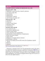
TABLE 93.1
CLASSIFICATION OF ANEMIA BY RED BLOOD CELL SIZE
Macrocytic (MCV >100)
• Megaloblastic anemia: folic acid or vitamin B12 deficiency
• Liver disease (target cells)
• Reticulocytosis
• Hypothyroidism
• Myelodysplastic syndrome
• Antiretrovirals, for example, AZT
Microcytic (MCV <80)
• Iron deficiency
• Thalassemia
• Anemia of chronic disease
• Sideroblastic anemia
• Lead poisoning
Normocytic (MCV 80–100)
• Low reticulocyte count
• Iron deficiency (especially early)
• Anemia of chronic disease
• Chronic renal disease (low erythropoietin)
• Hypothyroidism, adrenal insufficiency, or hypopituitarism
• Primary bone marrow disorder
• Aplastic anemia
• Malignancy, for example, leukemia, metastases
• Myelodysplastic syndromes
• Infection, for example, parvovirus B19 (causes red cell hypoplasia or aplastic
anemia)
• Blood loss
• Hemolysis
Adapted from Zeiger RF, McGraw-Hill’s Diagnosaurus 4.0. Available online at
/>
Alternatively, comparing the MCV level with age-appropriate normal values allows
for classification of anemia as macrocytic, normocytic, or microcytic ( Table 93.1 ). A
reticulocyte count indicates the reactivity of the marrow, and elevated levels are often
found in blood loss and hemolysis. Decreased levels of serum haptoglobin, elevated
lactate dehydrogenase, and increased unconjugated bilirubin along with the presence of
heme in the urine suggest hemolysis.
BLOOD LOSS
CLINICAL PEARLS AND PITFALLS
In acute hemorrhage, measured hemoglobin changes may lag behind blood
loss, and normal values should not provide reassurance against clinically
significant blood loss.
Clinical Considerations
Clinical Recognition
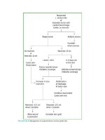
Anemia due to blood loss occurs from a variety of causes ( Fig. 93.1 ). Overall, these
conditions are divided into traumatic or atraumatic bleeding. The possibility of occult
nonaccidental trauma must always be considered, particularly in younger children.
Gastrointestinal hemorrhage is the most common cause of atraumatic blood loss, but
postsurgical (e.g., posttonsillectomy hemorrhage), renal, gynecologic, and other
etiologies may also present. In some cases, an anatomic lesion or process may combine
with a congenital or acquired coagulopathy to precipitate significant anemia. For
example, adolescent girls with unrecognized von Willebrand disease (VWD) may
present with anemia due to both acute and chronic blood loss during menses.
Assessment for anemia should be considered in any patient with pallor, jaundice, or
unexplained tachycardia. Asymptomatic chronic anemia may be detected as an
incidental finding that requires further evaluation.
Triage
Known or suspected anemia due to blood loss, hypotension, hypoxia, or evidence of
end-organ dysfunction is a medical emergency and warrants immediate intervention to
prevent progression to cardiopulmonary collapse. Patients with both acute and chronic
blood loss may become unstable. The chronicity of symptoms should not reassure the
clinician, as it may be the exhaustion of physiologic compensatory mechanisms that
prompts the patient to present to medical attention.
Initial Assessment
Initial assessment of a patient presenting with anemia secondary to blood loss includes a
focused history targeted at symptoms of compromise related to anemia/hypoxia as well
as potential etiologies. In suspected blood loss, the clinician must assess for evidence of
trauma including nonaccidental injury, postprocedure bleeding, symptoms of upper or
lower gastrointestinal bleeding, medication use that could precipitate gastrointestinal
bleeding, reports of epistaxis, hematuria or menorrhagia, and any concern for
complications of pregnancy or hemorrhagic ovarian cyst. Inquiries in the history related
to symptomatic anemia should include fatigue, exercise intolerance, syncope,
orthostasis, chest pain or dyspnea, decreased urine output, and any change in mental
status.
FIGURE 93.1 Causes of blood loss.
Assessment of hemodynamic parameters to identify signs of impending
cardiopulmonary collapse (e.g., severe tachycardia, hypotension, hypoxia) is critical,
remembering that hypotension is a late finding in shock in young children. Physical
examination should assess for location of blood loss and signs of systemic illness that
may cause anemia. Signs of end-organ dysfunction, such as change in mental status,
congestive heart failure, or renal insufficiency should be noted. In the trauma patient,
bleeding may be evident or occult, as in the case of femoral, pelvic, or abdominal
(including both intra- and retroperitoneal) hemorrhage. These may be hemodynamically
significant but not immediately obvious. The presence of trauma itself may be subtle in
nonaccidental injury. Consider gastrointestinal or gynecologic bleeding when the
etiology is unclear.
Diagnostic Testing
Laboratory testing for patients with suspected blood loss includes complete blood count
(CBC), reticulocyte count, coagulation studies, and a type and screen, or type and crossmatch if transfusion is anticipated. If the etiology of the anemia is unclear, obtain stool
guaiac for occult blood, hemolysis labs, and other studies as outlined below (see section
on Hemolytic Anemia). Send a pregnancy test if clinically indicated.
Management
Initial management steps for all patients include immediate vascular access,
cardiorespiratory monitoring, and administration of oxygen, regardless of oxygen
saturation to maximize oxygen-carrying capacity of the existing RBC mass and plasma.
Unstable patients require multiple sites of vascular access. Peripheral intravenous (IV)
catheters are typically more useful than central lines for rapid volume resuscitation;
intraosseous access may also be used. If transfusion is anticipated, the blood bank
should be notified promptly.
Trauma patients (see Chapter 7 A General Approach to the Ill or Injured Child )
require a multidisciplinary team to complete primary, secondary, and tertiary surveys.
Occult blood loss as discussed above should be considered in all trauma patients. The
use of ultrasound and FAST (focused assessment with sonography in trauma) may be
helpful in assessing for intra-abdominal hemorrhage in the multisystem trauma patient
(see Chapter 131 Ultrasound ). Management is simultaneously directed at immediate
hemodynamic stabilization and efforts to control blood loss through surgical and
catheter-guided embolization strategies as indicated. Patients in uncompensated shock
(hypotension, signs of end-organ dysfunction) require rapid fluid resuscitation via push–
pull, pressure bag, or rapid infuser. Crystalloid fluids are typically available most
quickly and should be used until O-negative blood is available. However, patients with
uncompensated shock due to hemorrhage ultimately require transfusion ( Table 93.2 ).
Hemoglobin changes typically lag behind acute blood loss and are not a useful guide to
initial management. Patients with isolated tachycardia but no other signs of end-organ
dysfunction such as altered mental status, decreased urine output, or poor perfusion
(compensated shock) should be managed based on the severity of the tachycardia,
anticipated trajectory of the blood loss, and the need for embolization or operative
management. Failure to respond to initial crystalloid resuscitation in these patients may
be an indication for transfusion. Traumatic hemorrhage may result in coagulopathy that
could worsen bleeding. Trauma patients requiring massive transfusion will need
additional blood product support with platelets and fresh-frozen plasma (see Table 93.2
) and careful monitoring for electrolyte derangements associated with these treatments.
Tranexamic acid (TXA) is a hemostatic agent used in adult patients presenting with
traumatic hemorrhage; while pediatric data are limited, it may be considered as an
adjunct in an unstable pediatric patient with significant acute hemorrhage.