Báo cáo y học: "Intrathecal siRNA against Toll-like receptor 4 reduces nociception in a rat model of neuropathic pain"
Bạn đang xem bản rút gọn của tài liệu. Xem và tải ngay bản đầy đủ của tài liệu tại đây (2.59 MB, 9 trang )
Int. J. Med. Sci. 2010, 7
251
I
I
n
n
t
t
e
e
r
r
n
n
a
a
t
t
i
i
o
o
n
n
a
a
l
l
J
J
o
o
u
u
r
r
n
n
a
a
l
l
o
o
f
f
M
M
e
e
d
d
i
i
c
c
a
a
l
l
S
S
c
c
i
i
e
e
n
n
c
c
e
e
s
s
2010; 7(5):251-259
© Ivyspring International Publisher. All rights reserved
Research Paper
Intrathecal siRNA against Toll-like receptor 4 reduces nociception in a rat
model of neuropathic pain
Fei-xiang Wu
1
, Jin-jun Bian
2
, Xue-rong Miao
1
, Sheng-dong Huang
3
, Xue-wu Xu
1
, De-jun Gong
3
, Yu-ming
Sun
1
, Zhi-jie Lu
1
, Wei-feng Yu
1
1. Department of Anesthesiology, Eastern Hepatobiliary Hospital, Second Military Medical University, Shanghai 200438,
China
2. Department of Anesthesiology, Changhai Hospital, Second Military Medical University, Shanghai 200433, China
3. Institute of Thoracic Cardiac Surgery, Changhai Hospital, PLA, Shanghai 200433, China
Corresponding author: Wei-feng Yu, Department of Anesthesiology, Eastern Hepatobiliary Hospital, Second Military
Medical University. Address: No. 225, Changhai Road, Shanghai 200438, P.R. China. E-mail: ,Tel:
86-21-65564166.
Received: 2010.07.19; Accepted: 2010.08.02; Published: 2010.08.02
Abstract
Background: Neuropathic pain is characterized by hyperalgesia, allodynia and spontaneous
pain. It often occurs as a result of injury to peripheral nerves, dorsal root ganglions (DRG),
spinal cord, or brain. Recent studies have suggested that Toll-like receptor 4 (TLR4) might
play a role in neuropathic pain. Methodology/Principal Findings: In this study, we inves-
tigated the role of TLR4 in a rat chronic constriction injury (CCI) model and explored the
feasibility of treating neuropathic pain by inhibiting TLR4. Our results demonstrated that in-
trathecal siRNA-mediated suppression of TLR4 attenuated CCI-induced mechanical allodynia
and thermal hyperalgesia through inhibiting the activation of NF-κB p65 and production of
proinflammatory cytokines (e.g., TNF-α and IL-1β). Conclusions/Significance: These
findings suggest that suppression of TLR4 mediated by intrathecally administered siRNA may
be a new strategy for the treatment of neuropathic pain.
Key words: Toll-like receptor 4; neuropathic pain; NF-κB; RNA interference; IL-1β; TNF-α.
Introduction
Neuropathic pain is characterized by hyperalge-
sia, allodynia and spontaneous pain. It often occurs as
a result of injury to peripheral nerves, dorsal root
ganglions (DRG), spinal cord, or brain. 7% to 8% of
the population suffer from neuropathic pain, and 5%
may be severely affected (1-2). Neuropathic pain re-
mains a prevalent and persistent clinical challenge
due to unknown pathogenesis. A variety of mechan-
isms have been proposed for the induction and/or
maintenance of neuropathic pain. Recently, investi-
gations have focused on the role of central nervous
system (CNS) immune responses after nerve injuries
that lead to behavioral hypersensitivity (3-5). A cur-
rent theory for the etiology of neuropathic pain in-
volves CNS immune activation with cytokine pro-
duction inducing the expression of final common pain
mediators such as TNF-α and IL-1β (6-8).
The Toll-like receptor 4 (TLR4) has recently been
implicated in chronic neuropathic pain (9-10). TLR4 is
a transmembrane receptor protein containing extra-
cellular domains with leucine-rich repeat and a cy-
toplasmic signaling domain. The role of TLR4 in in-
nate immune response has been well elucidated. The
binding of exogenous (e.g. Lipopolysaccharides, LPS)
or endogenous (e.g. members of heat shock protein
family and proteoglycans) ligands to TLR4 activates
Int. J. Med. Sci. 2010, 7
252
NF-κB and then releases proinflammatory cytokines
such as TNF-α, IL-1β and IL-6 (11-13). Previous stu-
dies have demonstrated that TLR4 is expressed in
microglia of CNS (14-16). Since microglial activation is
essential for the release of proinflammatory cytokines
(17), it is plausible that TLR4 might be a common
mediator through which different pain-inducing sig-
nals are linked to the production of proinflammatory
factors. Consistent with this notion,
N-methyl-D-aspartate (NMDA) receptor-modulated
innate immune responses were dependent on TLR4
(18), and mice with TLR4 deficiency demonstrated
decreased cytokine production and attenuated neu-
ropathic pain responses upon nerve injury (19).
In this study, we suppressed TLR4 expression
using siRNA in a rat CCI model. Knockdown of TLR4
in spinal cord inhibited pain response, and blocked
NF-κB activation and production of proinflammatory
cytokines (e.g. IL-1β and TNF-α).
Materials and methods
Ethics Statement
All animal experiments were approved by the
Administrative Committee of Experimental Animal
Care and Use of Second Military Medical University
(SYXK(Hu)2007-0003), and conformed to the National
Institute of Health guidelines on the ethical use of
animals.
Screening siRNA sequence with reporter vector
A scrambled sequence was designed as a mis-
match control (MM-siRNA) (5’-GGCGUGUCUCUCU
UACGAC-3”). SiRNAs targeting the cDNA sequence
of rat TLR4 (GenBank accession NM_019178) were:
5’-CUACCAACAGAGAGGAUAU-3” (siRNA1),
5’-GUCUCAGAUAUCUAGAUCU-3’ (siRNA2),
5’-GAGCCGGAAAGUUAUUGUG-3’ (siRNA3).
All siRNAs were chemically synthesized by
United Gene Company (Shanghai, China). The pri-
mers amplifying the full length cDNA of rat TLR4
were 5'-CGGGAGCTCTGAATGCTCTCTTGCATC
TGGCTGGC-3’ (forward) and 5'-CGGGTCGACGCG
ATACAATTCGACCTGCTG-3’ (reverse).
To construct a green fluorescent protein (GFP)
tagged TLR4 expressing vector, total RNA was ex-
tracted from rat lung tissues using Tri-Reagent (Ta-
KaRa, Japan). RT-PCR was used to obtain the full
length TLR4 fragment. After pEGFPC1 vector was
linearized by SacⅠ and SalⅠ, the fragment of TLR4
was inserted to construct the reporter vector,
pEGFPC1-TLR4. The reporter vector was verified by
RT-PCR using primers 5'-CGGGAGCTCTGAA
TGCTCTCTTGCATCTGGCTGGC-3’ and 5'-CGGGT
CGACGCGATACAATTCGACCTGCTG-3’.
To identify the knockdown efficacy of different
siRNA oligonucleotides, HEK-293 cells were cotrans-
fected with pEGFPC1-TLR4 and siRNA (iRNA1-3,
respectively) with lipofectamine2000 (Invitrogen,
USA). EGFP expression was observed under an in-
verted fluorescence microscope and the fluorescence
intensity was quantified by flow cytometry.
To identify the knockdown efficacy of different
siRNA oligonucleotides, HEK-293 cells were cotrans-
fected with pEGFPC1-TLR4 and siRNA (iRNA1-3,
respectively) using lipofectamine2000 (Invitrogen,
USA). An oligonucleotide sequence with no homolo-
gy to the sequence of TLR4 was used as a mismatch
controls. After 48 h, cells were visualized under an
inverted fluorescence microscope and the inhibitory
effects of different siRNA oligonucleotides were de-
termined by measuring EGFP expression using flow
cytometry.
Animals and chronic constriction injury
Male Sprague-Dawley rats (200-250g) were
purchased from Shanghai Experimental Animal Cen-
ter, Chinese Academy of Sciences. The chronic con-
striction injury (CCI) model was established as pre-
viously described (20). Briefly, rats were anesthetized
with sodium pentobarbital (40 mg/kg, i.p.). The
common sciatic nerve was exposed at the mid-thigh
level. The nerve was ligated loosely with 4-0 chromic
gut thread at 4 sites with an interval of 1 mm, so that
the nerve diameter was only slightly reduced.
Meanwhile, a sham surgery was performed with the
sciatic nerve exposed but not ligated. Upon recovery
from anesthesia, animals were housed individually in
clear plastic cages with the floor covered by 3-6 cm of
sawdust.
Lumbar subarachnoid catheterization
One week prior to CCI, a chronic indwelling
catheter was implanted into the subarachnoid space
of each rat. Briefly, rats were anesthetized with so-
dium pentobarbital (40 mg/kg, i.p.). A PE-10 catheter
(Becton Dickinson, Sparks, MD, USA) was inserted
into the lumbar subarachnoid space between 5
th
and
Int. J. Med. Sci. 2010, 7
253
6
th
lumbar vertebrae (L5) and L6 (21). The catheter
was chronically implanted and the external portion of
the catheter was protected according to Milligan’s
method (22).
Intrathecal delivery of siRNA
Rats were randomly divided into four groups
with 10 rats in each group: a sham group (Sham sur-
gery + Normal saline, NS), a CCI group (CCI + NS), a
MM group (CCI + MM siRNA), and a siRNA group
(CCI + TLR4-siRNA). 10 μg SiRNA dissolved in 30 μl
i-Fect transfection reagent (Neuromics, Edina, MN,
USA) was administered intrathecally once daily for 7
days, starting from 1 day before CCI surgery.
Evaluation of tactile allodynia and thermal
hyperalgesia
The paw withdrawal latency (PWL) to radiant
heat and paw withdrawal threshold (PWT) were used
to evaluate thermal hyperalgesia and mechanical al-
lodynia respectively as previously described (23-24).
To measure PWL, rats were placed in an inverted
clear plexiglass cage (23×18×13 cm) on a piece of
3-mm-thick glass plate and allowed to acclimate to
their surroundings for 30 minutes before testing. After
acclimation, the radiant heat source was positioned
under the glass floor directly beneath the hind paw.
The radiant heat source consisted of a high-intensity
projection lamp bulb (8V, 50W), located 40 mm below
the glass floor and projecting through a 5×10-mm
aperture in the top of a movable case. A digital timer
automatically recorded the duration between the start
of stimuli and the paw withdrawal (PWL). Three trials
were carried out in each rat with a 5-minute interval.
The cut-off was set at 20 seconds to avoid tissue
damage.
Mechanical allodynia was assessed with von
Frey filaments. Rats were placed on a wire mesh
platform, covered with a transparent plastic dome,
and allowed to acclimate for 30 minutes before test-
ing. The filament was applied perpendicularly to the
plantar surface of the hind paw (ipsilateral to the side
of CCI). The paw withdrawal threshold (PWT) was
determined by sequentially increasing and decreasing
the stimulus intensity (the ‘up-and-down’ method) (in
gram, g), and data were analyzed using the nonpa-
rametric method of Dixon (24). Tests were performed
1 day before CCI surgery, and 1, 3, 7, 10 and 14 days
after CCI surgery.
Enzyme linked immunosorbent assay (ELISA)
Dorsal spinal cord tissue and cerebrospinal fluid
(CSF) samples were prepared as previously described
(25). IL-1β and TNF-α in spinal cord tissues and CSF
were detected by ELISA (Peprotech, UK).
Spinal cord RNA extraction and real time PCR
Total RNA was extracted from L4–L5 spinal cord
tissues. Extracted RNA was pretreated with DNaseⅠ
at 37Ԩ for 30 minutes before reverse transcription
reaction was performed using a high capacity cDNA
archived kit (TaKaRa, Japan). A Real-Time PCR De-
tection System (Roche, Switzerland) was used to con-
tinuously monitor the intensity of fluorescence, which
was directly proportional to the PCR products.
Western blotting
Nuclear extracts were prepared from lumbar
spinal cord (L4-L5) tissues as previously described
(26). Proteins were separated on an 8% polyacryla-
mide SDS-PAGE gel and transferred onto a nitrocel-
lulose membrane. The nitrocellulose membrane was
blotted with a primary antibody recognizing the p65
subunit of NF-κB (1:100, Santa Cruz, CA, USA), fol-
lowed by a secondary antibody conjugated with
horseradish peroxidase. Protein signals were detected
with an ECL system (Amersham Pharmacia Biotech,
Uppsala, Sweden). Histone3 (Sigma, St. Louis, MO,
USA, 1:500) was used as an internal control.
Statistical analyses
Data are expressed as mean ± standard deviation
(SD). Statistical analyses were performed using Stu-
dent's t-test or multiple ANOVA followed by
least-significance difference post-hoc comparison.
P<0.05 was considered statistically significant.
Results
Identification of pGEFPC1-TLR4
The TLR4 fragment of 2376 bp was successfully
obtained by PT-PCR. Restriction analysis and se-
quencing results demonstrated that the recombinant
pEGFP-TLR4 indeed contained the TLR4 fragment.
Silencing of TLR4 transgene with siRNA in
HEK-293 cells
In order to select a siRNA oligonucleotide for an
efficient knockdown of TLR4, three siRNA oligonuc-
leotides targeting the rat TLR4 were used to cotrans-
fect pEGFPC1-TLR4 in HEK-293 cells in vitro and then
the level of TLR4 transgene expression was evaluated
by GFP fluorescence (Figure 1A). Meanwhile, the rel-
ative fluorescence intensity was also detected by flow
cytometry (Figure 1B). Flow cytometry and fluoresce
observation revealed that all 3 siRNAs demonstrated
effective inhibition on GFP fluorescence, and
TLR4-siRNA2 was the most potent. Therefore,
TLR4-siRNA2 was used for further in vivo study.
Int. J. Med. Sci. 2010, 7
254
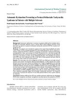
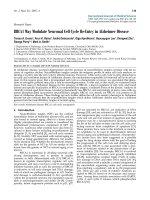
Figure 1. Screening siRNA for an efficient suppression of TLR4 expression in vitro. HEK-293 cells were co-transfected with
both pEGFRC1-TLR4 and either one of three independent siRNA oligonucleotides targeting TLR4 (TLR4-siRNA1-3) or a
control siRNA (MM-siRNA). Two days after transfection, EGFP fluorescence was observed under microscope (A) or
quantified by flow cytometry (B). (A) EGFP fluorescence under an inverted fluorescence microscope (×100) or cell density
under an optical microscope (×100). A, control; B, siRNA1; C, siRNA2; D, siRNA3. (B) The quantification of TLR4-EGFP
fluorescence intensity upon siRNA knockdown was evaluated by flow cytometry analysis. Immunofluorescence and flow
cytometry results revealed that all 3 siRNAs had efficient inhibition on GFP fluorescence, and TLR4-siRNA2 was the most
potent.
Effects of TLR4-siRNA on TLR4 and its down-
stream signaling in CCI rats
Real time RT-PCR showed a significant
up-regulation of TLR4 mRNA expression 1 day after
CCI compared to the sham group (P=0.0000). The
siRNA-TLR4 decreased TLR4 mRNA expression and
continued for 7 days (P=0.0003). However, there was
no significant difference in TLR4 mRNA expression
between 10-14 days after CCI (Figure 2A, 2B). The
TLR4 protein expression in spinal cord tissues was
detected by Western blotting. No statistical difference
was found between CCI group and MM siRNA group
for nuclear TLR4 protein expression (P=0.6062). Inte-
restingly, activation of NF-κB p65 was also blocked by
the TLR4-siRNA treatment (P=0.0070) (Figure 2C).
Thus, the TLR4 mRNA and protein in spinal cord
tissues were decreased by siRNA-TLR4, and inhibi-
tory effects of siRNA on TLR4 expression were con-
firmed at the mRNA and protein levels.
TNF-α and IL-1β were up-regulated in the dorsal
spinal cord tissues of CCI rats, and there were no sig-
nificant differences in TNF-α and IL-1β between the
Int. J. Med. Sci. 2010, 7
255
CCI group and MM group (P>0.05). However, com-
pared with the MM group, the production of TNF-α
and IL-1β in spinal cord tissues was significantly
lower in the CCI group during the course of
TLR4-siRNA treatment, indicating that intrathecal
administration of TLR4-siRNA significantly atte-
nuated TLR4 induction in the CCI rats (Figure 3).
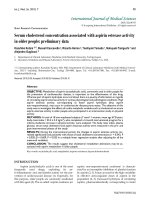
Figure 2. Inhibition of TLR4 signaling upon TLR4-siRNA in CCI rats. Either saline or 10μg of selected siRNA was admi-
nistered intrathecally once daily for 7 days as described in Materials and Methods. Tissue biopsy was performed from lumbar
L4-L5 spinal cord tissues at indicated time points as described. A RT-PCR analyses of TLR4 mRNA expression in rat lumbar
spinal cord tissues in four groups one day after CCI. Maker, DL200; Lane 1, CCI group; Lane 2, MM group; Lane 3, siR-
NA-TLR4 group; Lane 4, sham group. B Real-time quantitative RT-PCR analyses of TLR4 mRNA expression in rat lumbar
spinal cord tissues in four groups (* P<0.05 VS MM group), sham group (Sham surgery + NS), CCI group (CCI + NS), MM
group (CCI + MM siRNA), siRNA group (CCI + TLR4-siRNA). C Western blotting showed the levels of NF-κB P65 protein
in spinal cord of rat tissues in four groups. Interestingly, the expression of κB p65 protein in the TLR4-siRNA treatment
group was significantly lower than the MM group (P=0.0070).