Báo cáo y học: "Endovascular Treatment of Bilateral Carotid Artery Occlusion with Concurrent Basilar"
Bạn đang xem bản rút gọn của tài liệu. Xem và tải ngay bản đầy đủ của tài liệu tại đây (543.54 KB, 7 trang )
Int. J. Med. Sci. 2011, 8
263
I
I
n
n
t
t
e
e
r
r
n
n
a
a
t
t
i
i
o
o
n
n
a
a
l
l
J
J
o
o
u
u
r
r
n
n
a
a
l
l
o
o
f
f
M
M
e
e
d
d
i
i
c
c
a
a
l
l
S
S
c
c
i
i
e
e
n
n
c
c
e
e
s
s
2011; 8(3):263-269
Case Report
Endovascular Treatment of Bilateral Carotid Artery Occlusion with Con-
current Basilar Apex Aneurysm: A Case Report and Literature Review
Kan Xu
1,*
, Honglei Wang
1,*
, Qi Luo
1
, Ye Li
2
, Jinlu Yu
1,
1. Department of Neurosurgery, First Hospital of Jilin University, Changchun, 130021, PR China
2. Department of Radiology, First Hospital of Jilin University, Changchun, 130021, PR China
* Kan Xu and Honglei Wang contributed equally to the work.
Corresponding author: Jinlu Yu, +86043188782331, E-mail:
© Ivyspring International Publisher. This is an open-access article distributed under the terms of the Creative Commons License (
licenses/by-nc-nd/3.0/). Reproduction is permitted for personal, noncommercial use, provided that the article is in whole, unmodified, and properly cited.
Received: 2011.03.04; Accepted: 2011.03.23; Published: 2011.03.30
Abstract
We report a case of successful endovascular treatment of bilateral carotid artery occlusion
with concurrent basilar apex aneurysm. An elderly female patient with subarachnoid hem-
orrhage (SAH) onset was admitted to the hospital. Computed tomography (CT) and digital
subtraction angiography (DSA) confirmed the presence of bilateral carotid artery occlusion
with concurrent basilar apex aneurysm. Brain blood supply was provided by the bilateral
vertebral artery through the basilar artery. We treated the aneurysm with the endovascular
approach by embolizing the aneurysm with three coils. The patient recovered well after
surgery and showed no recanalization of the aneurysm on a one-year follow-up DSA. We also
reviewed six similar cases found with a PUBMED database search (1980-2010), including
those with bilateral common carotid artery occlusion. In conclusion, by using the endovas-
cular approach, bilateral carotid artery occlusion with concurrent basilar apex aneurysm was
efficiently treated.
Key words: carotid artery occlusion, basilar apex aneurysm, endovascular treatment
1. Introduction
Bilateral carotid artery occlusion with concurrent
basilar apex aneurysm is extremely rare
1
. When it
occurs, the brain blood supply mainly relies on the
vertebral artery through the basilar artery. The un-
natural reliance on this route is such that the pressure
inside the apex of the basilar artery makes it vulnera-
ble to aneurysm
2-4
. These need to be treated quickly,
as they may cause hemorrhaging and subsequent
death
3,5
.
Craniotomy is one approach to treat patients
with basilar apex aneurysm. However, in the case of a
bilateral carotid artery occlusion, the increased blood
pressure in the basilar artery leads to higher risks in
the surgical clipping of aneurysms
3,6
. The alternative
approach is the endovascular treatment that has been
developed since 1991
7,8
. It is performed with the
guidance of DSA, for accurate localization during
surgery with minimal tissue damage.
Here we report a case of successful treatment of
bilateral carotid artery occlusion with concurrent bas-
ilar apex aneurysm using the endovascular approach.
In addition, we reviewed six similar cases found
through a PUBMED database search for the years
1980-2010, including cases with bilateral common
carotid artery occlusion. These cases further sup-
ported the application of endovascular treatment for
bilateral carotid artery occlusion with concurrent bas-
ilar apex aneurysm.
Int. J. Med. Sci. 2011, 8
264
2. Case Report
The female patient aged 69 was admitted to the
hospital after reporting a sudden headache accompa-
nied by nausea and vomiting for ten days. The patient
had a history of hypertension for 4 years and diabetes
for 10 years, which were well-controlled with anti-
hypertensive and oral hypoglycemic medication.
Upon admission to the hospital, the patient presented
with Hunt-Hess grade III and positive Kernig’s signs.
CT scan showed that the hemorrhage was localized
on the pontine cistern and interpeduncular cistern,
extending to the right of the ambient cistern into the
posterior horn of the right ventricle. The patient was
diagnosed with SAH, diabetes and hypertension.
CTA showed an aneurysm at the apex of the
basilar artery with a diameter of 3.2 mm. There was
no signal at the bilateral internal carotid artery, and
the bilateral posterior communicating artery was
supplying the anterior circulation. This result led to a
diagnosis of bilateral carotid artery occlusion with
concurrent basilar apex aneurysm. DSA showed that
the bilateral internal carotid artery was occluded from
the beginning of the bifurcation, with the external
carotid artery system developed and no signs of
anastomosis or vascular reconstruction of the
branches of the external carotid and intracranial ar-
teries. The brain blood supply mainly relied on the
vertebral artery through the bilateral posterior com-
municating arteries. The angiograph of the vertebral
artery showed no delay in the blood flow of anterior
circulation. The saccular aneurysm with a diameter of
3.2 mm was observed at the apex of the basilar artery.
Under general anesthesia, three coils [3 mm × 5
cm Morpheus 3D CSR (Ev3), 2 mm × 1 cm Morpheus
3D CSR (Ev3), and 2 mm × 1 cm Helical (MicroVen-
tion)] were used to embolize the aneurysm, and the
patient recovered well. After one year, DSA showed
no aneurysm recanalization.
3. Literature review
We reviewed 6 similar cases of bilateral carotid
artery occlusion with concurrent basilar apex aneu-
rysm found with a PUBMED search for the years 1980
to 2010
3,5,9-12
. These studies included cases of bilateral
common carotid artery occlusion. The clinical data is
summarized in Table 1. The following is a summary
of the studies.
Table 1. Clinical data for all cases in this study.
Case Study Year Age/
gender
Cause Position of
Occlusion
Symptom Other aeurysms Collateral
circulation
Treatment Outcome
1 Kumagai et
al.
9
1981 41/F aortitis Begnning
of the
CCA
SAH PCA VA→ECA→IC
A
Clipping (BA
aneurysm)+
Clipping (others)
Good Re-
covery
2 Matsuzawa
et al.
5
1982 54/F aortitis Begnning
of the
CCA
SAH No Subclavian ar-
tery
→CCA→ICA
Conservative Death (re-
rupture)
3 Yamanaka
et al.
3
1987 71/F Arterio-
sclerosis
Begnning
of the ICA
SAH No ECA→ICrA Conservative Death (re-
rupture)
4 Ishibashi et
al.
10
1993 63/M Arterio-
sclerosis
Left
cavernous
sinus part
of the ICA,
right
beginning
of the ICA
Hydrocephalus No No VP shunt+ Con-
servative
Good Re-
covery
5 Konishi et
al.
11
1998 39/F Arterio-
sclerosis
Begnning
of the ICA
Thalamic he-
matoma
SCA+AICA+PIC
A
ECA→ICrA Coiling (basilar
apex aneurysm)+
Clipping (others)
Good Re-
covery
6 Meguro et
al.
12
2008 62/F Arterio-
sclerosis
Begnning
of the
CCA
SAH PCA VA→ECA→IC
A
Coiling assisted
by balloon (BA
aneu-
rysm+others)
Good Re-
covery
7 Present
case
2010 69/F Arterio-
sclerosis
Begnning
of the ICA
SAH No No Coiling Good Re-
covery
Abbreviations: F, female; M, male; SAH, subarachnoid hemorrhage; BA, basilar apex; VA, vertebral artery; ECA, external carotid artery;
ICA, internal carotid artery; ICrA, intracranial artery; CCA, common carotid artery
Int. J. Med. Sci. 2011, 8
265
3.1 General information.
1) Six patients (5 female, one male, aged 39 to 71
years old with a mean age of 55 years). 2) The causes
of carotid artery occlusion included aortic inflamma-
tion in two cases and atherosclerosis in four cases. 3)
Upon admission to hospital, there were 4 cases with
SAH, one case with thalamic hemorrhage and one
case with hydrocephalus.
3.2 Radiology features.
1) Location of occlusions: the beginning part of
the common carotid artery in 3 cases, the beginning
part of the internal carotid artery in 2 cases, the cav-
ernous sinus segment at left and beginning part at
right of the internal carotid artery in one case. 2) An-
eurysm: all cases were saccular; 3 cases single; 3 cases
combined with aneurysms in other regions. 3) Collat-
eral circulation: in 2 cases, the vertebral artery to the
external carotid artery to anastomosis of the internal
carotid artery; in one case, the subclavian artery to the
common carotid artery to anastomosis of the internal
carotid artery; in 2 cases, the external carotid artery to
anastomosis of the intracranial artery; in one case, no
anastomosis.
3.3 Treatment.
(1) Basilar apex aneurysm: conservative treat-
ment in 3 cases, clipping treatment by craniotomy in
one case, and endovascular treatment in 2 cases; (2)
Other concurrent aneurysms in the 3 cases: clipping in
2 cases, embolization in one case; (3) one case of hy-
drocephalus was treated with a ventriculoperitoneal
shunt.
3.4 Treatment results.
1) In the 3 cases of conservative treatment, 2
cases died of rupture, and the hydrocephalus case
showed good prognosis; 2) The case treated by clip-
ping showed good prognosis; (3) One case treated by
embolization showed good prognosis; (4) One case
treated by embolization of the basilar apex aneurysm
and clipping to the concurrent aneurysm showed
good prognosis.
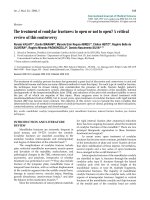
Figure 1. A: Head CT scan shows that the hemorrhage was localized on the pontine cistern and interpeduncular cistern,
extending to the right of the ambient cistern, into the posterior horn of the right ventricle. The patient was diagnosed with
subarachnoid hemorrhage (SAH). B: Head CT angiograph shows mound-like protuberances at the apex of the basilar artery
with a diameter of 3.2 mm, no signal at the bilateral internal carotid artery, and bilateral posterior communicating artery
supplying the circulation.
Int. J. Med. Sci. 2011, 8
266
Figure 2. Common carotid artery DS angiographs: occlusion at the beginning of internal carotid artery, with the remaining
external carotid artery. No formation of anatomosis between the external carotid artery and intracranial vessels is
observed. A, B: The right common carotid artery; C,D: The left common carotid artery.
Figure 3. A,B: Angiograph of the vertebral artery showing developed posterior circulation with blood supply through the
bilateral posterior communicating artery. No delay was observed in the anterior circulation angiograph, and from (B) a
basilar apex aneurysm of about 3.2 mm could be observed.
Int. J. Med. Sci. 2011, 8
267
Figure 4. A, B: DS angiographs taken after the aneurysm coil embolization. The aneurysm with dense embolization is not
seen.
Figure 5. A,B: One year after embolizing the aneurysm with the endovascular approach, embolization was still in good
condition, without recanalization.
4. Discussion
Bilateral carotid artery occlusion is rare but has
gained increased attention due to the improvement in
diagnostic techniques in recent years, especially in
non-invasive imaging
13,14
. Following the development
of a bilateral carotid artery occlusion, the posterior
circulation bears the brunt of the brain blood supply,
increasing blood pressure as well as the risk of saccu-
lar aneurysms
2,3
. The basilar artery supporting the
whole brain blood flow under high pressure has made
treatment relatively difficult
7
.
Two cases of bilateral carotid artery occlusion
with concurrent basilar apex aneurysm treated con-
servatively in the 1980’s failed and led to patient
deaths due to rerupture of the aneurysms
3,5
. Even
when the aneurysm is distant from the apex of the
internal carotid artery and located in the posterior
cerebral and communicating arteries, conservative
therapy can fail
2
. On the other hand, in craniotomy
with clipping of these aneurysms the transient occlu-
sion of the parent arteries on the cerebral perfusion
could lead to serious detrimental results
3,6
. In contrast
to these two treatment modes, the endovascular ap-
proach developed in recent years has fewer risks and
significantly less trauma to patients, as demonstrated
in previous studies as well as the present study.
Occlusion of the bilateral internal and common
carotid arteries can be caused by many factors, in-