Báo cáo y học: "An unusual case of gout in the wrist: the importance of monitoring medication dosage and interaction. A case report"
Bạn đang xem bản rút gọn của tài liệu. Xem và tải ngay bản đầy đủ của tài liệu tại đây (251.71 KB, 5 trang )
BioMed Central
Page 1 of 5
(page number not for citation purposes)
Chiropractic & Osteopathy
Open Access
Case report
An unusual case of gout in the wrist: the importance of monitoring
medication dosage and interaction. A case report
Craig L Jacobs*
†
and Paula J Stern
†
Address: Graduate Education and Research Programs, Canadian Memorial Chiropractic College, 6100 Leslie St., Toronto, ON M2H 3J1, Canada
Email: Craig L Jacobs* - ; Paula J Stern -
* Corresponding author †Equal contributors
Abstract
Background: Gouty arthritis of the wrist is uncommon although gout itself is the most common
inflammatory arthritis in older patients. Some known risk factors for the development of gout
include trauma, alcohol use, obesity, hyperuricaemia, hypertension and diabetes mellitus. As well,
certain medications have been shown to promote the development of gout. These include thiazide
diuretics, low dose salicylates and cyclosporine. We present a case of gouty wrist pain possibly
precipitated by a medication dosage increase as well as medication interactions.
Case presentation: A 77 year old male presented with right wrist pain. Redness and swelling was
present at the dorsal aspect of his wrist and range of motion was full with pain at end range upon
examination. One week prior, his anti-hypertensive medication dosage had been increased. The
patient's situation continued to worsen. Radiographic examination revealed changes consistent
with gouty arthritis.
Conclusion: It is important for clinicians treating joint conditions to be aware of patients'
comorbidities, medication usage and changes in dosages. Education of patients with gout is of prime
importance. Clinicians should educate patients that gout may occur at any joint in the body not only
the lower limb. Patients should be aware of the signs and symptoms of an acute gouty attack and
be made aware that changes in certain medication dosages may precipitate an attack. Awareness
of radiographic changes associated with gout is still of importance although these changes are not
seen as frequently as they have been in the past due to better control of the disease.
Background
Joint pain accompanied with swelling is a common com-
plaint seen in clinical practice. The challenge is to deter-
mine the underlying etiology and to provide the
appropriate treatment. Many joint diseases present as
acute monoarthritis with the most common causes due to
gout or calcium pyrophosphate dihydrate crystal deposi-
tion disease (CPPD) [1]. The peak incidence of gout is
between the ages of 30–50 with the prevalence increasing
with age [2]. Both the incidence and prevalence of gout
has been on the rise in recent years [3]. The increased prev-
alence is believed to be related to several factors which
include increased age and obesity in the population and
widespread diuretic use for hypertension treatment [3,4].
Gout is five times more common in men. Most acute
gouty attacks occur in a single joint in the lower limb with
the first metatarsal joint most commonly affected [2,5].
On clinical presentation, the joint often appears red, swol-
len and very tender. Some differentials to keep in mind
Published: 9 October 2007
Chiropractic & Osteopathy 2007, 15:16 doi:10.1186/1746-1340-15-16
Received: 8 May 2007
Accepted: 9 October 2007
This article is available from: />© 2007 Jacobs and Stern; licensee BioMed Central Ltd.
This is an Open Access article distributed under the terms of the Creative Commons Attribution License ( />),
which permits unrestricted use, distribution, and reproduction in any medium, provided the original work is properly cited.
Chiropractic & Osteopathy 2007, 15:16 />Page 2 of 5
(page number not for citation purposes)
include septic arthritis, rheumatoid arthritis, osteoarthri-
tis and erosive arthritis [1]. Some known risk factors for
the development of gout include trauma, alcohol use,
obesity, hyperuricaemia, hypertension and diabetes mel-
litus [2,5]. As well, certain medications have been shown
to promote the development of gout. These include thi-
azide diuretics, low dose salicylates and cyclosporine
[2,5].
We present an unusual case of gouty wrist pain possibly
precipitated by a medication dosage increase as well as
medication interactions.
Case presentation
A 77 year old male was treated at a chiropractic clinic for
low back pain resulting from lumbar facet arthrosis and
lateral canal stenosis. On a subsequent visit he reported
right wrist pain which began while lifting a heavy box. On
examination, redness and swelling was noted on the dor-
sal aspect of his right wrist. Range of motion was full with
pain at end range of flexion and extension. His health his-
tory included two hip replacements, two previous epi-
sodes of gout in both first metatarsophalangeal joints (2
and 5 years prior), and hypertension. Medications for
hypertension included perindopril (4 mg), hydrochloro-
thiazide (25 mg), and Norvasc (10 mg). In addition, he
was prescribed 80 mg of aspirin/day and took a daily mul-
tivitamin. One week prior, the patient's general practi-
tioner had increased his Norvasc dosage and also
prescribed Tylenol 3 to be taken as needed for his back
pain.
Two days later the swelling had increased in the dorsal
aspect of his right wrist and hand. Wrist flexion was lim-
ited by 80% with severe pain. Pain was present on palpa-
tion of the scaphoid bone. Due to the suspicion of
fracture, the patient was referred to his general practi-
tioner for radiographs. The radiologist who read the films
described tiny cysts at the distal radius and concluded that
these were most likely due to old trauma. Mild osteoar-
thritic changes were noted at the carpal-metacarpal joint
at the base of the thumb. The report stated that the radio-
graphs were otherwise normal.
The patient's symptoms worsened with increased pain
and swelling over the next few days. Due to the worsening
symptoms, repeat radiographs were performed five days
later and were read by a radiologist. The radiographs
revealed well-marginated juxta-articular bony erosions at
the radial styloid process and the dorsal rim of the distal
radius with soft tissue swelling. These findings were
deemed to be consistent with gouty arthritis. (See figure
1).
The patient was referred back to his general practitioner
with the radiologist's report and the patient was immedi-
ately put on colchicine. At follow up two days later, the
wrist swelling had decreased significantly. One week after
the initiation of colchicine, no swelling was present and
only mild pain was noted with flexion/extension of his
wrist and fingers. The patient was taken off colchicine due
to diarrhea. A recurrence of wrist pain occurred several
weeks later. The patient was referred to a rheumatologist
who prescribed colchicine for one month duration. When
the pain resolved completely, the patient was prescribed
allopurinol. Anti-hypertensive medications were not
altered. At one year follow up, no further gouty attacks
had been reported.
Discussion
A review of the literature reveals that gouty arthritis of the
wrist is rare in isolation although gout itself is the most
common inflammatory arthritis in older patients [4,6-9].
Gout at the wrist as the initial appearance of the condition
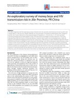
PA view of right wrist reveals a subchondral cyst in the distal radius (black arrow) and a well-marginated juxta-articular bony erosion at the radial styloid process (white crossed arrow)Figure 1
PA view of right wrist reveals a subchondral cyst in the distal
radius (black arrow) and a well-marginated juxta-articular
bony erosion at the radial styloid process (white crossed
arrow).
Chiropractic & Osteopathy 2007, 15:16 />Page 3 of 5
(page number not for citation purposes)
occurs between 0.8 to 2% of all gout cases [9]. Gout
patients who are not treated have a 19–30% chance of
developing gout in the wrist during their lifetime [9].
Reported cases of carpal tunnel syndrome, tendon entrap-
ment or rupture, and scapholunate dissociation have been
reported in the literature due to tophaceous deposits in
the wrist due to gout [6,8,9]. The prevalence of gout in the
USA ranges between 0.5–2.8% in men and 0.1–0.6% in
women [2]. The prevalence rises to 4.4% of men and 1.8%
of women over the age of 65 [4]. A two-fold increase in
incidence of gout has been reported in the USA and New
Zealand over the past 30 years, while the prevalence of
gout has been reported to have risen three-fold in the UK
over 20 years of follow-up [10]. This rise may be attrib-
uted in part to the continuous aging of the population as
well as the widespread use of diuretics for treatment of
hypertension [4].
Gout is a clinical syndrome caused by the deposition of
monosodium urate monohydrate crystals into synovial,
bursal, and cartilaginous tissue. The underlying metabolic
disorder is hyperuricemia. The exact trigger of an acute
attack of gout is poorly understood however predictors for
the development of gout in hyperuricemic individuals
have been identified. These include: increasing uric acid
level, alcohol consumption, hypertension, use of diuretic
drugs (thiazides and loop diuretics), increased body mass
index, and family history of gout [4,5,11]. These predic-
tors appear to have an additive effect on the risk of devel-
oping gout [11].
Hyperuricemia results from either decreased renal excre-
tion (which occurs in 90% of gout patients) or hyperpro-
duction of uric acid [4]. Drugs that may cause
hyperuricemia and gout include: diuretics, cyclosporine,
low-dose aspirin, ethambuthol, pyrzinamide, and nico-
tinic acid [4]. As some are commonly prescribed medica-
tions, it is imperative that health care practitioners dealing
with joint and musculoskeletal conditions be aware of the
medications that their patients are taking. In addition,
they should be prudent to be made aware of any changes
in prescribed dosages. In this case, the patient was taking
diuretics as well as other anti-hypertensive medications,
aspirin, and vitamin B3 (nicotinic acid).
A retrospective cohort study found a substantially
increased risk of receiving treatment for gout among eld-
erly hypertensive patients who were prescribed thiazide
diuretics when compared to those subjects who were
receiving non-thiazide antihypertensive medications [12].
The thiazide diuretic therapy subjects were almost twice as
likely to have undergone anti-gout therapy. This risk
increased even more so when thiazide diuretics were com-
bined with any other non-thiazide antihypertensive med-
ication. In this case, the patient's non-thiazide
antihypertensive medication was increased. This change
in blood chemistry may have contributed to the precipita-
tion of the acute gouty attack however the exact trigger of
this acute gouty attack cannot be determined and is most
likely multifactorial. The mechanisms by which diuretics
contribute to elevated serum uric acid levels are: decreased
filtration of uric acid, increased reabsorption, as well as
decreased secretion [12]. Ene-Stroescu and Gorbien state
that due to these mechanisms, diuretics are the most com-
mon cause of secondary gout with diuretic use being
reported in over 75% of patients with late-onset gout and
even approaching 100% in women [4]. The amount of
risk is also related to dosage. Thiazide diuretic dosages less
than 25 mg/day did not have a significant increase in risk,
whereas dosages ≥25 mg/day had a relative risk of
between 2.10–2.16 [12]. In this case, the patient was tak-
ing a dosage of 25 mg/day and thus had an increased risk
of gouty attack. Gurwitz et al state that low doses of thi-
azide diuretics can be just as efficacious as larger doses
with a reduced risk of metabolic disturbances. Doses as
low as 6.25 mg can be effective when combined with
another low dose anti-hypertensive medication [12].
Aspirin is prescribed widely in the elderly. Given that aspi-
rin is attainable without prescription, this leads to prob-
lems with self-prescription and dosage issues. Low-dose
aspirin, up to 2 g/day has the potential to increase uric
acid retention [11,13]. The combination of low-dose aspi-
rin and diuretics compounds this effect [13]. Clinicians
should inquire regarding aspirin usage in patients due to
this widespread and often unmonitored use.
The three clinical stages of gout are: acute gouty arthritis,
intercritical gout, and chronic tophaceous gout. Acute
gouty arthritis refers to acute inflammation due to the pre-
cipitation of urate crystals within a joint. Gouty attacks
may be precipitated by trauma, starvation, surgery, inges-
tion of high purine content food, excessive alcohol intake,
and drugs that affect urate concentration [4]. It should be
noted that drugs that reduce urate concentration may also
precipitate a gouty attack [4]. Initial attacks most com-
monly occur in the lower limbs and are usually monoar-
ticular with up to 50–60% occurring at the first
metatarsophalangeal joint [2,4,14]. Gout may occur in
any joint including the ankle, knee, hand, wrist, elbow,
sacroiliac joint and other joints of the spine, however
most commonly occurs in the lower extremity. In this
case, although the patient had suffered two previous gouty
attacks in the first metatarsophalangeal joint he was una-
ware that gout could occur in his wrist.
Typical presentation includes sudden onset of intense
pain, redness and swelling of the joint. Examination will
reveal a red, swollen, and extremely tender joint. Natural
history of an acute attack ranges from a few days to a few
Chiropractic & Osteopathy 2007, 15:16 />Page 4 of 5
(page number not for citation purposes)
weeks. Radiographs during early attacks may only reveal
soft-tissue swelling. Serum uric acid levels may be normal
during an attack due to pro-inflammatory cytokines [5].
The majority of untreated patients will experience another
acute attack within 2 years [4]. Prophylactic treatment is
usually recommended in patients who have more than 2–
3 gouty attacks per year [5]. Recent studies have advocated
the avoidance of diuretics, weight gain and alcohol con-
sumption. A low carbohydrate, high protein and unsatu-
rated fat diet has also been recommended as it enhances
insulin sensitivity and may reduce serum uric acid levels
[13].
Patients who experience multiple attacks of acute gouty
arthritis are predisposed to the development of polyartic-
ular gouty arthritis [14]. Attacks can then occur in more
than one joint simultaneously, especially in the lower
extremity. This emphasizes the unusual presentation in
this case of an isolated attack of gout in the wrist. Acute
onset of polyarticular gouty arthritis is more frequently
seen in older patients most of whom are receiving diuret-
ics for the management of hypertension [4]. Radiographic
findings also tend to lag behind the clinical manifesta-
tions of gout by 5–10 years [14]. This is an especially unu-
sual aspect of this case in that the patient had no previous
gouty attacks in the wrist and radiographic changes were
present during this first acute episode in his wrist.
The success of prophylactic measures has led to a signifi-
cant decrease in the numbers of patients developing
chronic tophaceous gout [14]. Chronic tophaceous gout
occurs after years of recurrent acute gouty attacks and is
characterized by persistent pain and swelling in the
affected joints. Classic radiographic features include soft
tissue densities (tophi) and para-articular bony erosions
[14]. Joint space is generally well maintained. Subchon-
dral cysts may be present as they were in this case. (See Fig-
ure 1) Due to the increasing rarity of these x-ray changes
because of better management, it is possible that clini-
cians may not be as familiar with these changes, especially
in the early stages of bone and joint destruction. Radio-
graphs still remain the imaging examination of choice for
gouty arthritis although advanced imaging techniques
may be used. The appearance of gout in MR imaging is
variable. Joint effusion and para-articular edema may be
present in an inflamed joint. Tophaceous deposits will
appear low to intermediate signal intensity on T1-
weighted images and range from low to high signal inten-
sity on T2-weighted images depending on the degree of
hydration of the tophi [2].
Differential diagnoses to consider include rheumatoid
arthritis, osteoarthritis, septic arthritis, calcium pyrophos-
phate dihydrate crystal deposition disease, erosive arthri-
tis, psoriatic arthritis, xanthomatosis, and amyloidosis.
The definitive diagnosis of gout is made by examination
of synovial fluid aspirated from the joint. Joint aspiration
is of prime importance in order to rule out infection.
Conclusion
This is an uncommon and unusual case of gout in the
wrist which occurred in isolation and which may have
been induced by a change in anti-hypertensive medica-
tion dosage. This case demonstrates several issues that cli-
nicians should keep in mind when assessing patients with
a history of gout. Patient education is very important and
patients who have had a previous attack of gout should be
made aware of common signs and symptoms, treatment
protocols during an acute attack, and that gout may occur
in any joint of the body, not only in the lower limb. Cli-
nicians should be aware of the various comorbidities
associated with gout which include hypertension, cardio-
vascular disease, and diabetes. Awareness of prescribed
medications and any dosage changes is important due to
the effects they may have on serum urate levels. Patients
should be made aware that dosage changes of certain
drugs may precipitate a gouty attack as well as bringing to
their attention the effect of aspirin on serum urate levels.
Awareness of radiographic changes associated with gout is
still of importance although these changes are not seen as
frequently as they have been in the past due to better con-
trol of the disease.
Competing interests
The author(s) declare that they have no competing inter-
ests.
Authors' contributions
CLJ and PJS both contributed substantially to the concep-
tion, writing and editing of the manuscript. Both authors
read and approved the final manuscript.
Acknowledgements
Written consent for publication was obtained from the patient.
The authors wish to thank Dr. William Hsu and Dr. Tania Pringle for their
contribution to the interpretation of the radiographs and providing key
information relevant to this case.
No funding was received for the publication of this manuscript.
References
1. Siva C, Velazquez C, Mody A, Brasington R: Diagnosing acute
monoarthritis in adults: a practical approach for the family
physician. American Family Physician 2003, 68:83-90.
2. Monu JUV, Pope TL: Gout: a clinical and radiologic review. Radi-
ologic Clinics of North America 2004, 42:169-184.
3. Hunter DJ, York M, Chaisson CE, Woods R, Niu J, Zhang Y: Recent
diuretic use and the risk of recurrent gout attacks: the online
case-crossover gout study. Journal of Rheumatology 2006,
33:1341-5.
4. Ene-Stroescu D, Gorbien MJ: Gouty arthritis: a primer on late-
onset gout. Geriatrics 2005, 60:24-31.
Publish with BioMed Central and every
scientist can read your work free of charge
"BioMed Central will be the most significant development for
disseminating the results of biomedical research in our lifetime."
Sir Paul Nurse, Cancer Research UK
Your research papers will be:
available free of charge to the entire biomedical community
peer reviewed and published immediately upon acceptance
cited in PubMed and archived on PubMed Central
yours — you keep the copyright
Submit your manuscript here:
/>BioMedcentral
Chiropractic & Osteopathy 2007, 15:16 />Page 5 of 5
(page number not for citation purposes)
5. Li EK: Gout: a review of its aetiology and treatment. Hong
Kong Medical Journal 2004, 10:261-70.
6. Ohishi T, Koide Y, Takahashi M, Miyata R, Kushida K: Scapholunate
dissociation caused by gouty arthritis of the wrist. Case
report. Scand J Plast Reconstr Surg Hand Surg 2000, 34(2):189-191.
7. Kamimura T, Hatakeyama M, Okazaki H, Minota S: Acute gout
attack in the wrist joint. Internal Medicine 2004, 43:641-2.
8. Schuind FA, van Geertruyden J, Stallenberg B, Remmelink M, Pasteels
JL: A rare manifestation of gout at the wrist--a case report.
Acta Orthop Scand 2002, 73(5):594-596.
9. Raimbeau G, Fouque PA, Cesari B, Le Bourg M, Saint-Cast Y:
Arthropathie goutteuse du poignet a propos de cinq cas.
Chirurgie de la Main 2001, 20:325-31.
10. Mikuls TR, Farrar JT, Bilker WB, Fernandes S, Schumacher HR, Saag
KG: Gout epidemiology: results from the UK General Prac-
tice Research Database, 1990–1999. Annals of the Rheumatic Dis-
eases 2005, 64:267-272.
11. Terkeltaub RA: Gout. The New England Journal of Medicine 2003,
349:1647-55.
12. Gurwitz JH, Kalish SC, Bohn RL, Glynn RJ, Monane M, Mogun H,
Avorn J: Thiazide diuretics and the initiation of anti-gout ther-
apy. Journal of Clinical Epidemiology 1997, 50:953-959.
13. Schlesinger N, Schumacher HR: Gout: can management be
improved? Current Opinion in Rheumatology 2001, 13:240-244.
14. Yochum TR, Rowe LJ: Essentials of Skeletal Radiology Volume 2. 2nd edi-
tion. Williams and Wilkins; 1996:929-936.