Normal pregnancy and delivery
Bạn đang xem bản rút gọn của tài liệu. Xem và tải ngay bản đầy đủ của tài liệu tại đây (294.47 KB, 62 trang )
II Normal pregnancy and delivery
10 ANATOMY OF THE SPINE AND PERIPHERAL NERVES
Although not exclusive to obstetric anaesthesia, a sound knowledge of the anatomy
pertinent to epidural and spinal anaesthesia is fundamental to obstetric anaesthe-
tists because of the importance of these techniques in this field. In addition, knowl-
edge of the relevant peripheral nerves is important in order to differentiate central
from peripheral causes of neurological impairment.
The structures involved in obstetric neuraxial anaesthesia comprise the vertebrae
and sacral canal, vertebral ligaments, epidural space, meninges and spinal cord.
The important peripheral aspects are the lumbar and sacral plexi and the muscular
and cutaneous supply of the lower part of the body.
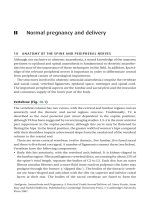
Vertebrae (Fig. 10.1)
The vertebral column has two curves, with the cervical and lumbar regions convex
anteriorly and the thoracic and sacral regions concave. Traditionally, T4 is
described as the most posterior part (most dependent in the supine position),
although T8 has been suggested by recent imaging studies. L3–4 is the most anterior
part (uppermost in the supine position), although this curve may be flattened by
flexing the hips. In the lateral position, the greater width of women’s hips compared
with their shoulders imparts a downward slope from the caudal end of the vertebral
column to the cranial end.
There are seven cervical vertebrae, twelve thoracic, five lumbar, five fused sacral
and three to five fused coccygeal. A number of ligaments connect them (see below).
Vertebrae have the following components:
• Body: this lies anteriorly, with the vertebral arch behind. It is kidney-shaped in
the lumbar region. Fibrocartilaginous vertebral discs, accounting for about 25% of
the spine’s total length, separate the bodies of C2 to L5. Each disc has an outer
fibrous annulus fibrosus and a more fluid inner nucleus pulposus (the latter may
prolapse through the former: a ‘slipped disc’). The bodies of the thoracic verteb-
rae are heart-shaped and articulate with the ribs via superior and inferior costal
facets at their rear. The bodies of the sacral vertebrae are fused to form the
Analgesia, Anaesthesia and Pregnancy: A Practical Guide Second Edition, ed. Steve Yentis, Anne
May and Surbhi Malhotra. Published by Cambridge University Press. ß Cambridge University
Press 2007.
sacrum, which encloses the sacral canal; the coccygeal vertebral bodies are fused
to form the triangular coccyx, the base of which articulates with the sacrum.
• Pedicles: these are round in cross-section. They project posteriorly from the
body and join the laminae. Each intervertebral foramen is formed by the pedicles
of the vertebra above and below.
• Laminae: these are flattened in cross-section. They complete the vertebral arch by
meeting in the midline at the spinous process. The superior and inferior articular
processes bear facets for articulation with adjacent vertebrae; those of the
thoracic vertebrae are flatter and aligned in the coronal plane, whereas those
of the lumbar vertebrae are nearer the sagittal plane.
• Transverse processes: in the lumbar region they are thick and pass laterally.
The transverse processes of L5 are particularly massive but short. The transverse
processes of thoracic vertebrae are large and pass backwards and laterally; they
bear facets that articulate with the ribs’ tubercles (except T11 and T12).
• Spinous process: these project horizontally backwards in the lumbar region; in the
thoracic region they are longer and inclined at about 60° to the horizontal.
The spinous process of T12 has a notched lower edge.
The cervical vertebrae have a number of features which distinguish them from the
others, including the foramen transverarium in the transverse processes, bifid
spinous processes and the particular characteristics of C1 and C2.
A line drawn between the iliac crests (Tuffier’s line) usually crosses the L3–4
interspace (slightly higher than in the non-pregnant state because of rotation of
the pelvis), although this is unreliable, and it has been shown that even experienced
anaesthetists can be one or more interspaces lower (or more commonly, higher)
than that intended.
Sacral canal (Fig. 10.2)
The sacral canal is 10–15 cm long, triangular in cross-section, runs the length of the
sacrum and is continuous cranially with the lumbar vertebral canal. The fused
Body
Spinous
process
Transverse
process
Vertebral
canal
Superior
articular facet
Superior
articular facet
Pedicle
Inferior
articular
process
Inferior
articular facet
Fig. 10.1 A lumbar vertebra, seen from superior and lateral aspects. Reproduced with
permission from Yentis, Hirsch & Smith: Anaesthesia and intensive care A-Z, 2nd edn,
Butterworth Heinemann, 2000.
10 Anatomy of the spine and peripheral nerves 19
bodies of the sacral vertebrae form the anterior wall, and the fused sacral laminae
form the posterior wall. The sacral hiatus is a deficiency in the fifth laminar arch, has
the cornua laterally and is covered by the sacrococcygeal membrane. Congenital
variants are common, possibly contributing to unreliable caudal analgesia.
Vertebral ligaments (Fig. 10.3)
• Anterior longitudinal ligament: this is attached to the anterior aspects of the
vertebral bodies, and runs from C2 to the sacrum.
• Posterior longitudinal ligament: this is attached to the posterior aspects of the
vertebral bodies, and runs from C2 to the sacrum.
• Ligamentum flavum (yellow ligament): this is attached to the laminae of adjacent
vertebrae, forming a ‘V’-shaped structure with the point posteriorly. It is more
developed in the lumbar than thoracic regions.
• Interspinous ligament: this passes between the spinous processes of adjacent
vertebrae.
• Supraspinous ligament: this is attached to the tips of the spinous processes from
C7 to the sacrum.
In addition, there are posterior, anterior and lateral sacrococcygeal ligaments.
Other ligaments are involved in the attachments of C1 and C2 to the skull.
The ligaments may become softer during pregnancy because of the hormonal
changes that occur.
Epidural space
• Boundaries: the space extends from the foramen magnum to the sacrococcygeal
membrane. It is triangular in cross-section in the lumbar region, its base anterior;
it is very thin anteriorly and up to 5 mm wide posteriorly. It lies external to the
dura mater of the spinal cord and internal to the ligamenta flava and vertebral
Sacral hiatus
Sacral foramen
Cornu
Articular process
Fig. 10.2 Sacrum. Reproduced with permission from Yentis, Hirsch & Smith: Anaesthesia and
intensive care A-Z, 2nd edn, Butterworth Heinemann, 2000.
20 Section 2 – Pregnancy
laminae posteriorly; the posterior longitudinal ligament anteriorly and the inter-
vertebral foramina and vertebral pedicles laterally. Magnetic resonance imaging
suggests the space is divided into segments by the laminae. The space may extend
through the intervertebral foramina into the paravertebral spaces.
• Contents: these include extradural fat, extradural veins (Batson’s plexus),
lymphatics and spinal nerve roots. The veins become engorged in pregnancy as
a result of the hormonal changes and any aortocaval compression. Connective
tissue layers have been demonstrated by radiology and endoscopy within the
extradural space, in some cases dividing it into right and left portions.
• Pressure: a negative pressure is usually found in the epidural space upon
entering it; the reason is unclear but may involve anterior dimpling of the dura
by the epidural needle, sudden posterior recoil of the ligamentum flavum when
it is punctured, stretching of the dural sac during extreme flexion of the back,
transmitted negative intrapleural pressure via thoracic paravertebral spaces and
Ligamentum flavum
Invertebral disc
Extradural
space
Dural sac
Posterior
longitudinal ligament
Interspinous ligament
Vertebral body
Anterior
longitudinal ligament
AB
Supraspinous ligament
Spinous process
Ligamentum flavumExtradural space
Dural sac
Posterior longitudinal ligamen
t
Anterior longitudinal ligament
Supraspinous ligament
Interspinous ligament
Vertebral body
(b)
(a)
Fig. 10.3 Vertebral ligaments: (a) longitudinal section and (b) transverse section
through A–B. Reproduced with permission from Yentis, Hirsch & Smith: Anaesthesia and
intensive care A-Z, 2nd edn, Butterworth Heinemann, 2000.
10 Anatomy of the spine and peripheral nerves 21
relative overgrowth of the vertebral canal compared with the dural sac.
Occasionally a positive pressure is found.
Meninges
• Pia mater: this delicate and vascular layer adheres closely to the brain and spinal
cord. Between it and the arachnoid mater is the cerebrospinal fluid (CSF) within
the subarachnoid space containing blood vessels, the denticulate ligament later-
ally along its length and the subarachnoid septum posteriorly. The pia terminates
as the filum terminale, which passes through the caudal end of the dural sac and
attaches to the coccyx.
• Arachnoid mater: this membrane is also delicate and contains CSF internally.
It lies within the dura externally, the potential subdural space containing vessels,
between them. It fuses with the dura at S2.
• Dura mater: this fibrous layer has an outer component, which is adherent to
the inner periosteum of the vertebrae and an inner one that lies against the
outer surface of the arachnoid. The dura projects into the extradural space,
especially in the midline. It ends at about S2.
Spinal cord
The spinal cord ends inferiorly level with L3 at birth, rising to the adult level of
L1–2 (sometimes T12 or L3) by 20 years. Below this level (the conus medullaris) the
lumbar and sacral nerve roots (comprising the cauda equina) and filum terminale
occupy the vertebral canal. The main ascending and descending tracts are shown
in Fig. 10.4.
Lateral corticospinal tract
Anterior corticospinal tract
Rubrospinal tract
Tectospinal tract
Vestibulospinal tract
Descending
Anterior spinothalamic tract
Spinotectal tract
Anterior
spinocerebellar tract
Posterior
spinocerebellar tract
Lateral
spinothalamic tract
Fasciculus cuneatus
Fasciculus gracilis
Ascending
Fig. 10.4 Ascending and descending tracts, spinal cord. Reproduced with permission from
Yentis, Hirsch & Smith: Anaesthesia and intensive care A-Z, 2nd edn, Butterworth Heinemann,
2000.
22 Section 2 – Pregnancy
The blood supply of the spinal cord is of relevance to obstetric anaesthetists, since
cord ischaemia may result in neurological damage:
• Anterior spinal artery: this descends in the anterior median fissure and supplies
the anterior two-thirds of the cord. The anterior spinal artery syndrome
(e.g. arising from profound hypotension) thus results in lower motor neurone
paralysis at the level of the lesion, and spastic paraplegia, reduced pain and
temperature sensation below the level and normal joint position sense and vibra-
tion sensation.
• Posterior spinal arteries: these descend along each side of the cord, one anterior
and one posterior to the dorsal nerve roots.
• Radicular branches: these arise from local arteries (from the aorta) and feed
the spinal arteries. Those at T1 and the lower thoracic/upper lumbar level
(artery of Adamkiewicz – usually unilateral) are the most important. The cord
at T3–5 and T12–L1 is thought to be most at risk from ischaemia. The conus
medularis and cauda equina are supplied by a vascular plexus arising from the
artery of Adamkiewicz above and pelvic vessels below. In 15% of the population,
Iliohypogastric nerve
Ilioinguinal nerve
Genitofemoral nerve
Obturator nerve
Femoral
nerve
Lateral cutaneous nerve of thigh
T12
L1
L2
L3
L4
Superior gluteal nerve
Inferior gluteal nerve
Sciatic nerve
L4
L5
S1
S2
S3
S4
Pudendal nerve
Perforating cutaneous nerve
Posterior cutaneous
nerve of thigh
(a)
(b)
Fig. 10.5 Plan of (a) lumbar and (b) sacral plexi. Reproduced with permission from Yentis,
Hirsch & Smith: Anaesthesia and intensive care A-Z, 2nd edn, Butterworth Heinemann, 2000.
10 Anatomy of the spine and peripheral nerves 23
the latter are the main source of arterial blood to the conus medularis and cauda
equina; compression during delivery may result in permanent paraplegia.
Venous drainage is via the internal iliac, intercostal, azygos and vertebral veins.
Peripheral nerves of the lower body
The lumbar and sacral plexi are shown schematically in Fig. 10.5. They form at the
posterior of the pelvis, and their branches pass round the interior of the pelvis where
they may be exposed to pressure during labour and delivery (Fig. 10.6; see also
Chapter 50, Peripheral nerve lesions following regional anaesthesia, p. 128).
Peripheral cutaneous innervation may be characterised according to the
dermatomal distribution or peripheral nerves (Fig. 10.7 and 10.8). Both representa-
tions may vary considerably between individuals. Peripheral motor innervation
may also be considered according to myotomal innervation or peripheral nerves
(Table 10.1).
Fig. 10.6 Major nerves of the pelvis. Adapted with permission from Holdcroft & Thomas:
Principles and practice of obstetric anaesthesia and analgesia, Blackwell Publishing, 2000.
24 Section 2 – Pregnancy
Table 10.1. Motor innervation of lower limbs by myotomes and peripheral nerves
Joint Movement Myotomes Nerve supply
Hip Flexion L1–3 Lumbar plexus
L2–4 Femoral nerve
Extension L5–S2 Sacral plexus
L5–S2 Sciatic nerve
Abduction L5–S2 Sacral plexus
Adduction L2–4 Obturator nerve
Knee Extension L2–4 Femoral nerve
Flexion L5–S2 Sciatic nerve.
S1–2 Tibial nerve*
Ankle/foot Dorsiflexion L4–5 Deep peroneal nerve
{
Eversion L5–S1 Superficial peroneal nerve
{
Plantar flexion S1–2 Tibial nerve*
Inversion L4–5 Tibial nerve*
*Branch of sciatic nerve
{
Branch of common peroneal nerve, itself a branch of the sciatic nerve
L5
S4
L3
L4
S5
S3
L1
L2
S2
S1
T12
L1
T10
T4
T2
C2
C3
L2
L3
L4
L5
C4
V
1
V
2
V
3
C7
C6
C5
T1
C8
S1
Fig. 10.7 Cutaneous innervation of lower body by dermatome. Reproduced with permission
from Yentis, Hirsch & Smith: Anaesthesia and intensive care A-Z, 2nd edn, Butterworth
Heinemann, 2000.
10 Anatomy of the spine and peripheral nerves 25
Dermatomal innervation of the upper body is also important when determining
the upper extent of regional blockade.
FURTHER READING
Broadbent CR, Maxwell WB, Ferrie R, et al. Ability of anaesthetists to identify a marked lumbar
interspace. Anaesthesia 2000; 55: 1122–6.
Capogna G, Celleno D, Simonetti C, Lupoi D. Anatomy of the lumbar epidural region using
magnetic resonance imaging: a study of dimensions and a comparison of two postures.
Int J Obstet Anesth 1997; 6: 97–100.
Harrison GR. Topographical anatomy of the lumbar epidural region: an in vivo study using
computerized axial tomography. Br J Anaesth 1999; 83: 229–34.
Render CA. The reproducibility of the iliac crest as a marker of lumbar spine level. Anaesthesia
1996; 51: 1070–1.
Femoral branch of
genitofemoral nerve *
Dorsal rami L1–3
Iliohypogastric nerve *
Ilio-inguinal nerve *
Subcostal nerve
(from T12 intercostal) *
Dorsal rami S1–3
Lateral cutaneous
nerve of thigh *
Lateral cutaneous
nerve of thigh *
Lateral cutaneous
nerve of calf §
Lateral cutaneous
nerve of calf §
Superficial
peroneal nerve §
Sural nerve ‡
Medial calcaneal
branches of
tibial nerve ‡
* From lumbar plexus
† From femoral nerve
‡ From tibial nerve (branch of sciatic nerve)
§ From common peroneal nerve (branch of sciatic nerve)
Saphenous
nerve †
Obturator
nerve *
Anterior and
medial cutaneous
nerves of thigh †
Sural nerve ‡
Deep peroneal
nerve §
Posterior cutaneous
nerve of thigh
(from sacral plexus)
Fig. 10.8 Cutaneous innervation of leg by peripheral nerve. Reproduced with permission from
Yentis, Hirsch & Smith: Anaesthesia and intensive care A-Z, 2nd edn, Butterworth Heinemann,
2000.
26 Section 2 – Pregnancy
11 PHYSIOLOGY OF PREGNANCY
Pregnancy is associated with major physiological changes throughout the
body. These are caused by both hormonal factors (influential from conception
onwards) and the mechanical changes caused by the enlarging uterus (of increasing
significance as pregnancy progresses). It is important to understand the normal
physiological changes occurring during pregnancy in order to predict the risks
and effects of analgesic and anaesthetic intervention, and also to anticipate the
impact of pregnancy on any coexisting medical condition.
Hormonal changes
Following fertilisation, the corpus luteum in the ovary secretes progesterone,
oestrogens and relaxin, and these hormones are secreted by the placenta when
it takes over the function of the corpus luteum from 6–8 weeks’ gestation onwards.
The placenta also secretes human chorionic somatomammotrophin (hCS;
previously known as human placental lactogen and chorionic growth hormone-
prolactin).
Human chorionic gonadotrophin (hCG) can be measured by radioimmunoassay
and detected in the blood 6 days after conception and in the urine 2–3 weeks
after conception. It is therefore a useful early diagnostic test of pregnancy. It is
produced by the syncytiotrophoblast, and levels rise rapidly during the first
8 weeks of pregnancy, falling to a plateau thereafter.
Progesterone is responsible for most of the hormonally mediated changes
occurring during pregnancy. It causes:
• Smooth muscle relaxation
• Generalised vasodilatation
• Bronchodilatation
• Dilatation within the renal tract
• Sluggish gastrointestinal tract motility and constipation.
It is thermogenic, causing an increase in basal temperature during
pregnancy. It may be responsible for the nausea and vomiting that are
common in early pregnancy. Progesterone is a neurotransmitter and, together
with increased endogenous endorphins, is implicated in the elevated pain
threshold experienced by pregnant women. It also decreases the minimum
alveolar concentration of inhalational anaesthetic agents. Progesterone has
also been demonstrated to enhance conduction blockade in isolated
nerve preparations, and it is therefore thought likely to play a role in the
decreased requirement for local anaesthetic agents for spinal and epidural
anaesthesia.
Progesterone levels return to pre-pregnancy values over a period of 3–4 weeks
after delivery, and thus hormonally mediated changes do not reverse immediately
in the puerperium.
11 Physiology of pregnancy 27
Mechanical changes
The uterus enlarges as pregnancy progresses. The fundus is palpable:
• Abdominally by the beginning of the second trimester
• At the umbilicus by 20 weeks’ gestation
• At the xiphisternum by 36 weeks.
If the fetal head engages in the maternal pelvis at the end of pregnancy, the fundal
height decreases and this may alleviate some symptoms attributable to mechanical
factors. In multiple pregnancies, the uterus expands to a greater extent and more
rapidly, and therefore the mechanical effects are usually greater.
Following delivery the uterus involutes rapidly, and should not be palpable above
the maternal umbilicus. It has usually returned to within the pelvis by 72 hours after
delivery.
Cardiovascular and haemodynamic changes
Pregnancy
• Blood volume increases throughout pregnancy, to approximately 45–50% more
than pre-pregnant values by term. This represents an increase in both red cell
volume and plasma volume with the latter being relatively greater, thus causing
the so-called ‘physiological anaemia’ of pregnancy. The magnitude of the
increase is greater in women with multiple pregnancy and greatly reduced in
women with pre-eclampsia.
• Cardiac output, heart rate and stroke volume all increase as pregnancy
progresses. Cardiac output increases by approximately 40–50% by term, with
most of the increase occurring by 20 weeks’ gestation. The increased blood
flow is distributed primarily to the uterus, where blood flow increases from
approximately 50 ml/minute at 10 weeks’ gestation to 850 ml/minute at term.
• Renal blood flow increases by 80% over non-pregnant levels, and this level is
achieved by the middle of the second trimester. Glomerular filtration rate and
creatinine clearance increase by 50% during pregnancy.
• Systemic vascular resistance falls (peripheral vasodilatation mediated by proges-
terone, prostacyclin and oestrogens), and there is a decrease in both systolic and
diastolic blood pressures, which reach a nadir during the second trimester
and then increase gradually towards term, although remaining lower than
pre-pregnancy values.
• Aortocaval compression can occur from the middle of pregnancy onwards if the
supine position is adopted. This is due to mechanical compression of the aorta
and inferior vena cava. Venous return is dependent on the competence of collat-
eral circulation via the azygos and ovarian veins. Recent studies have demon-
strated that uterine blood flow decreases primarily as a result of aortic rather
than venous compression.
• Central venous and pulmonary arterial pressures are unchanged during normal
pregnancy.
28 Section 2 – Pregnancy
Labour and delivery
• Cardiac output increases by 25–50% in labour, with an additional 15–30%
increase during contractions. This increase in cardiac output is mediated through
increased sympathetic nervous system activity, and is therefore significantly
attenuated by epidural analgesia.
• Central venous pressure increases during contractions, partly due to sympathetic
activity and partly from the transfer of up to 500 ml of blood from the intervillous
space. The latter is unaffected by epidural analgesia, as is the increase in central
venous pressure which occurs when the Valsalva manoeuvre is performed during
pushing.
• Autotransfusion of blood (from the placenta) occurs during the third stage.
The effect of this may be significant in women with cardiac disease.
• After delivery there is a sustained increase in cardiac output and central
venous pressure for several hours, which is associated with hypervolaemia. The
implications of these changes for women with cardiac disease are significant
(see relevant sections).
Respiratory changes
Pregnancy
• Progesterone increases the sensitivity of the respiratory centre to carbon dioxide
and also acts as a primary respiratory stimulant. These effects are enhanced by
oestrogens, and the combined hormonal effect causes an increase in minute
ventilation of 45–50%.
• The partial pressure of carbon dioxide in arterial blood (P
a
CO
2
) is reset to approxi-
mately 4 kPa during the first trimester and remains at that level throughout
pregnancy. A partially corrected respiratory alkalosis is found in normal pregnant
women.
• Functional residual capacity decreases to 80% of pre-pregnancy values as
pregnancy progresses, caused by increased intra-abdominal pressure and
upward displacement of the diaphragm by the enlarging uterus. Total lung capa-
city remains unchanged. Functional residual capacity remains greater than
closing capacity throughout pregnancy whilst the woman remains in an
upright position, but falls when a recumbent position is adopted. It has
been estimated that airway closure may occur within normal tidal ventilation
in as many as 50% of all supine pregnant women during the second half of
pregnancy.
• Oxygen consumption increases progressively during pregnancy to 35% above
pre-pregnancy levels.
Labour and delivery
• Massive hyperventilation occurs during labour (unless there is effective
analgesia), with minute ventilation increasing by up to 350% compared with
pre-labour values.
11 Physiology of pregnancy 29
• P
a
CO
2
falls to below 2 kPa in some women. This respiratory alkalosis is associated
with a metabolic acidosis, since maternal aerobic requirement for oxygen
(increased by hyperventilation, hyperdynamic circulation and uterine activity)
cannot be met, resulting in a progressive lactic acidosis.
• Effective epidural analgesia abolishes these effects during the first stage of
labour but not during the second, when the additional uterine activity
and work of pushing produce a further oxygen demand that cannot be met.
Gastrointestinal changes
Pregnancy
• Lower oesophageal sphincter pressure is reduced because of the smooth muscle
relaxant effect of progesterone.
• Intragastric pressure rises as a mechanical consequence of the enlarging
uterus.
• The overall effect of these changes is a decrease in gastro-oesophageal barrier
pressure, with a concomitant increase in risk of regurgitation and aspiration of
gastric contents.
• Some 75–85% of pregnant women complain of heartburn during the third
trimester, and a significant number will have a demonstrable hiatus hernia.
• Gastric emptying is not delayed during pregnancy.
• There is some evidence that gastric volume is increased, and the pH of the intra-
gastric volume may be lower than in the non-pregnant individual.
Labour and delivery
• Gastric emptying is now thought to be normal in labour in most cases, unless
opioids have been given.
• Opioid analgesia (regardless of route of administration) delays gastric emptying.
• Recent work suggests that gastric volume (but not acidity) may remain elevated
for 48 hours after delivery.
Management options
Positioning
• It is the anaesthetist’s responsibility to exercise vigilance, with special attention
being paid to the hips and back. The pregnant woman has increased ligamentous
laxity, and may be particularly at risk of musculoskeletal trauma if she has
received epidural analgesia. This risk is considerably increased if she has received
either regional or general anaesthesia, when she is unable to safeguard her
position.
• No pregnant woman should lie in the unmodified supine position at term
(it is rare to find a mother who will voluntarily adopt this position). The wedged
supine position and the use of lateral tilt are compromises and do not reliably
30 Section 2 – Pregnancy
relieve aortocaval compression. Women should be encouraged to remain sitting
upright or in the full lateral position whenever possible. Walking and standing in
labour should also be encouraged.
• Obstetricians and midwives should be asked to perform fetal scalp blood
sampling and vaginal examinations with the woman in the left lateral, or at
least tilted, position.
• Closing volume may occur within tidal volume when the semi-recumbent
position is adopted, and consideration should be given to continuous adminis-
tration of oxygen to women particularly at risk (e.g. those who are obese, and
those with respiratory disease).
General anaesthesia
• Pregnant women have increased oxygen consumption and decreased oxygen
reserves. They are therefore at greatly increased risk of hypoxia during periods
of apnoea.
• The risk of pulmonary aspiration of gastric contents means that rapid
sequence induction of general anaesthesia, preceded by measures to reduce
the acidity of the gastric contents, may be required, depending on the
gestation and severity of symptoms (see Chapter 56, Aspiration of gastric
contents; p. 138).
12 AORTOCAVAL COMPRESSION
Aortocaval compression (supine hypotensive syndrome) was first reported in
1931. The inferior vena cava and aorta become compressed by the pregnant
uterus (the vena cava may be totally occluded), causing reduction in venous
return and cardiac output and thus compromising the mother, fetus or both.
Vasovagal syncope may follow aortocaval compression. Maternal symptoms
and signs vary from asymptomatic mild hypotension to total cardiovascular
collapse, partly dependent on the efficacy of the collateral circulation bypassing
the inferior vena cava. Onset of symptoms and signs is associated with lying in
the supine or semi-supine position, and is relieved by turning to the full lateral
position in most cases.
Problems/special considerations
• Aortocaval compression is not confined to the woman at term. The condition
has been reported in the fifth month of pregnancy. Women with multiple
pregnancy or polyhydramnios are at increased risk because of the increased
uterine size.
12 Aortocaval compression 31
• It is important to appreciate that normotension and lack of maternal symptoms
do not exclude a significant fall in cardiac output and placental perfusion.
• Onset of symptoms may occur within 30 seconds, but may be delayed by
30 minutes. Severity of symptoms is not a reliable guide to severity of
hypotension.
• Slight changes in maternal position may cause significant change in symptoms.
A15° lateral tilt does not reliably relieve aortocaval compression, and even
a45° tilt does not guarantee abolition of hypotension.
• Catastrophic hypotension, and even cardiac arrest, may occur if general anaes-
thesia is induced in a woman who is experiencing severe aortocaval compression
(e.g. in the supine position). Even mild degrees of aortocaval compression can
lead to severe hypotension after spinal or epidural anaesthesia.
• It is impossible to perform effective cardiopulmonary resuscitation on the
undelivered woman in the supine position; use of a purpose-made resuscitation
wedge is recommended. If this is not available, the uterus must be displaced off
the vena cava and aorta by other means.
Management options
Women will not voluntarily adopt positions in which aortocaval compression
occurs, and therefore the condition is largely iatrogenic, occurring after a woman
has been placed in the supine position by her midwifery or medical attendants.
A history suggestive of aortocaval compression in late pregnancy may indicate an
increased risk of developing the condition during labour and delivery. All those
caring for pregnant women must be aware of aortocaval compression and of the
need to avoid the supine position. This is particularly important if the woman is
unable to change her own position because of administration of analgesia or
anaesthesia.
Uterine displacement (usually to the left, although occasionally improved
symptomatic relief will be obtained by displacement to the right) must be used
during all vaginal examinations and during both vaginal and operative delivery,
and is especially important if regional analgesia or anaesthesia is used. This can
be achieved manually or by use of table tilt or a wedge under the hip. Use of uterine
displacement rather than the full lateral position is a compromise between mater-
nal safety and obstetricians’ convenience. Use of the full lateral position for
Caesarean section has been reported.
Extreme vigilance is necessary when maternal symptoms are abolished by
induction of general anaesthesia. During regional anaesthesia for operative
delivery, complaints of faintness, dizziness, restlessness and nausea should alert
the anaesthetist to the onset of hypotension. Pallor, particularly of the lips, yawning
and non-specific feelings of anxiety are also warning signs of aortocaval compres-
sion. Continuous fetal monitoring may indicate signs of fetal distress when the
mother adopts the supine or semi-supine position, and occasionally this may be
the only indicator of the condition. Turning the mother into the full left lateral
32 Section 2 – Pregnancy
position should be the first step in the treatment of hypotension or cardiotoco-
graphic abnormalities.
Key points
• No pregnant woman should lie flat on her back beyond 16–18 weeks.
• The uterus must be displaced off the aorta and vena cava during vaginal examinations
and during Caesarean section. This can be done manually, with a wedge under the hip,
or by using lateral tilt of the operating table.
• Cardiopulmonary resuscitation will be ineffective if the mother is supine.
FURTHER READING
Kinsella SM, Lohmann G. Supine hypotensive syndrome – a review. Obstet Gynecol 1994;
83: 774–88.
13 NORMAL LABOUR
A large number of pregnant women are assessed as being ‘low risk’ and are
predicted to have normal labours, but the diagnosis of normal labour is
retrospective.
The parameters for normal labour are:
• Contractions once in every 3 minutes, lasting 45 seconds
• Progressive dilatation of the cervix
• Progressive descent of the presenting part
• Vertex presenting with the head flexed and the occiput anterior
• Labour not lasting less than 4 hours (precipitate) or longer than 18 hours
(prolonged)
• Delivery of a live healthy baby
• Delivery of a complete placenta and membranes
• No complications.
First stage of labour
During the latent phase, the cervix effaces then cervical dilatation begins. The rate
of cervical dilatation should be around 1 cm/h for a primiparous woman and
2 cm/h for a multigravid woman.
It is standard practice to perform a vaginal examination every 4 hours to assess
the dilatation of the cervix, or more frequently if there is cause for concern.
The following routine observations are charted on the partogram:
• Fetal heart rate quarter-hourly
13 Normal labour 33
• Maternal pulse rate half-hourly
• Blood pressure half-hourly
• Temperature 4-hourly
• Urinalysis at each emptying of the bladder.
The fetal heart may be monitored intermittently by auscultation using Pinard’s
stethoscope or by cardiotocographic monitoring. The cardiotocogram (CTG) is
recorded either intermittently or continuously depending on the condition of
the fetus. Continuous recording of fetal heart rate may be done using either an
abdominal transducer or a clip applied to the fetal head. Radiotelemetry is available
in some units and this allows the woman to be mobile while her baby is monitored.
Uterine contractions may be monitored externally by an abdominal transducer
or internally by an intrauterine catheter. The fetal heart rate and the uterine
contractions are recorded together.
Second stage of labour
The second stage of labour commences at full dilatation of the cervix and termi-
nates at the delivery of the baby.
At full dilatation of the cervix, the character of the contractions changes and they
are usually, but not invariably, accompanied by a strong urge to push. In normal
labour there is an increase in circulating oxytocin secondary to Ferguson’s reflex,
with consequent increased strength of uterine contractions at full dilatation.
Higher-dose epidural analgesia is thought to diminish the effect of this reflex.
The second stage of labour can be divided into passive and active stages and
this is particularly relevant when epidural analgesia is used. With epidural analge-
sia, especially using older, higher-dose techniques, the labouring woman may not
have the normal sensation at the start of the second stage of labour; therefore
the active stage of pushing should only commence when the vertex is visible or
the woman has a strong urge to push. In normal labour, the active stage usually
commences at full dilatation. Traditionally, the second stage is limited to 2 hours
because of the risk of fetal acidosis; up to 3 hours is often allowed in the presence
of epidural analgesia in recognition of the slower descent of the fetal head. It is
difficult for a woman to push efficiently for more than one hour, and after this
time fetal acidosis is felt to be more likely. If there is not good progress, the
advice of the obstetrician should be sought. At the delivery of the anterior shoulder,
intramuscular oxytocics (e.g. Syntometrine) are given to hasten the delivery of
the placenta and to stimulate uterine contraction.
Third stage of labour
The third stage of labour is the complete delivery of the placenta and membranes
and the contraction of the uterus. It is usually managed actively by administering an
oxytocic as above, but it may also be managed physiologically without oxytocics.
This may prolong the third stage and increase the risk of postpartum haemorrhage.
34 Section 2 – Pregnancy
During the third stage of labour there is a major redistribution of (and increase in)
maternal circulating blood volume. This is potentially dangerous to those women
who have cardiac disease and who may be precipitated into heart failure immedi-
ately postpartum.
Key points
• Normal labour can be anticipated but can only be diagnosed after delivery.
• The first stage comprises cervical effacement and dilatation.
• During the second stage, the baby passes through the birth canal.
• The placenta and membranes are delivered during the third stage.
FURTHER READING
Ferguson E, Owen P. The second stage of labour. Hosp Med 2003; 64: 210–13.
Steer P, Flint C. Physiology and management of normal labour. BMJ 1999; 318: 793–6.
14 GASTRIC FUNCTION AND FEEDING IN LABOUR
Physiological changes in pregnancy affect the volume, acidity and emptying of
gastric secretions as well as sphincter mechanisms in the lower oesophagus.
Interventions in labour such as analgesia may also affect these changes adversely.
General anaesthesia is occasionally necessary in emergency situations, and the
presence of a full stomach (and thus the risk of aspiration of gastric contents)
should always be assumed in such patients (see Chapter 56, Aspiration of gastric
contents, p. 138).
Problems/special considerations
Increased circulating progesterone associated with pregnancy relaxes smooth
muscle and causes relaxation of the lower oesophageal sphincter, whereas
placental gastrin increases the volume and decreases the pH of gastric contents.
The enlarging uterus increases intragastric pressure and there is an increase
in small and large bowel transit time. However, evidence suggests that gastric
emptying per se is not affected by pregnancy though it may be decreased in
labour if opioids are given.
Extradural analgesia with local anaesthetic solutions in labour is associated
with normal gastric emptying, whereas subarachnoid or extradural opioids
(fentanyl or diamorphine) in large doses cause a modest decrease in gastric
emptying. Systemic opioid analgesia causes a much greater and prolonged
decrease in gastric emptying. However, recent randomised studies have
14 Gastric function and feeding in labour 35
demonstrated large gastric volumes and a high incidence of vomiting in
women allowed to eat solid food, even when pain was adequately controlled with
a low-dose fentanyl/bupivacaine epidural.
Plasma progesterone concentrations return to non-pregnant values
within 24 hours of delivery, and gastroesophageal reflux is considerably
reduced within 48 hours of delivery. The period of risk of aspiration thus
extends to an ill-defined time after delivery, and appropriate general
anaesthetic management in the early postpartum period is thus somewhat
controversial.
Routine withholding of food and fluids in labour has been challenged by
a number of authors, particularly those who are not anaesthetists. They
point out that absolute starvation is not popular with mothers, that aspiration
associated with emergency general anaesthesia nowadays is uncommon and
that there may be risks associated with prolonged starvation. On the other
hand, there is little evidence that a period of starvation during labour is harmful,
although it may be unpleasant. Starvation is associated with ketosis, but this has
not been found to affect the duration or outcome of labour.
Management options
There are three approaches to the treatment of feeding in labour. The tradi-
tional approach is to assume that all women in labour are at risk of an event in
labour that will require emergency general anaesthesia and that they are therefore
at risk of aspiration of large volumes of acid gastric contents. As a consequence
of this assumption, many women in labour are starved, allowed only sips of water to
drink and given regular H
2
antagonists (e.g. ranitidine 150 mg orally 6-hourly,
or 50 mg intramuscularly 8-hourly) and regular sodium citrate (30 ml of
0.3 M orally).
Another approach is to assume that women in labour require food and fluid and
to give these liberally. Often no H
2
-blockers are given.
A more rational approach is to stratify management on the basis of risk. Women
at high risk of requiring general anaesthesia are advised to have only clear fluids and
receive regular H
2
-blockers. In addition, for those who do eat and drink during
labour, substances that are associated with slower gastric emptying (those with
high fat or sugar content) should be discouraged in favour of protein-based
snacks and isotonic drinks.
If intravenous water is required in labour, the most sensible fluid to provide
might be 5% or 10% dextrose. Unfortunately this has been associated with
fluid overload in the mother and hyponatraemia in the neonate. However,
modest volumes (51 litre) do not significantly affect neonatal plasma sodium
concentrations. Many units give relatively low volumes of intravenous saline,
dextrose saline or Hartmann’s solution when intravenous fluid is considered
necessary.
36 Section 2 – Pregnancy
Key points
• Women are being encouraged to eat in labour, especially by other professionals.
• Solid food ingested during labour is not predictably absorbed.
• Women treated with epidural analgesia may have normal gastric emptying unless
large boluses of opioid are given.
• Opioids given parenterally markedly decrease gastric emptying.
• Acid aspiration prophylaxis should be given to all women at risk of intervention in
labour.
FURTHER READING
Porter JS, Bonello E, Reynolds F. The influence of epidural administration of fentanyl infusion
on gastric emptying in labour. Anaesthesia 1997; 52: 1151–6.
Scrutton NJL, Metcalfe GA, Lowy C, Seed PT, O’Sullivan G. Eating in labour. A randomised
controlled trial assessing the risks and benefits. Anaesthesia 1999; 54: 329–34.
15 DRUGS AND PREGNANCY
Pregnancy may interact with drugs in a number of different ways. Firstly, the
pregnant state confers alterations in both pharmacokinetics and pharmacody-
namics; secondly, the fetus may be affected by drugs administered to the mother,
and in many cases this may restrict the use of certain drugs; and thirdly, there may
be further passage of certain drugs to the neonate in breast milk (see Chapter 149,
Drugs and breastfeeding, p. 337). Because of these considerations, special licensing
requirements exist for drugs to be used in pregnancy, which have not been met
by many drugs in current use.
Pharmacokinetics
Each of the traditional components of pharmacokinetics may be altered in the
pregnant, as opposed to the non-pregnant, state.
• Absorption of drug: this depends on the route of administration and, in general,
is little affected by pregnancy. However, absorption of enterally administered
drugs may be affected by pregnancy-associated gastrointestinal upsets, including
vomiting. Because of the increased minute ventilation and cardiac output,
absorption of inhalational agents is more rapid.
• Distribution of drug: this is affected by the increased blood volume and body
fluid and altered plasma protein profile. The former two result in a greater
volume of distribution. In addition, the fetus represents an additional compart-
ment to which drugs will distribute, depending on their lipid solubility, pKa
and protein binding. The increased cardiac output will tend to redistribute
15 Drugs and pregnancy 37
drugs more quickly unless they are extensively bound to the tissues. During
labour, acute changes in plasma pH (e.g. acidosis associated with maternal
exhaustion or alkalosis associated with pain-induced hyperventilation) may
affect both protein binding and degree of dissociation of drugs.
• Metabolism of drugs: drugs broken down in the major organs (usually the liver)
should be handled normally in pregnancy, unless there is hepatic impairment,
e.g. in HELLP (haemolysis, elevated liver enzymes and low platelet count)
syndrome. Some drugs are metabolised by plasma cholinesterases and may
thus have longer duration of action if the protein concentration is reduced,
e.g. suxamethonium.
• Elimination: since glomerular filtration rate is increased in pregnancy, clear-
ance of many drugs is increased unless renal function is impaired, e.g. in pre-
eclampsia. An extra route of elimination is in breast milk, although this represents
a relatively small amount of total drug elimination. Inhalational agents are
excreted via the lungs more rapidly in the pregnant than non-pregnant state.
Pharmacodynamics
The effects of most drugs are unchanged in pregnancy. However, notable and
important exceptions are anaesthetic agents. Thus the minimum alveolar concen-
tration of inhalational agents is reduced, as is the minimal blocking concentration
of local anaesthetics. The cause of this decrease in anaesthetic requirement is
thought to be progesterone and/or a metabolite thereof. In addition, a given
amount of epidural local anaesthetic solution produces a more extensive block
than in non-pregnant subjects, possibly related to the reduction in epidural space
caused by epidural venous engorgement, although progesterone has also been
suggested as being involved.
Fetal effects of drugs
Drugs may affect the fetus at any stage of pregnancy. During the first trimester
the developing organ systems and overall body structure are especially at risk,
particularly between the third and tenth weeks; administration of certain drugs
during this period may result in congenital malformations. During the second
and third trimesters, the growth and development of fetal tissues may be affected.
Finally, drugs given before delivery may affect fetal oxygenation indirectly
(e.g. by causing maternal hypotension or respiratory depression), may affect
labour (e.g. b-agonists), or may have neonatal effects after birth (e.g. opioids).
Many drugs are known to be harmful when given during pregnancy, but for
many others, precise information is not always available. Thus, in general, drugs
are not prescribed unless the benefits are felt to outweigh any possible risk,
especially during the first trimester. Where possible, older drugs of which clinicians
have greater experience are preferred over newer ones, and this is also true of
anaesthetic agents.
38 Section 2 – Pregnancy
Licensing of drugs in pregnancy
Many drugs, including anaesthetic agents, are not licensed for use in pregnancy,
mainly because of the prohibitive costs to the manufacturer of performing the
appropriate studies required and the relatively limited addition such licensing
would make to the market. For example, the data sheets of etomidate, alfentanil
and fentanyl contain the sentence ‘safety in human pregnancy has not been estab-
lished’ or words to that effect, whilst those of propofol and fentanyl specifically
warn against their use in obstetrics. Even in the case of thiopental, the data sheet
merely states that there is ‘epidemiological and clinical evidence’ of its safety in
pregnancy, whereas that of atracurium, vecuronium and suxamethonium state that
they should only be used in pregnancy ‘if the potential benefits outweigh any poten-
tial risks’.
Key points
• Pharmacokinetics and pharmacodynamics in pregnancy may be altered from those in
the non-pregnant state.
• Most drugs administered to the mother will pass to the fetus to a degree.
• Many drugs pass into breast milk.
• Most anaesthetic drugs are not licensed for use in pregnancy.
FURTHER READING
Howell PR, Madej T. Administration of drugs outside of product licence: awareness and
current practice. Int J Obstet Anesth 1999; 8: 30–6.
Rubin P. Drug treatment during pregnancy. BMJ 1998; 317: 1503–6.
16 PLACENTAL TRANSFER OF DRUGS
The placenta is a complex structure composed of both maternal and fetal tissues.
Nevertheless, it is basically a semi-permeable biological membrane and as such
obeys the laws that govern transport across such membranes. Virtually all transfer
of drugs across the placenta occurs by simple diffusion, and all drugs administered
to the mother will reach the fetus, albeit to a variable extent depending upon the
factors discussed below.
Factors determining placental transfer
Molecular weight and lipid solubility
The molecular weight of the drug, its degree of ionisation, its lipid solubility and
the degree to which it is protein bound will all affect the readiness with which it
16 Placental transfer of drugs 39
will cross the placenta. The majority of anaesthetic drugs are small (molecular
weights of less than 500) and lipid soluble; thus they cross the placenta readily.
The main exceptions are the neuromuscular blocking drugs, which are less lipid
soluble, more highly ionised quaternary ammonium compounds, and in the doses
used in normal clinical anaesthesia do not cross the placenta to any significant
extent. However, if used in large doses or over a prolonged period of time (e.g. to
facilitate artificial ventilation in the intensive care unit) they do reach the fetal
circulation in doses that may have a clinical effect necessitating ventilatory support.
Changes in maternal or fetal pH may alter the degree of ionisation and protein
binding of a drug, and thus alter its availability for transfer. This is most likely to
occur if the pKa of a drug is close to physiological pH, and becomes clinically
relevant in the acidotic fetus. Once drug transfer to the fetus has occurred, acidosis
results in increased ionisation of the drug, which is then unable to equilibrate
with the maternal circulation by diffusion back across the placenta. This results
in drug accumulation in the fetus (so-called ion trapping), and is particularly
relevant for local anaesthetics, which all have a pKa 4 7.4.
Maternal drug concentration
Drug transfer occurs down a concentration gradient (which is usually from mother
to fetus but can also occur from fetus to mother). The drug concentration on the
maternal side depends on the route of administration, total maternal dose, volume
of distribution and drug clearance and metabolism. The highest maternal blood
concentration of a drug will be achieved following intravenous administration;
epidural and intramuscular administration result in similar maternal blood
concentrations. Systemic drug absorption will be greater from more vascular
tissues, such as the paracervical region.
The increase in blood volume and cardiac output that accompanies normal preg-
nancy has an effect on maternal drug concentration; the volume of distribution and
plasma clearance of drugs such as thiopental is increased.
Placental factors
The area of placenta available for transfer is important. Physiological shunting
occurs in the placenta, and in maternal disease such as pre-eclampsia the placenta
itself may present an increased barrier to transfer. Although there is evidence that
some drug metabolism occurs within the placenta itself, this is not clinically
significant.
Fetal drug concentration
Once a drug has reached the fetus it is subject to redistribution, metabolism
and excretion. The fetus has less plasma protein binding capacity and less
mature enzyme systems than the mother, and will therefore eliminate drugs less
effectively. Some transfer of drugs occurs back across the placenta to the mother
if the maternal concentration falls below that in the fetus (unless ion trapping
occurs – see above).
40 Section 2 – Pregnancy
Uteroplacental blood flow
This is the other major factor influencing placental transfer. Any reduction in blood
flow to the placenta will inevitably reduce transfer of drugs (and nutrients) to the
fetus. Reduction in uteroplacental flow may occur as a result of generally reduced
maternal blood flow (hypotension, reduced cardiac output states, aortocaval com-
pression, generalised vasoconstriction) or direct obstruction of flow (aortocaval
compression, uterine contraction, umbilical cord compression).
Problems/special considerations
All general anaesthetic agents cross the placenta readily; and in normal clinical
practice their effects on the fetus are only of significance immediately after delivery.
The compromised fetus, or one in whom the uterine incision to delivery interval has
been prolonged, may be depressed at birth, but rarely requires more than simple
resuscitative measures.
Pethidine (and all other opioids) crosses the placenta readily. It has maximal
effect in the fetus 3–4 hours after maternal administration and minimal effect if
given to the mother within an hour of delivery. (This is contrary to traditional mid-
wifery teaching, which recommends that pethidine is not given if delivery is
expected within 2–3 hours.) Both pethidine and its active metabolite norpethidine
have prolonged half-lives in the fetus and cause respiratory depression and reduced
sucking ability. Opioid side effects are reversed by naloxone.
Local anaesthetics cross the placenta by simple diffusion, but the extent of pla-
cental transfer is also dependent on maternal plasma protein binding (bupivacaine
and ropivacaine are highly protein bound, and therefore cross less readily than
lidocaine, which is less protein bound.)
Key points
• The major determinants of transfer by simple diffusion are the maternal–fetal drug
concentration gradient, molecular weight of the drug, lipid solubility, degree of drug
ionisation and extent of protein binding.
• Uteroplacental blood flow is also important.
• Opioids given to the mother for labour analgesia cross the placenta freely and may
cause fetal respiratory and neurobehavioural depression, which are reversible with
naloxone.
FURTHER READING
Littleford J. Effects on the fetus and newborn of maternal analgesia and anesthesia: a review.
Can J Anaesth 2004; 51: 586–609.
16 Placental transfer of drugs 41
17 PRESCRIPTION AND ADMINISTRATION OF DRUGS BY MIDWIVES
In the UK, regulations for prescription and administration of drugs by midwives fall
under the responsibility of the Nursing and Midwifery Council (NMC; previously
the UK Central Council for Nursing, Midwifery and Health Visiting (UKCC)), which
issues codes and standards relating to the practical application of acts such as
the Medicines Act 1968, Misuse of Drugs Act 1971, and Medicinal Products:
Prescription by Nurses Act 1992, and their subsequent amendments. Many of the
the NMC’s publications on the matter are not legally binding but would be taken
into account if there were to be medicolegal or regulatory action concerning admin-
istration of drugs. Against this background of central control, the setting up of, and
adherence to, local policies is strongly encouraged, in recognition of the differing
requirements from unit to unit.
Problems/special considerations
A compromise must exist between (i) supporting the midwife’s role as an indepen-
dent practitioner; (ii) reducing the workload on, and requirement for, medical staff
to treat common and relatively minor conditions; (iii) permitting the rapid admin-
istration of drugs that may have real benefits to mothers and reduce morbidity or
mortality; and (iv) restricting the use of potentially harmful drugs or reducing the
incidence of adverse effects. Whether a particular drug should be allowed to be
given thus depends on the incidence, importance and potential severity of the
condition for which it is indicated and the efficacy, method of administration and
safety profile of the drug concerned.
Drugs that midwives can administer without medical prescription
There is regional variation according to local policies, and individual trusts bear
ultimate responsibility for approving drug policies within their maternity services.
However, the drugs that midwives are allowed to prescribe and administer generally
fall into a number of categories (Table 17.1). Local regulations are usually decided
by a panel including representatives of midwives, pharmacists and obstetricians;
anaesthetic staff may also be involved, e.g. in helping with analgesic or local
anaesthetic drug policies.
Midwives in different units may interpret the NMC’s guidelines differently,
especially with regard to epidural top-ups; thus, for example, midwives in certain
units may be prepared to administer epidural drugs prescribed by a doctor
(i.e. anaesthetist) whereas those in other units may not. This is not usually a prob-
lem with local anaesthetic drugs alone but has been problematic with mixtures
of local anaesthetics and opioids, e.g. fentanyl, which are, first, controlled drugs,
and second, unlicensed for epidural use. Recently, in the UK, there has been
stricter attention to the proper handling of all preparations containing controlled
drugs, even the dilute mixtures used for epidural analgesia. Interpretation of current
42 Section 2 – Pregnancy