Báo cáo y học: "A k2A-positive Klebsiella pneumoniae causes liver and brain abscess in a Saint Kitt’s ma"
Bạn đang xem bản rút gọn của tài liệu. Xem và tải ngay bản đầy đủ của tài liệu tại đây (541.9 KB, 4 trang )
Int. J. Med. Sci. 2009, 6
301
I
I
n
n
t
t
e
e
r
r
n
n
a
a
t
t
i
i
o
o
n
n
a
a
l
l
J
J
o
o
u
u
r
r
n
n
a
a
l
l
o
o
f
f
M
M
e
e
d
d
i
i
c
c
a
a
l
l
S
S
c
c
i
i
e
e
n
n
c
c
e
e
s
s
2009; 6(6):301-304
© Ivyspring International Publisher. All rights reserved
Short Research Communication
A k
2
A-positive Klebsiella pneumoniae causes liver and brain abscess in a
Saint Kitt’s man
Melissa S. Doud
1
, Reni Grimes-Zeppegno
4
, Enrique Molina
4
, Nancimae Miller
4
, Danajeyan Balachandar
3
,
Lisa Schneper
2
, Robert Poppiti
4
, and Kalai Mathee
2
1. Department of Biological Sciences, College of Arts and Sciences, Florida International University, Miami, Florida 33199;
2. Department of Molecular Microbiology and Infectious Diseases, College of Medicine, Florida International University,
Miami, Florida 33199;
3. Department of Agricultural Microbiology, Tamil Nadu Agricultural University, Coimbatore 641 003, India,
4. Department of Pathology, Mount Sinai Medical Center – FIU College of Medicine, Miami Beach, FL33140, USA
Correspondence to: Kalai Mathee, Ph.D., Department of Molecular Microbiology and Infectious Diseases, Florida Inter-
national University, College of Medicine, University Park, HLS 673A, Miami, Fl 33199. Tel. No: 305 348 0629; FAX: 305 348
2913; Email:
Rec
eived: 2009.06.29; Accepted: 2009.09.11; Published: 2009.09.15
Abstract
Klebsiella pneumoniae isolated in community-acquired pneumonia is increasingly found in
primary pyogenic liver abscesses. The presence of magA in K. pneumoniae has been impli-
cated in hypermucoviscosity and virulence of liver abscess isolates. The K2 serotype has
also been strongly associated with hypervirulence. We report the isolation of non-magA,
K2 K. pneumoniae strain from a liver abscess of a Saint Kitt’s man who survived the invasive
syndrome.
Key words: Klebsiella pneumoniae, magA, 16S rRNA, k
2
A, dengue fever
Klebsiella pneumoniae is often isolated in hospi-
tal-acquired urinary tract infections, septicemias,
pneumonia, and intra-abdominal infections [1]. Al-
though bacterial liver abscesses are rare, K. pneumoniae
is one of the leading etiologic agents [2]. Since 1981,
a distinct clinical syndrome of septicemia with liver
abscess and metastatic infections due to K. pneumoniae
has emerged, with a predominance of cases in Taiwan
[3-6]. The invasive K. pneumoniae liver isolates asso-
ciated with this syndrome more often exhibited a
hypermucoviscosity and belong to either the serotype
K1 or K2 [5, 6]. The presence of a mucoviscosity as-
sociated gene A (magA) has also been shown to more
prevalent in strains isolated from human liver ab-
scesses and are only associated with the K1 serotype
[7]. The presence of the K2 capsule-associated gene A
(k
2
A) is associated with K2 isolates which are also
hypervirulent [8]. We report the first case of a hy-
pervirulent non-magA, K2 K. pneumoniae liver isolate
found in a man from Saint Kitts, a Caribbean island.
Case Report
A 49 year-old Afro-Caribbean man from the is-
land of Saint Kitts presented to a Saint Kitts hospital
with a two-day history of fever, left shoulder pain,
mild headache, vomiting and watery diarrhea. The
patient had no significant medical history including
no diagnosis of diabetes mellitus, no recent travel
history, and denied alcohol, tobacco, or drug use.
There had been a recent outbreak of dengue fever in
Saint Kitts. On admission, the physical examination
revealed dehydration and mild right basilar rales by
pulmonary auscultation. Laboratory studies showed
a normal white blood cell count, hemoglobin, plate-
Int. J. Med. Sci. 2009, 6
302
lets, (5,900/mm
3
, 13.1 g/dL, and 146,000/mm
3
, re-
spectively), but elevated serum glutamic oxaloacetic
transaminase (SGOT) 88 U/L (normal levels: 15-37
U/L), and serum glutamic pyruvic transaminase
(SGPT) 79 U/L (normal levels: 30-65 U/L). While in
the hospital, the patient had daily spiking fevers.
Four days after admission, he developed acute respi-
ratory distress that required intubation. Chest X-ray
showed bilateral pulmonary infiltrates, mainly in the
upper lobes. The patient was started empirically on
vancomycin and cefepime. Follow-up laboratory
studies demonstrated a rising white blood cell count
of 13,200/mm
3
with an increase in immature neutro-
phils (a left shift) suggesting acute inflammation. He
developed thrombocytopenia with a platelet count of
36,000/mm
3
(normal levels: 150,000 to 400,000/mm
3
)
and prolonged coagulation studies with a prothrom-
bin time (PT) and partial thromboplastin time (PTT) of
34 (normal: 11-13.5) and 47 (normal: 25-35) seconds,
respectively. The patient’s IgM and IgG serology for
dengue fever were positive. His condition continued
to worsen. He was transfused multiple units of fresh
frozen plasma and platelets to maintain hemody-
namic stability.
On day eight, he was airlifted to Mount Sinai
Medical Center, a tertiary care facility in Miami Beach,
Florida, for further management. Upon arrival, he
was afebrile (T 98.0°F), bradycardic (38 beats per
minute), intubated, and unresponsive. Physical ex-
amination revealed diffuse rhonchi bilaterally and
right upper quadrant tenderness. Endopthalmitis
was specifically sought and was not present in this
patient. The remainder of the physical examination
was unremarkable. Blood cultures drawn at this
time were negative. The initial working diagnosis
was dengue fever. Subsequently, a computed to-
mographic (CT) scan of the abdomen revealed multi-
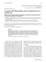
ple liver abscesses with possible necrosis with the
largest measuring abscess (Figure 1a). Entamoeba
titers were negative. Blood cultures drawn three
days after admission grew K. pneumoniae. The sus-
ceptibility pattern for the K. pneumoniae was not un-
usual. It was resistant to ampicillin and intermediate
to ampicillin/sulbactam. It was sensitive to the
cephalosporins (cefazolin, cefotaxime, and cefepime),
the fluoroquinolones (ciprofloxacin, levofloxacin), the
aminoglycosides (amikacin, gentamicin, and tobra-
mycin), meropenem, pipercillin/tazobactam and
trimethoprim/sulfa. At this point, the patient was
started on meropenem. The initial CT scan of the
brain done on arrival to Mount Sinai was negative.
However, follow-up CT of the brain seven days later
showed a 2.7 cm abscess located in the frontal lobe.
Two days later an additional lesion developed in the
left parietal region (Figure 1c).
Figure 1: A and B are computed tomography (CT) scans of the abdomen. (a) Before treatment showing multiple liver
abscesses with the largest measuring 3.9 cm. (b) After treatment showing improved resolution. C and D are CT scans of
the brain. (c) Nine-days after arrival an additional lesion developed in the left parietal region. (d) After treatment showing
improved resolution.
Int. J. Med. Sci. 2009, 6
303
The patient underwent CT-guided drainage and
biopsy of the largest liver abscess. The biopsy dem-
onstrated abundant acute and chronic inflammation
with surrounding necrosis consistent with a liver ab-
scess. A sample aspirated from the liver was submit-
ted for culture. The Gram stain of the material
showed many neutrophils, but no organisms. On the
third day of culture, there was growth of a mucoid
Gram-negative lactose-fermenting bacillus identified
as Klebsiella pneumoniae. This isolate, henceforth re-
ferred to as FIUMS1, had a characteristic hyperviscous
phenotype as demonstrated by the formation of
elongated (>5 mm) mucoviscous strings when a loop
was passed through a colony. Subsequently, his an-
tibiotic was changed to ceftriaxone. Three weeks after
admission, he became afebrile, was extubated, and the
brain and liver lesions improved radiologically (Fig-
ure 1b and 1d). He returned to Saint Kitts where he
remained clinically stable and completed six weeks of
antibiotic treatment.
Background
Klebsiella pneumoniae, a member of the Entero-
bacteriaceae family, is a Gram-negative enteric bacil-
lus that forms large mucoid colonies. Though rare,
this organism has been associated with bacterial liver
abscesses and metastatic infections with a predomi-
nance of cases in Taiwan [2]. This syndrome has
several distinguishing characteristics from traditional
liver abscesses including its community-acquired ori-
gin, absence of underlying hepatobiliary diseases, and
the presence of other invasive complications includ-
ing endophthalmitis, suppurative meningitis, brain
abscess, necrotizing fasciitis, and osteomyelitis [4].
Fang et al. reported that the invasive K. pneumoniae
strains associated with this syndrome more often ex-
hibited a hypermucoviscosity phenotype as demon-
strated by “extreme stickiness of these colonies on
agar plate” producing a positive string test [9]. The
hypermucoviscosity phenotype is highly associated
with community-acquired K. pneumoniae bacteraemia
that leads to distinctive invasive syndromes such as
liver abscess, meningitis, pleural empyema or
endophthalmitis [5].
Liver isolates belong to either serotype K1 or K2
[6, 10]. Recently, a number of genes have been iden-
tified as potential markers of virulence, including
magA, which has a high association with the K1 sero-
type and is more prevalent in strains isolated from
human liver abscesses [7]. The K2 serotype can be
detected by the presence of k
2
A [8].
Molecular Analysis
The presence of magA from the K. pneumonia
FIUMS1 (16S rRNA sequence deposited as GenBank
accession number FJ436718) was determined using
PCR. The 1,282 bp gene was amplified using the magA
Forward Primer (5’-GGT GCT CTT TAC ATC ATT
GC-3’) and magA Reverse Primer (5’-GCA ATG GCC
ATT TGC GTT AG-3’) [7]. A magA product was not
detected in the strain. To test for the K2 serotype, k
2
A
(523 bp) was amplified using the forward primer
(5’-CAA CCA TGG TGG TCG ATT AG-3’) and the
reverse primer (5’-TGG TAG CCA TAT CCC TTT
GG-3’) [8]. The k
2
A fragment of 523 bp was detected
(data not shown). These results demonstrate that
this pathogenic strain has a K2 serotype [11].
Though magA has been implicated in the hypermu-
coviscosity phenotype, the magA gene has only been
identified in 24% of clinical isolates [5]. Thus, it is not
surprising to find the K. pneumoniae FIUMS1 is
magA-negative. In addition, the K2 serotype of K.
pneumoniae is the second most common type of strain
isolated from liver abscesses [8].
In conclusion, through culture methods and 16S
rRNA sequencing, the strain isolated from the liver
abscess was verified as K. pneumoniae. The dengue
viral pneumoniae may have predisposed the St. Kitts
patient to the subsequent K. pneumoniae infection [12,
13]. The absence of magA gene and the presence of k
2
A
in this isolate indicate that the hypervirulence of the
strain is due to the K2 capsule. This form of K.
pneumoniae has not been previously noted on the is-
land of St. Kitts. Earlier identification of an infection
caused by this hypervirulent serotype should result
better patient prognosis.
Acknowledgements
Sequencing was performed at the Florida Inter-
national University DNA Core (Miami, Fl). MSD is
funded by MBRS Research Initiative for Scientific
Enhancement (RISE) Program NIH\NIGMS R25
GM061347. We would like to thank Steve Libby
(University of Washington, Seattle, WA) for the K.
pneumoniae control strain. We are extremely grateful
to Dr. J. Patrick O’Leary for his invaluable editorial
suggestions.
Conflicts of Interest
The authors have declared that no conflict on
interests exists.
References
1. Podschun R, Ullmann U. Klebsiella spp. as nosocomial patho-
gens: epidemiology, taxonomy, typing methods, and patho-
genicity factors. Clin Microbiol Rev. 1998; 11: 589-603.
2. Han SH. Review of hepatic abscess from Klebsiella pneumoniae.
An association with diabetes mellitus and septic
endophthalmitis. West J Med. 1995; 162: 220-4.
Int. J. Med. Sci. 2009, 6
304
3. Keynan Y, Karlowsky JA, Walus T, Rubinstein E. Pyogenic liver
abscess caused by hypermucoviscous Klebsiella pneumoniae.
Scand J Infect Dis. 2007; 39: 828-30.
4. Keynan Y, Rubinstein E. The changing face of Klebsiella pneu-
moniae infections in the community. Int J Antimicrob Agents.
2007; 30: 385-9.
5. Lee HC, Chuang YC, Yu WL, Lee NY, Chang CM, Ko NY,
Wang LR, Ko WC. Clinical implications of hypermucoviscosity
phenotype in Klebsiella pneumoniae isolates: association with
invasive syndrome in patients with community-acquired bac-
teraemia. J Intern Med. 2006; 259: 606-14.
6. Yeh KM, Kurup A, Siu LK, Koh YL, Fung CP, Lin JC, Chen TL,
Chang FY, Koh TH. Capsular serotype K1 or K2, rather than
magA and rmpA, is a major virulence determinant for Klebsiella
pneumoniae liver abscess in Singapore and Taiwan. J Clin Mi-
crobiol. 2007; 45: 466-71.
7. Fang CT, Chuang YP, Shun CT, Chang SC, Wang JT. A novel
virulence gene in Klebsiella pneumoniae strains causing primary
liver abscess and septic metastatic complications. J Exp Med.
2004; 199: 697-705.
8. Yu WL, Fung CP, Ko WC, Cheng KC, Lee CC, Chuang YC.
Polymerase chain reaction analysis for detecting capsule sero-
types K1 and K2 of Klebsiella pneumoniae causing abscesses of
the liver and other sites. J Infect Dis. 2007; 195: 1235-6.
9. Nadasy KA, Domiati-Saad R, Tribble MA. Invasive Klebsiella
pneumoniae syndrome in North America. Clin Infect Dis. 2007;
45: e25-8.
10. Wu KM, Li LH, Yan JJ, Tsao N, Liao TL, Tsai HC, Fung CP,
Chen HJ, Liu YM, Wang JT, Fang CT, Chang SC, Shu HY, Liu
TT, Chen YT, Shiau YR, Lauderdale TL, Su IJ, Kirby R, Tsai SF.
Genome Sequencing and Comparative Analysis of Klebsiella
pneumoniae NTUH-K2044, a strain causing liver abscess and
Meningitis. J Bacteriol. 2009; 191: 4492-501.
11. Chuang YP, Fang CT, Lai SY, Chang SC, Wang JT. Genetic
determinants of capsular serotype K1 of Klebsiella pneumoniae
causing primary pyogenic liver abscess. J Infect Dis. 2006; 193:
645-54.
12. Lee IK, Liu JW, Yang KD. Clinical characteristics and risk fac-
tors for concurrent bacteremia in adults with dengue hemor-
rhagic fever. Am J Trop Med Hyg. 2005; 72: 221-6.
13. Wang CC, Liu SF, Liao SC, Lee IK, Liu JW, Lin AS, Wu CC,
Chung YH, Lin MC. Acute respiratory failure in adult patients
with dengue virus infection. Am J Trop Med Hyg. 2007; 77:
151-8.