Báo cáo y học: "Revision of late periprosthetic infections of total hip endoprostheses: pros and cons of different concepts"
Bạn đang xem bản rút gọn của tài liệu. Xem và tải ngay bản đầy đủ của tài liệu tại đây (378.34 KB, 9 trang )
Int. J. Med. Sci. 2009, 6
287
I
I
n
n
t
t
e
e
r
r
n
n
a
a
t
t
i
i
o
o
n
n
a
a
l
l
J
J
o
o
u
u
r
r
n
n
a
a
l
l
o
o
f
f
M
M
e
e
d
d
i
i
c
c
a
a
l
l
S
S
c
c
i
i
e
e
n
n
c
c
e
e
s
s
2009; 6(5):287-295
© Ivyspring International Publisher. All rights reserved
Review
Revision of late periprosthetic infections of total hip endoprostheses: pros
and cons of different concepts
Bernd Fink
Department of Joint Replacement, General and Rheumatic Orthopaedics, Orthopaedic Clinic Markgröningen gGmbH,
Kurt-Lindemann-Weg 10, 71706 Markgröningen, Germany
Correspondence to: Prof. Dr. med. Bernd Fink, M.D., Department of Joint Replacement, General and Rheumatic Ortho-
paedics, Orthopaedic Clinic Markgröningen gGmbH, Kurt-Lindemann-Weg 10, 71706 Markgröningen gGmbH,
Kurt-Lindemann-Weg 10, 71706 Markgröningen, Germany, Phone: ++49-7145-912201, Fax: ++49-7145-912922, E-mail:
Rec
eived: 2009.04.16; Accepted: 2009.09.02; Published: 2009.09.04
Abstract
Many concepts have been devised for the treatment of late periprosthetic infections of total
hip prostheses. A two-stage revision with a temporary antibiotic-impregnated cement spacer
and a cemented prosthesis appears to be the most preferred procedure although, in recent
times, there seems to be a trend towards cementless implants and a shorter period of anti-
biotic treatment. Because of the differences in procedure, not only between studies but also
within studies, it cannot be decided which period of parenteral antibiotic treatment and
which spacer period is the most suitable. The fact that comparable rates of success can be
achieved with different treatment regimens emphasises the importance of surgical removal
of all foreign materials and the radical debridement of all infected and ischaemic tissues and
the contribution of these crucial procedures to the successful treatment of late peripros-
thetic infections.
Key words: periprosthetic infections, hip endoprostheses
Introduction
Periprosthetic infections occur with an incidence
of less than 1% of patients but nevertheless are a se-
rious complication of hip arthroplasties [1,2]. When
early infections occur, within 4 weeks of implantation,
the implant can be left in place with a high probability
of cure whereas late infections require prosthesis re-
vision to eradicate the infection [3,4]. In such cases,
one can differentiate between one-stage and two-stage
revisions. In the former a new prosthesis is implanted
immediately after the removal of all foreign material
in one operation. Two-stage revision involves an ini-
tial operation to remove all foreign materials and this
is followed by an interim phase of 6 – 10 weeks, either
left as a Girdlestone situation or with the implantation
of a cement spacer. Individual aspects of both forms
of revision have been treated very differently in the
past so, in the following paragraphs, the different
concepts are summarized and their respective ad-
vantages and disadvantages discussed.
One stage revision
The advantage of the one-stage revision is that
only one operation is required and functional prob-
lems associated with a Girdlestone situation, such as
leg shortening and instability, or, in the case of a ce-
ment spacer, spacer fracture, abraded particles from
the spacer or bone resorption, can be avoided. Most
surgeons have used bone cement laden with antibiot-
ics during the re-implantation whereby the antibiotic
contained in the cement or added to it is specific for
the pathogen involved [5-7]. A prerequisite for this
procedure is the isolation of the organism(s) from
Int. J. Med. Sci. 2009, 6
288
previously obtained aspirated fluid or biopsied mate-
rial and the determination of their antibiotic suscepti-
bility so that an organism-specific mixture of antibi-
otics can be added to the bone cement and a specific
local antibiotic treatment initiated [5,6]. Here it is
necessary for the fluid or tissue sample to be incu-
bated for 14 days [6,8,9]. This long incubation period
is necessary because the pathogens causing the pe-
riprosthetic infection usually occur in very small
numbers in the form of a biofilm and are also often in
a sessile state that is characterized by a slow rate of
reproduction [8,10-13]. An analysis we carried out of
110 infected hip and knee endoprostheses showed
that the culture detection rate after 7 days, the longest
incubation period reported in most studies, was a
mere 73.6%. To identify all infections it was necessary
to cultivate for 13 days [14]. If the incubation period is
of sufficient duration an accuracy of approximately
90% can be achieved with the aspiration method
[15,16]. We believe that a lack of sufficient incubation
led to the poor sensitivity of the pre-operative aspira-
tion reported in other studies (for example, 46.1%
reported by Hoffmann et al. [17]). The degree of suc-
cess of one-stage revision of prostheses with antibiot-
ics added to the cement led to 88% eradication re-
ported by Steinbrink et al. [6], to 91% reported by
Wroblewski et al. [7] and to 93,7% in a newer report
by Rudelli et al. [18].
Mixing antibiotic into the cement affects the
quality of the cement, which is why only antibiotic
powder to a maximum of 10% of the total cement
amount should be used [19]. Not all antibiotics can be
used because they have to be available in powder
form, be water-soluble and be thermostable. The most
commonly used are gentamicin, clindamycin, van-
comycin, tobramycin, aztreonam, ampicillin and
ofloxacin [1,19-21]. There is little data available that
addresses the release of antibiotics from spacers in
vivo over a period of several weeks although the level
of released antibiotic has been suggested by several
authors to be sufficient for at least 4 months [21-23].
Furthermore, it has been found that the antibiotics
affect each other's elution from the cement whereby
the use of two antibiotics results in a synergistic effect
and the release of the individual components is higher
than that of the single antibiotics on their own [24-28].
It has also been demonstrated that the elution of anti-
biotic from hand-mixed cement is higher than that
from cement mixed under vacuum because of the
presence of air bubbles and their greater surface area.
However the mechanical characteristics of
hand-mixed cement are not as good [19].
Some newer studies of one-stage cementless re-
vision of septic prostheses described the use of can-
cellous allografts that had been impregnated with
antibiotics. Winkler et al. [29] reported 37 such cases
of one-stage cementless revisions and demonstrated
an eradication rate of 92% after a follow-up period of
4.4 years.
A one-stage revision can be indicated irrespec-
tive of the concept involved when a microorganism
has been identified but spacer implantation is not
possible because of a severely defective acetabulum
and a Girdlestone situation is undesirable.
Two-stage revision
Two-stage septic revision surgery is the most
common method for treating infected endoprostheses.
A general advantage of the two-stage concept is that
the surgical debridement is carried out twice whereby
the second operation allows for the eradication of
residual organisms following the initial debridement.
The cement of the spacer is not intended as a means of
fixing the prosthesis so the mechanical characteristics
of the cement is not of primary importance at this
stage. Thus, large amounts of antibiotics can be mixed
into the cement before the spacer is formed. It has
been possible to achieve a survival rate using
two-stage revision concepts for infected hip arthro-
plasties of between 90% and 100% [1,30-32].
In most two-stage revisions an antibi-
otic-containing spacer is usually placed in position for
a certain period of time before the final prosthesis is
implanted [17,20,30,33,34]. The function of the spacer
is on the one hand to release the antibiotic into the
infected bed of the prosthesis and on the other to
minimize soft-tissue contractures, retain soft tissue
tension and so maintain reasonable functionality until
a prosthesis can be re-implanted [30]. There are sev-
eral different types of spacer: monoblock and
two-part spacers, commercially available and cus-
tomized spacers made in the operating theatre. The
potential disadvantages of the monoblock spacers are
spacer fracture and bone resorption while the
two-part spacer can produce abraded cement particles
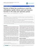
[35-37]. In order to avoid spacer fractures we use a
two-part spacer where the cup-shaped acetabulum
spacer is formed out of antibiotic loaded cement (with
a specific mixture of antibiotics recommended by the
microbiologist). The spacer stem component consists
of old prosthesis stem models, monoblock devices in
most cases and no longer used for primary implanta-
tions, that are encased in antibiotic-supplemented
cement and, just before implantation, coated in the
patient's own blood in order to facilitate easier re-
moval. The two spacer components are connected by
a metal headpiece (Figure 1) [20]. However, a recent
analysis of synovial membranes obtained during the
Int. J. Med. Sci. 2009, 6
289
operation to remove the spacer and to implant the
new prosthesis revealed the presence of abraded ce-
ment debris, in particular, zirconium dioxide particles
[unpublished data].
Figure 1: Radiograph of a hip spacer of a 63year old man
with late periprosthetic infection of the left hip
Another concept involves the use of antibi-
otic-laden beads although a disadvantage of this
procedure is that ready-manufactured beads are usu-
ally employed and these only contain gentamicin or
vancomycin [38,39]. Leg shortening and instability
still occur and cause problems with mobilization.
Re-implantation of a prosthesis is also often made
more difficult because of scarring, tissue shrinkage
and osteoporosis caused by inactivity [37,40,41]. In
addition, abrasion of zirconium dioxide particles is to
be expected during mobilization and this could lead
to third-body-wear following re-implantation of the
prosthesis. Disch et al. [35] decided therefore not to
use local antibiotic carriers following removal of the
prosthesis during two-stage revisions and found a
reinfection rate of 6.3% in 32 hips and 41.3 months
after re-implantation although there was a consider-
able reduction in the quality of life during the Girdle-
stone phase which lasted 13 months on average.
There are many questions pertaining to both
one-stage and two-stage revisions that still have to be
answered and existing procedures are based more on
empirical findings than on data from prospective
studies with a high level of evidence. It is for this
reason that the following aspects of two-stage revision
have been treated very differently by different groups:
the type of antibiotic used in the spacer, the duration
of the spacer period, the duration of systemic antibi-
otic treatment, aspiration before re-implantation and
the type of re-implantation (cemented or cementless).
Type of antibiotic used in the spacer
Most published studies always include the same
antibiotics in the cement. Some authors use vanco-
mycin and tobramycin as local antibiotics on a regular
basis because they have a broad spectrum of activity
[38,42]. However, not all bacteria can be successfully
treated with these agents (e.g., some gram-negative
organisms), so this is an argument for investigating
the antibiotic resistance pattern of the isolated bacteria
and selecting a specific antibiotic for the treatment.
Masri et al. [43] reported a success rate of 89.7% in
their retrospective study involving bacteria-specific
antibiotic mixed into the cement of a PROSTALAC®
spacer (DePuy Orthopaedics, Inc, Warsaw, IN) and
we saw no reinfection of 36 cases with a minimum
follow-up of 2 years using this concept for handmade
spacers [20].
Duration of antibiotic treatment
While most authors carry out a 6 week period of
intravenous antibiotic therapy, there is a great variety
of treatment regimens (Tables 1 and 2). In more recent
studies, very much shorter periods of antibiotic
treatment have been employed. Whittaker et al [44]
reported a 92.7% eradication of infection for 41
re-implanted hip endoprostheses over a follow-up
period of 4 years following a short, intravenous
treatment with vancomycin alone in combination
with cement spacers containing vancomycin and
gentamicin. McKenna et al. [45] only found one rein-
fection after an average of 35 month's follow-up of 30
patients with infected hip arthroplasties who as part
of the two-stage revision procedure, only received a 5
day systemic treatment with antibiotics. The design of
the antibiotic administration after re-implantation of
the prosthesis is even more variable and range from
no antibiotic treatment at all to three months of
post-surgery treatment (Tables 1 and 2).
The fact that there are differences in procedure
not only between studies but also within studies
means it cannot be decided which period of par-
enteral antibiotic treatment is the most suitable. That
different durations of antibiotic therapy lead to simi-
lar clinical results emphasizes the fact that treatment
with antibiotics is only a form of support therapy for
the periprosthetic infection and that the crucial fea-
Int. J. Med. Sci. 2009, 6
290
tures of all concepts are the rigorous surgical removal
of foreign material and the radical debridement of all
infected and ischaemic tissues. These procedures are
vital for the success of the revision process. However,
in cases of haematogenous infection the systemic an-
tibiotic therapy is essential for treating the focus and
preventing of septic metastases.
Duration of the spacer period and antibiotic
therapy
The period of time between the two operations
of a two-stage revision is also very variable, ranging
from a few days to several years (Tables 1 and 2).
Many authors determine the time of re-implantation
of a prosthesis according to clinical parameters and
clinical chemistry data and carry out an aspiration of
the area before surgery is carried out [32,36,43,46].
Other authors have a more or less rigid procedural
plan [31,33,39]. These differences in procedure, not
only between studies but also within studies, means
that it cannot be decided which time period between
the two steps and spacer period is the most suitable.
This also appears to underscore the importance of the
surgical debridement for therapeutic success of the
two-stage revision.
Aspiration before re-implantation
Many authors recommend aspiration before the
re-implantation operation in order to check whether
or not the joint is free of infection [43,47]. The disad-
vantage of this concept is that the second aspiration
requires a pause in the antibiotic therapy for at least 2
weeks, if not 4 weeks [48]. This is then followed by a
2-week incubation period so the second operation can
be delayed by up to 4 or 6 weeks. Moreover, the local
levels of antibiotic released by the spacer would likely
influence the detection of viable bacteria [3]. For these
reasons we do not perform an aspiration before
re-implantation and rather make a decision based on
clinical findings and CRP values as described by
Hsieh et al. [41,49].
Cemented re-implantation
The fixation method chosen for the final pros-
thesis in the two-stage technique usually involves the
use of cement because this allows the surgeon to add
antibiotics to the cement to help prevent recurrent
infection [1,31-33,50]. Rates of eradication between
84% and 100% have been described for this procedure
(Table 1).
Table 1: Results of two-stage cemented revision of periprosthetic infection of the hip.
Author N Fol-
low-up
Spacer/
Beads
Local
anti-biotics
Duration of
intravenous
antibiotics
Interval until
re-implan-tation
Antbiotics
after im-
planta-tion
Eradi-cation
rate
Aseptic loos-
ening
McDonald
[46]
82 5.5 years Resection
arthroplasty
No 26.1 (4 – 59
days)
1.5 years (6 days
– 6.2 years)
No antibiot-
ics in cement
87 % n.r.
Colyer [51] 37 2.7 years Resection
arthroplasty
No 6 weeks par-
enteral
6 weeks (4 – 214
weeks)
2 weeks par-
enteral, 3
months oral
84 % n.r.
Garvin [31] 32 ≥ 2 years,
4.1 years
Beads Gentamicin 6 weeks par-
enteral
6 weeks n.r. 91 % 0 %
Lieberman
[32]
32 40
(24-80)
mo
Beads
Spacer
Gentamicin
Tobramycin
Vancomycin
6 weeks (20 – 49
days)
8,8 weeks (3
weeks – 32
months)
n.r. 91 % n.r.
Younger [52] 48 43
(24-63)
mo
Spacer Gentamicin 3 weeks par-
enteral, 3 weeks
oral
13 weeks (5 – 42
weeks)
3 weeks par-
enteral, 3
weeks oral
94 % 0 %
Leunig [37] 12 2.2 years Spacer Gentamicin n.r. 4 (2-7) months 100 % n.r.
Evans [33] 23 Spacer Gentamicin 6 weeks 12 weeks No 95.7 % n.r.
Hsieh [36] 24 4.2 years Spacer Specific:
Vancomycin
Piperacillin
Aztreonam
Teicoplanin
2 weeks par-
enteral,
4 weeks oral
11 – 17 weeks,
when CRP
normal
1 week par-
enteral
100 % 0 %
Cementless re-implantation
The disadvantage of the cemented revision
technique is related to the fact that the osseous bed of
the prosthesis has not only been enlarged by the
loosening of the primary prosthesis but also become
thinner and sclerotic. This reduces the ability of the
cement to adhere to the bone. Dohmae et al. [53] re-
ported the resistance of the bone-cement interface to
shear force-related failure is reduced by 79% when
comparing a cemented revision implant to a cemented
primary implant. Wirtz and Niethard [54] reported a
higher revision rate associated with aseptic loosening
of cemented revision prostheses compared to ce-
mentless components (i.e., 15.1% versus 4.3% for the
acetabular cup and 12.7% versus 5.5% for the stem).
Therefore, the advantage of cementless revision may
also exist for implant fixation in two-stage septic re-
Int. J. Med. Sci. 2009, 6
291
visions although exact data concerning mid- and
long-term survival rates of cemented and cementless
implants in septic revision are rare in the literature
[40]. Sanchez-Sotelo et al. [55] reported a 10-year in-
fection-free survival rate of 87,5% and a mechanical
survival rate of only 75,2% for re-implanted femoral
components mostly fixed with cement.
Nevertheless, because the use of cementless
components at the second stage does not allow the
surgeon to add local antibiotics to the cement to help
prevent recurrent infection, there is some concern that
recurrent infection rates will be higher with cement-
less fixation [50,56]. A few retrospective studies have
reported promising results with two-stage revision
operations using cementless implants with rates of
eradication between 82% and 100% (Table 2)
[38,39,43,56-59].
Table 2: Results of two-stage cementless revision of periprosthetic infection of the hip.
Author N Fol-
low-up
Spacer/
Beads
Local
anti-biotics
Duration of
intravenous
antibiotics
Interval until
re-implan-tation
Antibiotics
after im-
planta-tion
Eradi-cation
rate
Aseptic loos-
ening
Wilson [56] 22/
13**
≥ 3 years,
48
months
Resection
arthroplasty
no 3 weeks par-
enteral
6-12 weeks 3 days par-
enteral
91 % /
100 % ce-
mentless
7.6 % stem
loose
Nestor [58] 34 47
(24-72)
mo
Resection
arthroplasty
no ≥ 4 weeks par-
enteral
8 (3-19) months different 82 % 18% stem
loose
Fehring [38] 25 41
(24-98)
mo
Beads Tobramicin in
16 cases
6 weeks par-
enteral
4.8 months 92 % 0 %
Haddad [39] 50 5.8
(2-8.7)
years
Beads + ce-
ment ball
Gentamycin 5 days par-
enteral and than
oral
3 weeks ≥ 3 months 92 % 8% stem
subsidence
Koo [57] 22 41
(24-78)
mo
Spacer
Beads
Vancomycin
Gentamicin
Cefotaxime
6 weeks 6-12 weeks n.r. 95 % 5%cup loose
30% stem
subsid.
Hofmann
[17]
27 76
(28-148)
mo
Old stem and
new poly-
ethy-lene cup
Tobramicin 6 weeks par-
enteral, in 17
cases additional
oral for 6 weeks
n.r. n.r. 94 % 0 %
Kraay [42] 33 ≥ 2 years Spacer in 16
cases
Tobramicin in
16 cases
≥ 6 weeks par-
enteral
7.4 (3-37)
months
n.r. 92 % 9 % cup
0% stem
Masri [43] 29 ≥ 2 years Prostalac
spacer
Tobramicin
Vancomycin
Cefuroxime
Penicillin*
6 weeks par-
enteral or in
combina-tion
with oral
12 weeks 5 days in-
tra-venous
90 % 0 %
Yamamoto
[60]
17 38 mo Spacer Gentamicin
Vancomycin
> 3 weeks n.r. 1 week par-
enteral, oral
until CRP
normal
100 % n.r.
Fink [20] 36 ≥ 2 years Spacer Specific:
Gentamicin
Clindamycin
Vancomycin
Ampicillin
Ofloxacin
2 weeks par-
enteral,
4 weeks oral
6 weeks 2 weeks par-
enteral,
4 weeks oral
100 % 6% stem
subsidence
0%
loose-ning
* = combination of another local antibiotic with tobramycin, mo = months, ** = 13 of 22 re-implantations without cement; stem subsid = stem
subsidence; nm = non-modular; pf = proximal fixation
Some reports describe the stability of cementless
fixation after septic revision surgery using mostly
non-modular implants: Fehring et al. [38] achieved
stable bone-ingrown fixation in 96% of their cases
using non-modular and modular cementless pros-
theses with proximal fixation, while Nestor et al. [58]
reported an implant stability of 79% using
non-modular, proximal porous-coated stems. Wilson
and Dorr [56] on the other hand, only achieved a 38%
bone-ingrown fixation after 3 years in, admittedly, a
small group of 13 patients using a cementless
non-modular stem with proximal fixation. Moreover,
the rate of early loosening of cementless revisions
stems varies from 0% to 18% (Table 2). We found low
rates of subsidence (6%) and loosening (0%) and a
high rate of bone-ingrown fixation (94%) of a ce-
mentless modular revision stem system (Revitan
curved, Zimmer GmbH, Winterthur, Switzerland),
which we believe is due to the distal fixation proce-
dures in viable bone on the one hand and to the