Báo cáo y học: "surgical Management of Hidradenitis Suppurativa"
Bạn đang xem bản rút gọn của tài liệu. Xem và tải ngay bản đầy đủ của tài liệu tại đây (1.07 MB, 8 trang )
Int. J. Med. Sci. 2010, 7
240
I
I
n
n
t
t
e
e
r
r
n
n
a
a
t
t
i
i
o
o
n
n
a
a
l
l
J
J
o
o
u
u
r
r
n
n
a
a
l
l
o
o
f
f
M
M
e
e
d
d
i
i
c
c
a
a
l
l
S
S
c
c
i
i
e
e
n
n
c
c
e
e
s
s
2010; 7(4):240-247
© Ivyspring International Publisher. All rights reserved
Research Paper
Surgical Management of Hidradenitis Suppurativa
Adnan Menderes, Ozgur Sunay
, Haluk Vayvada, Mustafa Yilmaz
Plastic Rec. and Aesthetic Surgery, Medical Faculty, Dokuz Eylul University, Inciraltı-Izmir, Turkey
Corresponding author: Ozgur Sunay, M.D., Dokuz Eylul Universitesi Tıp Fakultesi Plastik Rek. ve Estetik Cerrahi A.D.
Inciraltı-Izmir Turkey. Tel: +90 5055250343; e-mail: ;
Received: 2010.06.04; Accepted: 2010.07.12; Published: 2010.07.19
Abstract
Background: Hidradenitis suppurativa (HS) is a chronic, relapsing inflammatory disease of
skin, characterized by recurrent draining sinuses and abscesses, predominantly in skin folds
carrying terminal hairs and apocrine glands.
Method: This study reviewed 54 sites in 27 patients with moderate to extensive chronic
inflammatory skin lesions treated surgically in our hospital from 2004 through 2009, with a
follow-up of at least 6 months.
Result: A total number of 54 operative procedures were performed during the study period
with 42% (23 sites) involving the axilla, 20% (11 sites) involving the gluteal area, %24 (13 sites)
involving the perineal area and 12% (7 sites) involving the inguinal region.
Conclusion: Conservative treatment methods have little or no effects especially on gluteal,
perineal/perianal, axillary hidradenitis suppurativa. The morbidity associated with the estab-
lished form of this disease is significant, and the only successful treatment is wide surgical
excision.
Key words: Hidradenitis Suppurativa, Surgery, Treatment, Gluteal, Axillary, Perianal
Introduction
Hidradenitis suppurativa (HS) is a chronic, re-
lapsing inflammatory disease of skin, characterized
by recurrent draining sinuses and abscesses, predo-
minantly in skin folds carrying terminal hairs and
apocrine glands.
1
The incidence may be as high as one
in 300.
2
Hidradenitis suppurativa (from the Greek hi-
dros, sweat and aden, glands), also known as acne
inversa, was first described by Velpeau, a French
physician in 1839, who reported a peculiar inflamma-
tion of the skin with the formation of superficial ab-
scesses in the axillary, mammary and perianal areas.
3
In 1854, this condition was termed ‘hidrosade´nite
phlegmoneuse’ by Verneuil, a French surgeon who also
suggested an association between HS and sweat
glands, which had been described by Purkinje in
1833.
1
Hidradenitis suppurative may affect any area of
the body surface where apocrine glandular tissue is
found, but most often it affects the skin of the axillae
and inguinoperineal regions.
2
Although the pathophysiology is understood
poorly, it generally is believed that obstruction of the
apocrine and/or follicular pores results in glandular
dilatation and bacterial superinfection with subse-
quent gland rupture disseminating infection
throughout the subcutaneous tissue plane.
3
Conse-
quently, hidradenitis is associated with chronic pain-
ful abscesses, multiple odiferous draining sinus tracts,
and chronic fibrosis with range-limiting scar forma-
tion.
4
Anogenital involvement most commonly affects
the groins with extension to inguinal regions, mons
pubis, inner thighs and sides of scrotum. The peri-
neum, buttocks and perianal folds are often included.
Int. J. Med. Sci. 2010, 7
241
The sinuses can dissect deep into tissue, involving
muscle, fascia and bowel forming a labyrinth of tracts
in advanced cases.
8
However, as the abscesses extend deeper into the
subcutaneous tissue, intercommunicating sinus tracts
develop, resulting in irregular hypertrophic scars.
6
Rarely, the chronic inflammation results in malignant
transformation to squamous cell carcinoma.
10
In such
a developed phase, antibiotics are usually ineffective
alone and surgical treatment is required.
4,9,14
The exact etiology of HS still remains unclear,
genetic factors may play a role as a positive family
history has been elicited in 26% of patients with HS.
The role of endocrine factors in the etiology of HS has
been controversial.
1
There is no consensus about the relationship
between HS and sex, race, and site of the lesions.
Axillary location seems to be more frequent in wom-
en. The gluteal, inguinal, perineal, and perianal zones
are more frequently involved in men. HS appears
more commonly in young adults and is observed after
puberty.
3
In women, the condition frequently flares pre-
menstrually and following pregnancy and it some-
times eases during pregnancy and after the meno-
pause; these observations incriminate sex hormones.
Children are never affected unless they have preco-
cious puberty.
8
Although exogenous factors such as the use of
deodorants and shaving are thought to be causal, they
have not been shown to be significantly responsible in
a retrospective comparison of 40 patients with HS.
13
Smoking is more common in patients with HS but the
aetiological basis is unknown. From the exceedingly
high rate of smokers among patients with this condi-
tion one may conclude that cigarette smoking is a
major triggering factor of hidradenitis suppurativa.
15
Wiltz et al. reported an association between smoking
and perianal HS in 70% of patients.
1
Obesity is an ex-
acerbating factor, and weight loss can help control the
disease severity.
1,4
Early-stage treatment consists primarily of topi-
cal (clindamycin) or systemic antibiotics (tetracyc-
lines, clindamycin, rifampicin), topical antiseptics and
intralesional corticosteroids (triamcinolone aceto-
nide). Systemic retinoids (isotretinoin, etretinate) an-
tiandrogen therapy (cyproterone acetate, finasteride),
immunotherapy (TNF alfa inhibitors), oral immuno-
suppressive agents (cyclosporin) have also shown a
positive effect on disease progression.
4,12
Radiothera-
py and laser treatment applied cases available on li-
terature.
1
Currently available medical treatments are,
however, insufficient and their efficacy is only tran-
sient. As a result, advanced-stage severe HS requires
invasive surgical removal of all the involved tis-
sue.
1,5,8,11
In this report, we present our experience with
moderate and extensive perineal, perianal, axillary
and gluteal hidradenitis suppurativa cases, including
our treatment methods and outcomes.
Patients and Methods
This study reviewed 54 sites in 27 patients with
moderate to extensive chronic inflammatory skin le-
sions treated surgically in our hospital from 2004
through 2009, with a follow-up of at least 6 months.
Nineteen (%70) patients were men and eight patients
were women. The mean age at the time of presenta-
tion for operative management was 41.2 years (range,
24-58 y) and the average duration of symptomatic
disease was 7.3 years (range, 0.9-30 y). None of these
patients were detected to have any comorbid or asso-
ciated conditions. According to answers about clean-
ing habits; personal hygiene was poor in most of the
patients. 18 of the 21 (85%) male patients and 3 of the 8
(37%) female patients were smokers. 3 patients (2 fe-
male, 1 male) had insulin-dependent diabetes melli-
tus. (See Table 1) Most of the included patients had
previously been prescribed a treatment by non sur-
gical or inadequate surgical treatment modalities such
as short term antibiotic treatments, local wound care
and abscess drainage for long periods (up to 20 years).
Seven patients previously were treated by limited
local excisions and primary closure.
Total surgical excisions under general anaesthe-
sia were performed on all patients. All patients were
operated on in the lithotomy, jackknife, supine or
prone positions. Rectal tubes were used for preven-
tion of the surgical field from contamination with
stool in the patients with perianal or gluteal lesions,
colostomy was not used in any patients for this pur-
pose. The operative technique was based on the com-
plete excision of the entire diseased skin and subcu-
taneous fatty tissue and down to the muscular fascia
on aggressive cases. Patients with limited disease in-
volving the axilla or inguinal region were selected for
excision and primary closure if the skin and soft tissue
could be mobilized adequately. The size of the defects
created after excision of the lesions ranged from 15
cm
2
up to 1680 (42X40) cm
2
. Preoperative and post-
operative antibiotherapy is administered for all pa-
tients according to wound tissue culture tests results.
Culture results were Proteus Mirabilis, Staphylococ-
cus Aureus and E. Coli predominantly, and were
sensitive to Ciprofloxacin and Seftazidim antibiotic
protocol mostly.
Int. J. Med. Sci. 2010, 7
242
Table 1. Patient characteristics.
Patient
No.
Sex Age Smoking,p/y Site Defect Size,cm Duration
of ill-
ness
/y
Surgical Method Follow-up,mo Recurrence
1 M 50 - Gluteal 10X15 5 Skin grafting 11 -
2 F 42 - Axilla 8X12 0.9 Parascapular fasciacutaneous
fla
p
33 -
3 M 39 0.5/20 Inguinal 3X10 3 Local fasciacutaneous flap 15 +
4 M 47 1/30 Bilateral Axilla 8X3,6X4 2 Primary closure 48 -
5 F 24 - Axilla 3X5 1 Primary closure 10 -
6 M 38 1/12 Gluteal 9X13 1.5 Skin grafting 17 -
7 M 49 1.5/22 Gluteal 14X11 6 Skin grafting 16 -
8 M 56 1/30 Gluteal 14x12 20 Skin grafting 24 -
Bilateral İnguinal 3X15, 4X10 Primary closure -
Bilateral Perineal 20X20,18X20 Skin grafting -
9 M 47 1/23 Gluteal,perianal 30X20 3 Skin grafting 36 -
Perineal 5X15 Primary closure -
10 F 57 - Gluteal,perianal 42X40 30 Skin grafting 40 -
Bilateral Perineal 20X20,15X20
Skin grafting -
Bilateral axilla 18X10,15X12 Primary closure
11 F 55 - Bilateral Peri-
neal,
15X10 2 Primary closure 36 -
Bilateral İnguinal
9X12
Primary closure -
Bilateral Axilla
3X5
Primary closure
12 M 51 2/25 Gluteal,perianal 21X38 12 Skin grafting 21 -
13 M 24 1/5 Axilla 3X10 3 Primary closure 6 -
14 M 24 - Bilateral Axilla 20X15,15X15 6 Parascapular fasciacutaneous
12 -
15 M 32 2/10 Axilla
10X10,
4 Parascapular fasciacutaneous 20 -
İnguinal
2X10
Primary closure
-
Perineal 10X18 Gracilis musculocutaneous flap -
16 M 42 1/20 Axilla 12X12 16 Parascapular fasciacutaneous
flap
18 -
17 M 39 1/15 Axilla
5x8
4
Primary closure 10 -
Gluteal 16X20 Skin grafting -
Perineal 2X8 Primary closure -
18 F 45 - Axilla 10X5 5 Parascapular fasciacutaneous
flap
20 -
19 M 58 1/30 Gluteal,perianal 15X20 20 Fasciacutaneous V-Y advance-
ment
10 -
20 M 40 2/20 Gluteal,perianal
20X42
7 Skin grafting
48 -
Perineal 15x12 Fasciacutaneous flap -
21 M 45 3/22 Bilateral axilla 15X8,15x10 12 Parascapular fasciacutaneous
flap
20 -
22 M 55 1/30 Gluteal 10X12 7 Local transposition flap 8 -
23 F 30 1/12 Axilla 3X9 15 Primary closure 16 -
İnguinal 4X10 Primary closure -
24 F 32 0.5/5 Bilateral axilla 4X8,3X8 8 Primary closure 9 -
25 M 45 1/22 Bilateral axilla
3X7,12X6
5
Parascapular fasciacutaneous
fla
p
7 -
Bilateral perineal 20X10,8X15 Fasciacutaneous flap +
26 M 24 - Perineal 4x5 2 Primary closure 12 -
27 F 25 - Axilla 3X7 3 Primary closure 10 -
Int. J. Med. Sci. 2010, 7
243
Patient reports
Patient 8
A 56-year old male patient had recurrent and
progressive lesions on his perineal, inguinal, perianal
and gluteal regions for 20 years. Drainage, local
wound care and antibiotherapy was used previously
every time the lesions were exacerbated. Perineal le-
sions also affected the scrotal skin. (See Figure 1) The
lesions were excised widely and the scrotal skin down
to the dartos fascia anal sphincter was preserved. The
Inguinal defect was closed primarily and the perineal
lesions were reconstructed by split thickness skin
grafts. Gluteal lesions were excised and skin grafted 8
months later at a second stage.
Figure 1: Patient 8 (56 years). (A) Preoperative view:
Preineal area with putrid productive infection. (B) Post-
operative result after 1 year, no scar contracture, no re-
currence.
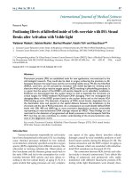
Patient 10
57-year old female patient. 30 years ago a lesion
was excised from the coccygeal region but recurred 10
years later and treatment with retinoids was started.
However the lesions relapsed and disseminated to a
wide area over time (Figure 2). The lesions of this pa-
tient were very extensive and drastic which are rare in
the literature and especially the effects of the disease
on the perineal area was dramatic. Both gluteal areas,
perianal, perineal, inguinal and axillary regions were
affected. The lesion on right labia majora was en-
larged to 10x15 cm and left labia majora was 10x20
cm. Surgical treatment was performed in 3 stages. At
the first stage all perineal and gluteal lesions were
excised with at least 1 cm margins. The size of the
excised material from the gluteal region was 42x40
cm. Split thickness skin grafts from the thigh were
used for reconstruction. Excision and primary closure
of the axillary lesions were done in second and third
stages. We obtained excellent functional results (con-
venience during urination etc.) and moderate cos-
metic results.
Figure 2: Patient 10. A 57–year-old female patient with
very severe widespread Hidradenitis suppurativa. (A,B)
Preoperative view of inguinal,perineal and gluteal areas.
(C,D) Postoperative results after 6 month. (E) View of
postoperative 3 year.
Patient 14
24-year old male patient had bilateral axillary
hidradenitis suppurativa for the last 6 years. Both
axillae were treated in the same stage. Excised area
was 20x15 cm on the right side and 15x15 cm on the
left side. Thoracodorsal perforator based fasciocuta-
neous flaps were used for reconstruction. (See Figure
3).
Int. J. Med. Sci. 2010, 7
244
Figure 3. Patient 14. A 24-year-old male. (A,B) Preo-
perative view of axillary region. (C,D) The axillary defect is
covered with toracodorsal perforator based fasciocuta-
neous flaps. (Intraoperative view)
Patient 19
58-year old male patient had draining lesions in
his intergluteal and perianal regions for the last 20
years. Abscess drainage was performed at least 3-4
times every year and he had frequent use of various
oral antibiotics. We performed total excision of a
15x20 cm lesion with 1 cm surgical margins down to
the muscle fascia. The external anal sphincter was
protected. A right sided gluteal V-Y advancement flap
was used for the closure of the defect. (Figure 4)
Figure 4. Patient 19. A 58-year-old male patient with
severe hidradenitis suppurativa of the gluteal and perianal
area. (A) Preoperative view with fistulas in intergluteal
area.(B) After excision of the inflammatory region and
closed with V-Y advancement fasciacutaneous flap.
Results
A total number of 54 operative procedures were
performed during the study period with 42,6% (23
sites) involving the axilla, 20,3% (11 sites) involving
the gluteal area, %24,2 (13 sites) involving the perineal
area and 12,9% (7 sites) involving the inguinal region.
Hurley’s clinical staging was used for the classifica-
tion of patients (Table 2). Excision and primary clo-
sure was used only for mild and moderate (Hurley
stage I) axillary and inguinal disease, whereas wide
local excision and split-thickness skin grafting or fas-
ciocutaneous flap was the mainstay of treatment in
patients with diffuse disease (Hurley stage II and III).
Table 2. Hurley Staging System.
Stage Characteristics
I Solitary or multiple isolated abscess formation without
scarrin
g or sinus tracts.
II Recurrent abscesses, single or multiple widely separated
lesions, with sinus tract formatio
n.
III Diffuse or broad involvement across a regional area with
multi
ple interconnected sinus tracts and abscesses.
Surgical margins were at least 0.5-1 cm in the
axillary region and 1-1.5 cm in the gluteal region and
down to the muscle fascia. Affected labia majora and
scrotal skin was also excised widely. In the perianal
region lesions were excised by protecting the external
anal sphincter and none of the patients required en-
doanal excision, Colostomy was not performed for
any patient.
Treatment was performed in 2 stages in three
patients and 3 stages in one patient. For the recon-
struction of the glutal region Split thickness skin graft
(STSG) was used in 9 patients (Figure 5,6), fasciocu-
taneous V-Y advancement flap in one patient and
transposition flap in 1 patient. Primary closure was