Báo cáo y học: "Identification of clinical and simple laboratory variables predicting responsible gastrointestinal lesions in patients with iron deficiency anemia"
Bạn đang xem bản rút gọn của tài liệu. Xem và tải ngay bản đầy đủ của tài liệu tại đây (330.56 KB, 9 trang )
Int. J. Med. Sci. 2011, 8
30
I
I
n
n
t
t
e
e
r
r
n
n
a
a
t
t
i
i
o
o
n
n
a
a
l
l
J
J
o
o
u
u
r
r
n
n
a
a
l
l
o
o
f
f
M
M
e
e
d
d
i
i
c
c
a
a
l
l
S
S
c
c
i
i
e
e
n
n
c
c
e
e
s
s
2011; 8(1):30-38
© Ivyspring International Publisher. All rights reserved.
Research Paper
Identification of clinical and simple laboratory variables predicting re-
sponsible gastrointestinal lesions in patients with iron deficiency anemia
Songul Serefhanoglu
, Yahya Buyukasik, Hakan Emmungil, Nilgun Sayinalp, Ibrahim Celalettin Hazne-
daroglu, Hakan Goker, Salih Aksu, Osman Ilhami Ozcebe
Hacettepe University Hospital, Department of Internal Medicine, Division of Hematology, Ankara, Turkey
Corresponding author: Songul Serefhanoglu, Hacettepe University Hospital, Department of Internal Medicine, Division
of Hematology, Ankara, Turkey. E-mail: ; Tlf: +903123051543.
Received: 2010.08.29; Accepted: 2010.12.20; Published: 2010.12.28
Abstract
I r o n d e f i c i e n c y a n e m i a ( I D A ) i s a f r e q u e n t d i s o r d e r . A l s o , i t m a y b e a s i g n o f u n d e r l y i n g s e rious
d i s e a s e s . I r o n d e f i c i e n c y p o i n t s t o a n o c c u l t o r f r a n k b l e e d i n g l e s i o n w h e n o c c u r r e d i n m e n o r
p o s t m e n o p a u s a l w o m e n . I n t h i s s t u d y , w e a i m e d t o e v a l u a t e t h e d i a g n o s t i c y i e l d o f e n d o s c o p y
in patients with IDA and to define predictive factors of gastrointestinal (GI) lesions causing
IDA. Ninety-one patients (77 women, 14 men; mean age: 43 years) who were decided to have
esophago-duodenoscopy and/or colonoscopy for iron deficiency anemia were interviewed
and responded to a questionnaire that included clinical and biochemical variables. The en-
doscopic findings were recorded as GI lesions causing IDA or not causing IDA. Endoscopy
r e v e a l e d a s o u r c e o f I D A i n 1 8 . 6 % o f c a s e s . T h e r i s k f a c t o r s f o r f i n d i n g G I l e s i o n s c a u s i n g I D A
were as follows: male gender (p= 0.004), advanced age (> 50 years) (p= 0.010), weight loss
(over 20% of total body weight lost in last 6 month) (p= 0.020), chronic diarrhea (p= 0.006),
change of bowel habits (p= 0.043), epigastric tenderness (p= 0.037), raised carcinoembryonic
antigen (CEA) level (normal range: 0-7 ng/mL) (p= 0.039), < 10 gr/dl hemoglobin (Hb) level
(p=0.054). None of these risk factors had been present in 21 (23%) women younger than 51
years. In this group, no patient had any GI lesion likely to cause IDA (negative predictive
value= 100%). In multivariate analysis, advanced age (p=0.017), male gender (p< 0.01) and
weight lost (p=0.012) found that associated with GI lesions in all patients. It may be an ap-
propriate clinical approach to consider these risk factors when deciding for gastrointestinal
endoscopic evaluation in iron deficiency anemia.
Key words: Iron deficiency anemia, gastrointestinal lesions, predictive risk factors, endoscopic in-
vestigation.
Introduction
Iron deficiency anemia (IDA) remains the most
common cause of anemia and affects about 5–12% of
non-pregnant women and 1–5% of men have IDA
[1-2]. It is a result of blood loss from the gastrointes-
tinal tract or the uterus and is a requiring further in-
vestigation due to sign of serious underlying disease.
While menstrual blood loss is the commonest cause of
IDA in pre-menopausal women, blood loss from the
gastrointestinal (GI) tract is the commonest cause in
adult men and post-menopausal women [3-6].
Laboratory tests used to make the diagnosis
have not changed in many decades, their interpreta-
tion has, and this is possibly due to the availability of
extensive testing in key populations. A l o s s o f 1 0 m l o f
blood per day is usually required for a positive based
fecal occult blood test (FOBT), although FOBT posi-
tivity is highly dependent on the locus of the bleeding
source. Bleeding lesions in the GI tract are identified
in about 50% of patients with IDA [7-8]. Laboratory
findings in IDA include elevated total iron-binding
Int. J. Med. Sci. 2011, 8
31
capacity (TIBC), low transferrin saturation, and low
serum iron level [9]. Those with a mixed diagnosis (an
addition vitamin B
12
, folic acid deficiency or chronic
disease anemia), the use of transferrin saturation in
the diagnosis of IDA have been discouraged [9].
When the diagnosis remains ambiguous after labora-
tory results are analyzed, a bone marrow biopsy
should be considered in order to make a definitive
diagnosis. The absence of stainable iron is the “gold
standard”, for diagnosis of IDA. Marrow examination
shows, in addition to the absence of hemosiderin iron,
a decrease in the proportion of sideroblasts, because
too little iron is available to support siderotic granule
formation.
Lower and upper GI tract evaluation is recom-
mended to diagnose the cause of IDA, particularly in
men >50 and in post-menopausal women, in whom
IDA is suspected to occur from a bleeding lesion. GI
evaluation can be endoscopic and radiographic.
Asymptomatic colonic and gastric carcinoma may
present with IDA and exclusion of these conditions is
of prime concern. The upper endoscopic evaluation
should include random gastric antral and fundic bi-
opsies in addition to duodenal biopsies in order to
assess the histological changes of atrophic gastritis
and celiac disease [10]. Upper GI endoscopy can be
expected to reveal a cause in between 30 and 50% of
patients. Small bowel biopsies should be taken during
this endoscopy as 2–3% of patients presenting with
IDA have coeliac disease [3-6, 11]. Iron deficiency
anemia is considered as an alarm sign for the presence
of possible GI malignancies, and inadequate evalua-
tion of patients with IDA may delay the diagnosis of
GI tumors especially colorectal cancer [12].
In this study, we aimed to evaluate the diagnos-
tic yield of endoscopy in patients with IDA and to
define predictive factors of gastrointestinal (GI) le-
sions causing IDA and identify clinical and biochem -
ical variables that predict the outcome of up-
per/lower endoscopy in outpatients with iron defi-
ciency anemia. The aim of our study was to investi-
gate the incidence of GI pathological findings in
symptomatic and asymptomatic patients with IDA
and to identify the predictive factors for such lesions.
Patients and Methods
From March 2006 to July 2007, 91 patients who
visited our hematology or gastroenterology
out-patient clinics with a diagnosis of IDA were con-
secuti v e l y e n r o l l e d i n t o t h e p r e s e n t s t u d y a f t e r p a t i e n t
consent was obtained. Our study is prospective.
The criteria for enrollment were as follows:
1. Hemoglobin concentration ≤13 g/dl for men
and ≤12 g/dl for women.
2. Age > 18 years.
3. With at least one of the following laboratory
values consistent with iron deficiency: a serum iron
concentration < 1 0 µ g / m l with a transferrin saturation
≤ 2 0 p e r c e n t , m e a n c o r p u s c u l a r v o l u m e ( M C V ) < 8 0 f L
and a serum ferritin concentration ≤ 30 ng/ml.
4. No other associated disease that could con -
tribute to anemia other than iron deficiency.
All patients were interrogated and examined
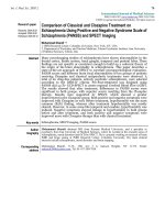
according to Form-1 in Figure 1. This form developed
by us for this study. The presence of dyspeptic com-
plaint and its severity calculated by presence of ab -
dominal pain, abdominal pain with hungry and an -
xiety, abdominal distension, nausea, vomiting, poor
appetite and symptoms of gastroesophageal reflux.
All patients were graded 1 to 5 for these symptoms.
Dyspepsia score of patients were minimally 12 and
maximally 60. The patients investigated previous
smoking history, coronary artery disease, diabetes
mellitus and malignancy history in their family.
Statistical Analysis
Data files were analyzed initially with Access
and SPSS (version 13.0). Chi-square (x
2
) tests were
performed to determine whether the clinical and bi-
ochemical variables were associated with a GI lesion.
A multivariate analysis was applied to identify va-
r i a b l e s s i g n i f i c a n t l y r e l a t e d w i t h t h e o u t c o m e o f t h e G I
lesions. Multiple analyses were performed with Cox
regression analysis. P < 0.05 was considered signifi-
cant in statistical analysis.
Results
Ninety-one patients fulfilled the entry criteria
and were enrolled. Their mean age was 43.3 (19-81)
years. 71 were patient a g e d u n d e r 5 0 a n d 2 0 w e r e o v e r
50 years. 77 were female and 14 were male. Sixty-six
of women were pre-menopausal and 11 were
post-menopausal. Presence or absence of GI symp -
toms was evaluated in every patient. Table 1 describes
the frequency predictive signs for possible gastroin-
testinal lesions in iron deficiency anemia patients.
Int. J. Med. Sci. 2011, 8
32
Figure 1. F orm -1 used in patients.
Table 1. Frequency predictive signs for possible gastrointestinal lesions in iron deficiency anemia patients.
Yes (%) No (%)
Hematemesis 0 (0) 91 (100)
Melena 4 (4.4) 87 (95.6)
Hematochezia 8 ( 8.8) 83 (91.2)
Hematuria 2 ( 2.2) 89 (97.8)
Menorrhagia 20 (30.7) 46 (69.7)
Diarrhea 3 (3.3) 88 (96.7)
Constipation 39 (42.9) 52 (57.1)
Change of bowel habits 5 (5.5) 86 (94.5)
Lost weight 4 (4.4) 87 (95.6)
Int. J. Med. Sci. 2011, 8
33
Frequently of NSAID
1
use 3 (14.3) 88 (96.7)
Intestinal parasite infection 7 (7.7) 84 (92.3)
Previous IDA
2
history 45 (49.5) 46 (50.5)
Smoking 23 (25.3) 68 (74.7)
Cancer in first degree relatives 22 (24.2) 69 (75.8)
Cancer in family 28 (30.7) 63 ( 69.3)
1
Nonsteroidal anti-inflammatory drugs,
2
Iron deficiency anemia
Clinically, significant predictive signs for possi-
ble gastrointestinal lesions were demonstrated in 11
patients. 8 patients had hematochezia and 4 had me-
lena. Only 2 patients had hematuria, 39 had constipa-
tion, 3 had diarrhea. 45 patients had been found to be
iron deficiency anemia previously. 20/66
pre-menopausal women had heavy menstrual bleed-
ing. 28/91 patients had cancer in their family. At ad-
mission, significant physical examination findings of
91 patients; 2 had hepatomegaly, 1 had splenomegaly
and 8 had epigastric sensitivity (Table 2).
18 of 89 patients had fecal occult blood test posi-
tive, 6 patients had parasite in feces, 7 had micro-
scopic hematuria and 3 had positive sprue serological
(antiendomysium antibodies IgA and tissue trans-
glutaminase antibodies). 55 patients had no additional
systemic disease, 13 patients had thyroid diseases (8
had hypothyroidism, 5 had hyperthyroidism), 9 pa-
tients had diabetes mellitus (7 had diabetes mellitus
type 2, 2 had diabetes mellitus type 1), 8 patients had
hypertension, 3 had coronary artery disease, 2 had
collagen tissue disease, 2 had immune thrombocyto-
penic purpura, 2 had hypophysial adenoma and 1 had
Parkinson disease (Table 4). Table 5 shows biochemi-
cal characteristics of patients. Their mean hemoglobin
level was 10.2 g/dl (range 6.4–12.7), mean white
blood cell count was 7095 l/mm
3
(range 3100-16900),
mean platelet count was 326x10
3
/mm
3
(range 74-669),
mean ferritin level was 7.5 ng/ml (range 1.38-28).
Table 2. Significant physical examination findings in iron
deficiency anemia patients
Yes (%) No (%)
Hepatosplenomegaly 3 (3.3) 88 (96.7)
Abdominal mass 0 (0) 91 (100)
Epigastric sensitivity 8 (8.8) 83 (91.2)
Table 3. Laboratory findings related to iron deficiency in
IDA patients.
Positive
(Positive / totally, %)
Negative (%)
Fecal Occult Blood 16 (16/89, 18) 73 (82)
Parasite in feces 6 (6/81, 7.4) 75 (92.6)
Microscopic hema-
turia
7 (7/91, 7.6) 84 (93.4)
Lungs film 2 (2/88, 2.2) 86 (97.8)
Sprue serological 3 (3/82, 3.6) 79 (96.4)
Table 4. The additional systemic disease in IDA patients
Patients
number
%
Absent 55 60.4
Thyroid diseases 13 14.2
Diabetes Mellitus 9 9.8
Hypertension 8 8.8
Coronary artery disease 3 3.2
Collagen tissue disease 2 2.1
Immune thrombocyto-
penic purpura
2 2.1
Hypophysial adenoma 2 2.1
Parkinson disease 1 2.1
Table 5. Biochemical variables of patients with iron defi-
ciency anemia
Patients
Number
Mean Range
Normal lab.
range
Hb
1
(gr/dl) 91 10.2 6.4–12.7 12-14/women
14-15/men
WBC
2
(l/mm
3
)
91 7095 3100–16900 4.8-10.8
Plt
3
(x10
3
/mm
3
)
91 326 74–669 150-400
Ferritin
(ng/ml)
91 7.5 1.38–28 10-291/women
22-322/men
CRP
4
(gr/dl)
83 0.66 0.1–7.6 0-5
ESR
5
(mm/h)
80 17.2 2–75 0-20
CEA
6
(ng/ml)
77 3.4 0.25–97 0-7
1
Hemoglobin,
2
White blood cell,
3
Platelets,
4
C reactive protein,
5
Erythrocyte sedimentation rate,
6
Carcinoembryonic antigen
86 patients underwent upper gastrointestinal
tract endoscopies and 62 patients underwent upper
and lower gastrointestinal tract endoscopies. An up-
per GI finding, mainly antral gastritis was the most
common pathologic finding (n=23, 26.7 %). The ab -
normalities considered as possible causes of upper
gastrointestinal lesions were Helicobacter pylori (HP)
gastritis (n=18), duodenitis (n=12), pangastritis
(n=11), coeliac disease (n=3), gastric ulcer (n=2), du-
odenal ulcer (n=2), erosive gastritis (n=1) and gastric
tumor (n=1). The lower gastrointestinal tract lesions
regarded as possible causes of IDA included he-
morrhoid (n=19), chronic colitis (n=2), inflammatory
Int. J. Med. Sci. 2011, 8
34
intestinal disease (n=2), interstitial colitis (n=1) and
colorectal cancer (n=1) (Table 6).
Table 6. Pathological conditions of the GI tract in ir on
deficiency anemia patients
Diagnosis Frequency Result/Number of
process, (%)
Non-diagnostic 12 12/86, (13.9)
Antral gastritis 23 23/86, (26.7)
Hemorrhoid 19 19/66, (28.7)
H.
1
pylori gastritis 18 18/86, (20.9)
Duodenitis 12 12/86, (13.9)
Pangastritis 11 11/86, (12.7)
Anal fissure 5 5/66, (7.5)
Colonic polyp 4 4/66, (6.0)
Diverticulitis 3 3/66, (4.5)
Coeliac disease 3 3/86, (3.4)
Gastric ulcer 2 2/86, (2.3)
Duodenal ulcer 2 2/86, (2.3)
Chronic colitis 2 2/66, (3.0)
IID
2
2 2/66, (3.0)
Atrophic gastritis 2 2/86, (2.3)
Interstitial colitis 1 1/86, (1.1)
Gastric polyp 1 1/86, (1.1)
Erosive gastritis 1 1/86, (1.1)
Gastric cancer 1 1/86, (1.1)
Colonic cancer 1 1/66, (1.5)
1
Helicobacter,
2
Inflammatory intestinal disease
A list of the upper and lower GI pathological
conditions associated with IDA is included in Table 7.
The patients were interviewed and responded to a
questionnaire that included clinical and biochemical
variables. Table 8 is shown that rate of clinically sig-
nificant lesions in IDA with positively symptoms-sign
or laboratory results. The presence of advanced age
(>50 years), male gender, diarrhea, lost weight,
change of bowel habits, epigastric tenderness, posi-
tively serological sprue, hemoglobin levels less than
10 g/dl and high CEA level (>5 pg/ml) were asso-
ciated with an increased likelihood of significant ga-
strointestinal lesions (p<0.05); melena, constipation,
cancer in first degree relatives, fecal occult test posi-
tivity, high C-reactive protein (CRP) and erythrocyte
sedimentation rate (ESR) level were associated with
limited positively findings (p≤ 0.19).
The risk factors for finding GI lesions causing
IDA were as follows: male gender (p= 0.004), ad-
vanced age (p= 0.010), weight loss (p= 0.020), chronic
diarrhea (p= 0.006), change of bowel habits (p= 0.043),
epigastric tenderness (p= 0.037), raised CEA level ( p =
0.039), < 10 gr/dl Hb level (p=0.054). None of these
risk factors had been present in 21 (23%) women
younger than 51 years. In this group, no patient had
any GI lesion likely to cause IDA (negative predictive
value= 100%). In multivariate analysis, advanced age
(p=0.017), male gender (p< 0.01) and weight lost
(p=0.012) found that associated with GI lesions in all
patients.
In addition, we determine the yield of endosco-
py evaluations in pre-menopausal and age < 50
women with iron deficiency anemia but without any
clinically significant sign-symptoms and laboratory
findings. There were 21 patients had these criteria but
none of them had any endoscopic significant lesions.
Table 7. Pathological conditions of the GI tract associated
with iron deficiency (Clinically meaningful lesions)
Patients number
Celiac disease (villous atrophy) 3
Erosive gastritis 1
Peptic ulcer 3
IID
*
/chronic colitis 4
Diverticulitis 3
Gastric cancer 1
Colon cancer 1
Familial polyposis 1
Helicobacter pylori gastritis 18
*
Inflammatory intestinal disease (Not: Hemorrhoid did not consi-
derate due to coincidentally lesions.
Table 8. Rate of clinically significant lesions in IDA with
positively symptoms-sign or laboratory results
Symptoms, sign or labor-
atory
results
Existence of
significant
lesion
Absence of
significant
lesion
P value
Age> 50 8 12 0.010
Sex (Male) 7 7 0.004
Diarrhea 3 0 0.006
Lost weight 3 1 0.020
Change of bowel habits 3 2 0.043
Epigastric tenderness 4 4 0.037
Serological of sprue 2 1 0.074
Hb level 7 50 0.054
High CEA level 3 2 0.039
Melena 2 2 0.157
Constipation 10 29 0.178
Cancer in first degree
relatives
7 15 0.112
Fecal occult blood test
positiviy
5 11 0.178
High CRP level 3 6 0.173
High ESR level 6 13 0.174
Whatever positively in
general evaluation
17 53 0.010
Discussion
Iron deficiency is the most common hematolog-
ical disorder encountered in general practice and
iron-deficiency anemia is the most frequently cause of
anemia worldwide [13]. Blood loss is a major cause of