Báo cáo y học: "Current Status of Methods to Assess Cancer Drug Resistance"
Bạn đang xem bản rút gọn của tài liệu. Xem và tải ngay bản đầy đủ của tài liệu tại đây (430.46 KB, 9 trang )
Int. J. Med. Sci. 2011, 8
245
I
I
n
n
t
t
e
e
r
r
n
n
a
a
t
t
i
i
o
o
n
n
a
a
l
l
J
J
o
o
u
u
r
r
n
n
a
a
l
l
o
o
f
f
M
M
e
e
d
d
i
i
c
c
a
a
l
l
S
S
c
c
i
i
e
e
n
n
c
c
e
e
s
s
2011; 8(3):245-253
Review
Current Status of Methods to Assess Cancer Drug Resistance
Theodor H. Lippert
1
, Hans-Jörg Ruoff
1
and Manfred Volm
2
1. Medical Faculty, University of Tübingen, Tübingen, Germany
2. Medical Faculty, University of Heidelberg, Heidelberg, Germany
Corresponding author: Prof. Theodor H. Lippert, Erlenweg 38, 72076 Tübingen, Germany. Tel.: 49 7071 62199; Fax: 49 7071
61234; e-mail:
© Ivyspring International Publisher. This is an open-access article distributed under the terms of the Creative Commons License (
licenses/by-nc-nd/3.0/). Reproduction is permitted for personal, noncommercial use, provided that the article is in whole, unmodified, and properly cited.
Received: 2010.10.06; Accepted: 2011.03.14; Published: 2011.03.23
Abstract
Drug resistance is the main cause of the failure of chemotherapy of malignant tumors, re-
sistance being either preexisting (intrinsic resistance) or induced by the drugs (acquired re-
sistance). At present, resistance is usually diagnosed during treatment after a long period of
drug administration.
In the present paper, methods for a rapid assessment of drug resistance are described. Three
main classes of test procedures can be found in the literature, i.e. fresh tumor cell culture
tests, cancer biomarker tests and positron emission tomography (PET) tests. The methods
are based on the evaluation of molecular processes, i.e. metabolic activities of cancer cells.
Drug resistance can be diagnosed before treatment in-vitro with fresh tumor cell culture
tests, and after a short time of treatment in-vivo with PET tests. Cancer biomarker tests, for
which great potential has been predicted, are largely still in the development stage. Individual
resistance surveillance with tests delivering rapid results signifies progress in cancer therapy
management, by providing the possibility to avoid drug therapies that are ineffective and only
harmful.
Key words: cancer drug resistance, in vitro cancer drug resistance tests, in vivo cancer drug re-
sistance tests, cancer biomarker tests
Introduction
Since the beginning of cancer chemotherapy the
frequent lack of drug response in solid tumors has
been a major problem. The main cause of failure to
respond to cytostatics is drug resistance. In nearly
50% of all cancer cases, resistance to chemotherapy
already exists before drug treatment starts (intrinsic
resistance), and in a large proportion of the remaining
half resistance develops during treatment (acquired
resistance) [1]. All efforts to overcome resistance to
chemotherapy so far have failed, owing to the enor-
mous heterogeneity and complex biology of cancer
cells, with wide individual variations [2]. Meanwhile,
the knowledge of various resistance mechanisms has
increased over the years [3], leading to the develop-
ment of new drugs that can be specifically targeted.
However, the new "targeted" drugs also suffer from a
considerable failure rate and from toxicity [4]. The
increasing number of new anticancer drugs has not
efficiently reduced the occurrence of drug resistance
up to now.
Diagnosis of drug resistance in individual pa-
tients would improve cancer treatment by the avoid-
ance of inefficient treatment. The aim of the present
paper is to discuss the possibilities for realizing this
goal. The following three methods are available to
assess cancer drug resistance: fresh tumor cell culture
assays, cancer biomarker tests, and positron emission
tomography tests.
Int. J. Med. Sci. 2011, 8
246
Fresh tumor cell culture tests
As early as the 1950s, research teams started to
develop laboratory tests in order to predict tumor
reaction to cytostatic drugs [5]. They used fresh cancer
tissue and examined the effect of the drugs on tumor
cell growth. At the beginning laboratory techniques
were still in their infancy. Short term cell cultures of
cancer tissues were difficult to perform and proce-
dures varied from laboratory to laboratory. However,
the cancer cell assays were, thanks to better tech-
niques, continuously improved over the last few
decades and brought to a certain perfection. There are
two steps in the preparation of the tests, first the fresh
cell culturing and then, when this is successful the
examination of the drug effect. Cell cultures in medi-
cine are now established laboratory tools. Whereas
immortalized cancer cell lines used for research pur-
poses have lost a large part of individual tumor char-
acteristics the preparation of fresh tumor tissue is
necessary in order to obtain cancer cells with still
highly preserved individual tumor properties [6].
Special arrangements have to be made before the bi-
opsy is taken by the oncologist to garantee a rapid and
safe transport of the probe i.e. a specialized laboratory
must be contacted, the means of transport and
transport medium arranged and precaution taken that
the probe is immediately placed in the transportme-
dium. Extensive descriptions of special laboratory
techniques for fresh cancer cell cultures are now
available [7, 8]. The cell preparation may vary de-
pending on the tumor type in test. Table 1 shows
frequently tested tumor types for which special cell
preparations were published [9-27]. For the examina-
tion of the drug effect after incubation several meth-
ods are in use. In the 1970s the method of measuring
the thymidine incorporation into cancer cell DNA [28]
was developed by one of us (M.V.). It estimates the
inhibitory effect on cell growth. This technique as well
as some others have found their way into laboratory
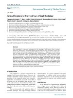
practice. Fig. 1 is a schematic illustration of the pro-
cedure of fresh tumor cell culture assays. Although
various assays have been developed, the principal
steps, i.e. isolation of cells, incubation of cells with
drugs and assessment of cell survival are the same.
Usually a range of drug doses is applied in order to
find a dose-response relationship. Drug concentra-
tions in the tests are similar to drug concentrations
usually found in-vivo during treatment. All methods
measure molecular processes of cancer cells, revealing
cell activity and thus indicating cell growth or death
[29, 30]. Frequently used methods are the thymidine
incorporation into cell DNA [31] and the loss of cell
ATP [32]. Drug resistance can be recognized by no
decrease of thymidine uptake into cell DNA or no
decrease of cell ATP. Fresh tumor cell culture assays
are applicable to many types of cancer, since they
register the integral cell reaction. The predictive value
of the assays, depending on cancer tissue, which is
usually only available before treatment, consists in
indicating intrinsic resistance.
Table 1. Tumor types for which short term primary cell cultures are used to test tumor response to cancer drug therapy.
Tumor
References
Colorectal cancer Paraskeva C et al [9], Park J-G et al [10], Whitehead [11]
Testicular Cancer Pera MF [12]
Skin cancer Parkinson EK et al [13], Halaban R [14]
Lung cancer Twentyman PR [15], WU R [16]
Brain cancer Darling JL [17]
Ovarian cancer Whelan RDH et al [18], Wilson AP [19]
Prostate cancer Harper ME [20], Bright RK et al [21]
Breast cancer O’Hare MJ [22], Speirs V [23]
Cervical cancer Stern P et al [24]
Bladder cancer Fu VX et al [25]
Head and neck cancer Edington KG et al [26]
Pancreatic cancer Iguchi H [27]
Int. J. Med. Sci. 2011, 8
247
Fig. 1. Schematic procedure of fresh tumor cell culture assays.
None of the tests developed has been adopted so
far in clinical routine practice, mainly because of the
lack of general recognition. Critical comments pub-
lished in the renowned New England Journal of
Medicine in the 1980s [33] on test artefacts causing
false results dramatically reduced interest in further
research. The verdict which arose then that assays for
drug response are unreliable is still widely accepted.
This opinion ignores the fact that assay techniques
have improved and that test results of drug resistance
and drug sensitivity should not be confused: drug
resistance is considered as highly predictable, which
is not the case with drug sensitivity. Results of sensi-
tive drugs obtained by the net effect of drug action on
cancer cells are not very reliably, since many steps in
the body are required to reach the target. However,
their effectivity may be increased by the fact that cases
of ineffective drugs can be eliminated [34].
With the recent recognition, that cancer therapy
can be optimized by personalized i.e. individualized
drug treatment, interest has again arisen in fresh tu-
mor cell culture assays. Recently ASCO felt induced to
publish an assessment of the assays reviewing the
literature [35]. It came to the conclusion that the tests
are still investigational but asserted that an in-vitro
approach has great potential to spare patients the
morbidity of ineffective chemotherapy regimens. The
ASCO judgment was criticized for the fact that only 12
studies were taken for the evaluation and that no dis-
tinction was made between sensitivity and resistance
results. Many studies, showing good correlation be-
tween in-vitro resistance with in-vivo outcome re-
mained unnoticed [36]. In the 1980s a review, cover-
ing 27 studies already showed excellent correlations
in different chemotherapy-treated tumor types
(>90%) [37]. Similar correlations were found in other
comprehensive reviews [34, 38]. In the meantime
many more studies on different tumor types have
been published, some with variable results. It has
been pointed out that the labor-intensive assays
should only be carried out by experienced, highly
specialized laboratories. Standardization of the tests
would make it easier to compare the results of dif-
ferent studies.
Ovarian cancer is now one of the best investi-
gated cancer types with promising results for indi-
vidualized assay-assisted chemotherapies. In a recent
review earlier results have been corroborated, i.e.
most tumor response tests showed excellent correla-
tion with clinical resistance but varied in their ability
to predict sensitivity [39]. Another recent study
demonstrated that assay-assisted chemotherapy in
ovarian cancer may result in reduced costs compared
to empiric therapy [40]. A novelty may be added here:
the National Comprehensive Cancer Network
(NCCN) in the USA [41], which provides “Clinical
Practice Guidelines for Oncology” mentioned chem-
otherapy-resistance assays for the first time in a recent
update on ovarian cancer treatment (2010). It declared
that such tests are being used in some NCCN centers
to aid in selecting chemotherapy in situations where
there are multiple equivalent chemotherapy options
available. In another recent publication [42], discuss-
ing the question of chemosensitivity testing for ad-
Int. J. Med. Sci. 2011, 8
248
vanced gastric cancer, it was cited that pre-treatment
testing is already approved by the Japanese Ministry
of Health in 11 institutes. This shows that interest in
further research on fresh tumor cell culture assays has
now considerably increased.
In-vitro diagnosis of drug resistance has not only
been carried out on solid tumors; it has been demon-
strated that patients with haematological neoplastic
diseases can also profit [43]. Recent publications cer-
tify the usefulness of such assays in the rapid recog-
nition of resistance which allows treatment modifica-
tion shortly after [44, 45].
Cancer biomarker tests
Tumor markers - also called cancer biomarkers -
already attracted attention as diagnostic tools for
cancer detection and growth indicators early in the
last century [46]. The search concentrated on specific
cancer-derived molecules occurring in the blood.
Several markers, such as the carcinoembryonic anti-
gen (CEA) and alpha-fetoprotein (AFP), found their
way at an early stage into clinical laboratories. Many
others have followed in the meantime. However, most
of them are not tumor specific. The use of changes of
serum markers as a measure of tumor response to
therapy seems appealing because it is non-invasive
and can be frequently repeated. No special efforts
have been made so far to carry out studies to investi-
gate their practical value for this purpose. Only a few
tumor markers were used in clinical trials e.g. pros-
tate-specific antigen (PSA) in prostate cancer, CA 125
in ovarian cancer, thyroglobulin in thyroid cancer and
human chorionic gonadotropin (HCG) in chori-
onepithelioma. In these cases it has been shown that
the markers fell to very low levels after successful
treatment. However, it is still not known to what ex-
tent markers can reliably reflect the viable tumor
mass. The pathobiology of tumor markers is still not
well understood. It remains hard to understand why
tumor markers have not been investigated to a greater
degree in the huge number of previous chemotherapy
studies.
Only recently have cancer biomarkers gained
wider recognition. The American National Cancer
Institute launched the project “Early Detection Re-
search Network” (EDRN) as a new field of cancer
research, focused on identifying markers both for the
early detection of cancer and of cancer risk. The main
aim is creating validated biomarkers for early thera-
peutic intervention in malignant diseases [47, 48]. A
large number of organizations are now participating
in cancer biomarker research [49]. Unfortunately the
program does not engage in investigation of markers
for drug response testing.
The pharmaceutical industry now uses overex-
pressed growth factors, i.e. their cell receptors as
cancer biomarkers to develop new targeted anticancer
drugs with better tumor response. However, tumor
concentrations of growth factor receptors do not reli-
ably predict their therapeutic effect in individual cas-
es. Only in some small subgroups of patients detected
by special biomarkers could a major therapeutic suc-
cess be demonstrated. Examples are: for trastuzumab
breast cancer with overexpressed HER2, for imatinib
gastrointestinal stroma cell tumor (GIST) with over-
expressed C-kit and chronic myeloid leukaemia
(CML) with BCR-ABL fusion protein, and for gefitinib
and erlotinib non small cell lung cancer (NSCLC) with
mutations in the EGFR gene [50]. Another subgroup
which benefits from EGFR inhibitor treatment is col-
orectal cancer with Kras wild type [51]. The search for
biomarkers to find new subgroups of cancer patients
for treatment with targeted drugs goes on.
Potential biomarkers for the prediction of drug
response are several proteins which play a role in
drug resistance mechanisms. Such cellular factors are
resistance proteins, which can be determined by im-
munohistochemistry. Laboratory experiments with
short-term cell cultures of lung cancers have shown
that excellent correlations exist between
drug-resistant cells and several of the resistance pro-
teins [52]. The determination of resistance proteins in
cancer cell biopsies seems a feasible way to detect
intrinsic drug resistance. So far no test based on re-
sistance protein determination has been adopted in
clinical practice.
In a wider sense, pharmacogenetics is part of
cancer biomarker research. Tests examine the influ-
ence of genetic factors on drug action. New laboratory
techniques, for instance genomics, proteomics, and
transcriptomics (omics), make it possible to determine
a great number of biological molecules whose com-
position is considered to provide information about
the effectiveness and toxicity of drugs. Since investi-
gations using omics are dependent on cancer tissue,
which is often only available before the commence-
ment of therapy, only intrinsic resistance can be veri-
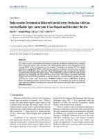
fied. In order to detect predictive biomarkers highly
sophisticated data analytical methods have now been
developed. In Fig. 2 a schematic illustration of the
main steps for such data analysis, algorithms for fin-
gerprint detection of cancer biomarkers, is shown.
Mathematics and Computer Sciences play an im-
portant part in observing essential markers compar-
ing biological material from patients with drug re-
sistance with material from patients without drug
resistance. Algorithms have to deal not only with the
giant mass of data, but also with their dynamic
Int. J. Med. Sci. 2011, 8
249
change. Thus it is well known that an individual pro-
teome changes quite dramatically during a day, de-
pending on a variety of factors. Only a large enough
group of patients allows to identify components that
do not differ much between individuals from the
same group.
Fig. 2. Different fields with sub-areas necessary for data
analysis algorithms for fingerprint detection of cancer bi-
omarkers
There are already several publications which
describe new biomarkers, detected by sensitive anal-
ysis algorithms. However, the clinical significance of
these substances, such as Let-7i, a biomarker for
therapy of epithelial ovarian cancer [53] or beta III
tubulin, a biomarker for chemoresistance in non-small
cell lung cancer [54] has still to be proven. A recent
review of biomarkers of chemotherapy resistance in
breast cancer discusses the difficulties of clinical bi-
omarker validation [55]. Prediction of cancer drug
action with pharmacogenetic assays is still in its in-
fancy. Results still have to be judged critically, since
misinterpretations are possible [56]. The microarrays
used for the tests are not standardized, which makes it
difficult to compare the results of different studies
[57].
Positron emission tomography tests
Diagnosis of drug resistance during drug treat-
ment was difficult in the past. The only method
available was tumor size control. A solution was
found recently by using a nuclear medicine technique,
positron emission tomography (PET). Already in
clinical use for many years for the detection of cancer
localisation, the method can now also be applied to
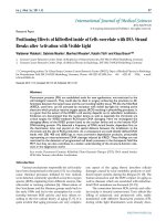
determine the metabolic activity of neoplastic tissue.
Fig. 3 shows the schematic illustration of quantitative
cancer image analysis in positron-emission tomogra-
phy.
Fig. 3. Schematic illustration of quantitative cancer image analysis in positron-emission tomography.