Báo cáo y học: "Spinal Intramedullary Cysticercosis: A Case Report and Literature Review"
Bạn đang xem bản rút gọn của tài liệu. Xem và tải ngay bản đầy đủ của tài liệu tại đây (432.15 KB, 4 trang )
Int. J. Med. Sci. 2011, 8
420
I
I
n
n
t
t
e
e
r
r
n
n
a
a
t
t
i
i
o
o
n
n
a
a
l
l
J
J
o
o
u
u
r
r
n
n
a
a
l
l
o
o
f
f
M
M
e
e
d
d
i
i
c
c
a
a
l
l
S
S
c
c
i
i
e
e
n
n
c
c
e
e
s
s
2011; 8(5):420-423
Case Report
Spinal Intramedullary Cysticercosis: A Case Report and Literature Review
Bin Qi,Pengfei Ge,Hongfa Yang,Chunhua Bi,Yiping Li
Department of Neurosurgery, The First Hospital of Jilin University, Changchun 130021, China
Corresponding author: Yiping Li, e-mail:
Received: 2011.04.12; Accepted: 2011.06.14; Published: 2011.07.06
Abstract
Neurocysticercosis, involvement of the central nervous system by taenia solium, is one of
the most common parasitic diseases of the CNS. However, spinal involvement by neu-
rocysticercosis is uncommon. Here, we reported a 40-year-old woman with intramedul-
lary cysticercosis in the thoracic spinal cord. MRI revealed two well-defined round in-
tramedullary lesions at T4 and T5 vertebral levels, which were homogeneously hy-
pointense on T1WI and hyperintense on T2WI with peripheral edema. Since the patient
had progressive neurological deficits, surgery was performed to decompress the spinal
cord. Histopathology examination of the removed lesion proved it was intramedullary
cysticercosis. In this report, we also discussed the principles of diagnosis and treatment
of intramedullary cysticercosis in combination of literature review.
Key words: intramedullary, cysticercosis, spinal cord
Introduction
Neurocysticercosis, caused by Taenia solium, is
the most common parasitic infection affecting the
central nervous system. However, the spinal cysti-
cercosis is rare, representing 1.2% to 5.8% of all cases
of neurocysticercosis
19, 20
. According to the cysticercus
location in spine, Cysticercosis has been classified
anatomically as extraspinal (vertebral) or intraspinal
(epidural, subdural, arachnoid, or intramedullary), of
which the intramedullary type is quite rare and only
fifty-three cases have been reported until 2010
1-3,8,13
.
Here, we reported a case of intramedullary cysticer-
cosis at T4 and T5 vertebral level and discussed its
diagnosis and treatment with literature review.
Case Report
A 40-year-old female patient was transferred to
our department from a local hospital for progressive
weakness in both lower limbs for one month, and anal
sphincter and bladder dysfunction for two days.
Neurological examination disclosed spastic parapare-
sis with decreased motor power of grade 3/5 in both
lower limbs, impaired sensations below T4 derma-
tome, brisk tendon jerks and positive Babinski signs
on both sides. Non-contrast MRI revealed two
well-defined round intramedullary cystic lesions at T4
and T5 vertebral levels, which were homogeneously
hypointense on T1WI and hyperintense on T2WI with
slightly peripheral edema. The subarachnoid space
from T4 to T5 was narrow due to the marked expan-
sion of spinal cord. There were no abnormalities at
cervical or lumbar levels or within the brain paren-
chyma. The diagnosis of intramedullary mass lesion
was made. There is no use of dexamethasone in the
perioperative period.
The patient underwent laminectomy from T4 to
T5, and the spinal cord was found swollen. When a
midline myelotomy was performed, a white cystic
lesion was seen and clear fluid was then aspirated.
The cyst wall of which slightly stuck to the sur-
rounding spinal cord. In order to dissect the cyst with
minimal injury to the peripheral tissue, the cystic liq-
uid was partly withdrawn first and the slackened cyst
was removed totally. The liquid was yellowish and
transparent. Histological examination of the resected
sample showed cysticercosis.
Postoperatively, the patient refused to be treated
with anticysticercal agents and steroids. The patient's
neurological function postoperatively was not un-
Ivyspring
International Publisher
Int. J. Med. Sci. 2011, 8
421
changed from his preoperatively status and she was
discharged 2 weeks later. At six months of follow-up,
the motor power of her lower limbs recovered to
grade 4/5, and she could ambulate without special
support. The function of anal sphincter and bladder
regained without compromise of the activities of her
daily living. However, her hypoesthesia over the T4
dermatome still existed.
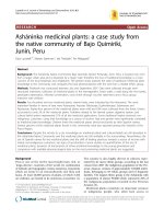
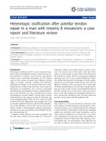
Figure 1. Sagittal T1, T2-weighted MR image of thoracic spine showing a relatively well defined cystic intramedullary
lesion with hypointense on T1WI and hyperintense on T2WI.
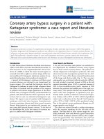
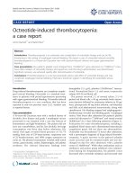
Figure 2. Photomicrographs of the histological specimen showing the cysticercosis cyst wall with neutrophile gran-
ulocyte.lymphocyte and necrosis cell. (H&E×100)
Discussion
Cysticercosis is widely endemic in Brazil, Peru,
Mexico, Korea and India
19-20
.
Intramedullary cysti-
cercosis often presents in the patients between 20 to 45
years old, with the youngest one 5 years old and the
oldest one 45 year’s old
15
. Most patients experienced a
progressively worsened course from a week to 10
years
20
. The common clinical manifestations included
pain, paraparesis, spasticity, bowel and bladder in-
continence, and sexual dysfunction
1,20
. However, in-
flammatory reaction against the dead parasite is as-
sociated with perilesional edema, which can damage
medullar parenchyma and therefore, worsen symp-
toms
2
. Inside the spinal cord, cysticercus usually dis-
tributes in the thoracic cord, with a few cases involv-
ing the cervical and the lumbar cord. This distribu-
tional mode of cysticercus supports the hypothesis
that intramedullary cysticercus comes from the blood
circulation, because thoracic cord has much more
blood supply than the other parts of the spinal
cord
6,20
. However, it is also thought that intramedul-
lary cysticercus could migrate to the spinal cord via
the ventriculo-ependymal pathway. On MRI, in-
tramedullary cysticercosis usually show a cystic le-
sion within the spinal cord, which of appears hy-
Int. J. Med. Sci. 2011, 8
422
pointense on T1WI with hyperintense scolex identi-
fied inside the cyst cavity, hyperintense on T2WI in
vesicular stage, a subtle hypointense rim may sur-
round the intramedullary cyst on T2WI. In the col-
loidal stage the thickened cyst capsule is hyperintense
on T1WI and hypointense on the T2WI. Cyst contents
appear hyperintense on T1WI resulting in scolex is
not seen. There is an amount of surrounding edema. If
cyst degeneration is present peripheral ring en-
hancement may be present
1,2,15,17
. The differential di-
agnosis of an intramedullary cystic lesion is extensive,
including some other cysts such as arachnoid cyst
14
,
ependymal cyst
10
, neurenteric cyst
18
, sarcoidosis
4
,
neoplasms such as ependymoma, and infections such
as abscess
21
.
When a patient had a history of cysticercosis or
came from an endemic region and MRI revealed a
cystic spinal cord lesion, the diagnosis of intramedul-
lary cysticercosis could be suspected and be further
verified by serologic alterations, subcutaneous nod-
ules, and changes in the cerebrospinal fluid. The CSF
examination often shows increased proteins, a low or
normal glucose, moderate lymphocytic pleocytosis
and eosinophilia
7
. Cysticercal antibodies found in CSF
either by ELISA or in serum by enzyme-linked im-
munoelectric transfer bolt assay have good sensitivity
and specificity in cysticercosis diagnosis
7,22
. However,
the patient lacked of neurocysticercosis history and
was not from an endemic region. Therefore, it was
difficult to clinically suspect intramedullary cysticer-
cosis prior to treatment. The diagnosis of neurocysti-
cercosis was established based on pathological ex-
amination. In our case, owing to increasing neurolog-
ical deficit, surgical treatment is a good choice for
removing the mass which produces progressive spi-
nal compression and confirm the diagnosis. Our pa-
tient showed improvement in motor power. Bowel
movements and urinary sphincters was better control.
However, the results of surgical outcome are mixed.
Mohanty
16
reported only a 75% satisfactory outcome
after surgery and cysticidal treatment. Early diagnosis
and treatment can improve the outcome. Outcomes
reported in other series have not been favorable.
Sharma
1
reported that 60% patients acquired im-
provement after surgery, 25% did not improve, and
15% died. In the reports published in recent years
1,2,9,12,15
, surgical outcome was significantly improved;
no death case and majority of patients could live a life
without special support. Surgery is procedure of
choice only when diagnosis is in doubt otherwise
medical treatment has its advantages. Albendazole is
a medicine that has been proved to be effective in the
patients with intramedullary cysticercosis since 1996
5
.
Preoperative adjunctive treatment with albendazole is
thought to be helpful to consolidate the lesion and
thus induce a clear plane of dissection during surgery.
Albendazole is normally used postoperatively as a
regular treatment (15mg/kg/day) for 4 to 6 weeks,
according to the idea that cysticercosis is a general-
ized disease with focal manifestation. Moreover, Al-
bendazole is often used with corticosteroids, because
its blood level could be synergistically increased by
the latter
11
. Except for being used after surgery,
Abendazole also could be used independently in the
conservative treatment for the patients whom are
highly suspected as intramedullary cysticercosis and
whose clinical courses are stable. The potential ad-
vantages of medical therapy alone include avoidance
of surgery and treatment of surgically unreachable
and multifocal cysticercus
2,3,5,7,17
.
Conclusions
In conclusion, we think that intramedullary cys-
ticercosis represents a diagnostic challenge and neu-
rocysticercosis should also be strongly considered for
intramedullary cystic lesions, even in a non-endemic
area. Surgery is required to facilitate extirpation of the
lesion, decompress the cord, confirm the pathological
diagnosis and provide a route for definitive therapy.
Conflict of Interest
The authors have declared that no conflict of in-
terest exists.
References
1. Agrawal R, Chauhan SP, Misra V, et al. Focal spinal intrame-
dullary cysticercosis. Acta Biomed. 2008;79(1): 39-41.
2. Ahmad FU, Sharma BS. Treatment of intramedullary spinal
cysticercosis: report of 2 cases and review of literature. Surg
Neurol. 2007;67(1): 74-7.
3. Chhiber SS, Singh B, Bansal P, et al. Intramedullary spinal cys-
ticercosis cured with medical therapy: case report and review of
literature. Surgical Neurology. 2009;72(6):765-9.
4. Clifton AG, Stevens JM, Kapoor R, et al. Spinal cord sarcoidosis
with intramedullary cyst formation. Br J Radiol.
1990;63(754):805-8.
5. Corral I, Quereda C, Moreno A, et al. Intramedullary cysticer-
cosis cured with drug treatment. A case report. Spine.
1996;21(19):2284-7.
6. De Souza Queirz L, Filho AP, Callegardo D, et al. Intramedul-
lary cysticercosis: case report, literature review and comments
on pathogenesis. J Neurol Sci. 1975;26(1):61-70.
7. Garg RK, Nag D. Intramedullary spinal cysticercosis: response
to albendazole: case reports and review of literature. Spinal
Cord. 1998;36(1):67-70.
8. Goncalves FG, Neves PO, Jovem CL, et al. Chronic myelopathy
associated to intramedullary cysticercosis. Spine. 2010;35(
5):159-62.
9. Homans J, Khoo L, Chen T, et al. Spinal intramedullary cysti-
cercosis in a five-year-old child: case report and review of liter-
ature. Pediatr Infect Dis J. 2001;20(9):904-8.
10. Iwahashi H, Kawai S, Watabe Y, et al. Spinal Intramedullary
ependymal cyst: a case report. Surg Neurol. 1999;52(4):357-61.
Int. J. Med. Sci. 2011, 8
423
11. Jung H, Hurtado M, Medina MT, et al. Dexamethasone in-
creases plasma levels of Albendazole. J Neurol.
1990;237(5):279-80.
12. Kasliwal MK, Gupta DK, Suri V, et al. Isolated spinal neuro-
cysticercosis with clinical pleomorphism. Turkish Neurosur-
gery. 2008;18(3):294-7.
13. Kumar S, Handa A, Chavda S, et al. Intramedullary cysticerco-
sis. J Clin Neurosci. 2010;17(4):522-3.
14. Lmejjati M, Aniba K, Haddi M, et al. Spinal Intramedullary
arachnoid cyst in children. Pediatr Neurosurg. 2008;44(3):243-6.
15. Mathuriya SN, Khosla VK, Vasishta RK, et al. Intramedullary
cysticercosis: MRI diagnosis. Neurol India. 2001;49(1): 71-4.
16. Mohanty A, Venkatrama SK, Das S. Spinal intramedullary
cysticercosis. Neurosurgery. 1997;40(1):82-7.
17. Parmar H, Shah J, Patwardhan V, et al. MR imaging in in-
tramedullary cysticercosis. Neuroradiology. 2001;43(11):961-7.
18. Riviérez M, Buisson G, Kujas M, et al. Intramedullary neuren-
teric cyst without any associated malformation. one case eval-
uated by RMI and electron microscopic study. Acta Neurochir.
1997;139(9):887-90.
19. Sawhney IM, Singh G, Lekhra OP, et al. Uncommon presenta-
tion of neurocysticercosis. J Neurol Sci. 1998;154(1):94-100.
20. Sharma BS, Banerjee AK, Kak VK. Intramedullary spinal cysti-
cercosis: case report and review of literature. Clin Neurol
Neurosurg. 1987;89(2):111-6.
21. Tacconi L, Arulampalam T, Johnston FG, et al. Intramedullary
spinal cord abscess: case report. Neurosurgery.
1995;37(4):817-9.
22. Tsang VC, Brand JA, Boyer AE. An enzyme-linked immunoe-
lectro transfer blot assay and glycoprotein antigens for diag-
nosing human cysticercosis. J Infect Dis. 1989;159(1):50-9.