- Trang chủ >>
- Sư phạm >>
- Sư phạm hóa
Chitosan immobilized porous polyolefin as sustainable and efficient antibacterial membranes
Bạn đang xem bản rút gọn của tài liệu. Xem và tải ngay bản đầy đủ của tài liệu tại đây (5.73 MB, 9 trang )
Research Article
pubs.acs.org/journal/ascecg
Chitosan Immobilized Porous Polyolefin As Sustainable and Efficient
Antibacterial Membranes
Prasanna Kumar S Mural,† Banothu Kumar,‡ Giridhar Madras,‡ and Suryasarathi Bose*,§
†
Center for Nano Science and Engineering, ‡Department of Chemical Engineering, and §Department of Materials Engineering, Indian
Institute of Science, Bangalore-560012, Karnataka, India
S Supporting Information
*
ABSTRACT: Polyolefinic membranes have attracted a great deal of interest
owing to their ease of processing and chemical inertness. In this study, porous
polyolefin membranes were derived by selectively etching PEO from PE/PEO
(polyethylene/poly(ethylene oxide)) blends. The hydrophobic polyolefin
(low density polyethylene) was treated with UV-ozone followed by dip
coating in chitosan acetate solution to obtain a hydrophilic-antibacterial
surface. The chitosan immobilized PE membranes were further characterized
by Fourier transform infrared spectroscope (FTIR) and X-ray photoelectron
spectroscope (XPS). It was found that surface grafting of chitosan onto PE
membranes enhanced the surface roughness and the concentration of
nitrogen (or amine) scaled with increasing concentration of chitosan (0.25 to
2% wt/vol), as inferred from Kjeldahl nitrogen analysis. The pure water flux
was almost similar for chitosan immobilized PE membranes as compared to
membranes without chitosan. The bacterial population, substantially reduced
for membranes with higher concentration of chitosan. For instance, 90 and 94% reduction in Escherichia coli (E. coli) and
Staphylococcus aureus (S. aureus) colony forming unit respectively was observed with 2% wt/vol of chitosan. This study opens
new avenues in designing polyolefinic based antibacterial membranes for water purification.
KEYWORDS: PE/PEO blends, Chitosan, UV-ozone, Antibacterial membrane
■
INTRODUCTION
There is a great need for the development of water treatment
technologies.1 Water purification by polymeric membranes is
preferred due to their low/no thermal inputs and low cost.
Among other conventional techniques, polymeric membranes
designed using melt blending of polymers and selectively
etching out one of the phases to create a porous structure has
gained a lot of attention recently.2 Commercially available
membranes are made of materials such as Teflon, cellulose
acetate, polyacrylonitrile, polyvinylidene fluoride, polyvinyl
chloride, polyamides, etc.3,4 In this context, polyolefin based
membranes are preferred due to their good chemical resistance,
low cost, and ease of processability.5,6
Melt blending of polymer may lead to heterogeneous
morphologies.7,8 In this case, selective etching of one of the
phases can lead to membranes with controlled porosity. The
bacteria form a biofilm on the surface that leads to increase in
resistance over a period of time. This can be avoided by
incorporating bactericidal agents or surface coating/modification to render an antimicrobial surface. In our previous studies,
we have reported antibacterial effects by incorporating various
antibacterial agents5,6,9 as the surface modification/coating is
governed by the stability and adhesiveness property of the
membranes. In this context, controlling the reaction parameters
that develop different functional moieties on the surface can be
an effective alternative to develop an antibacterial surface.
© 2015 American Chemical Society
Various surface modification techniques are available such as
chemical modification using acid/base,10 grafting polymer
chains on the surface, UV, or plasma treatment.11−20 It has
also been reported that a further addition of a layer of polymer
chains on the surface of the membrane will offer additional
resistance to water flow. But the conversion of hydrophobic
surface to hydrophilic surface will tend to reduce the resistance,
fouling, and membrane performances over a period of time.21,22
The presence of functional moieties such as phenolic, amine
groups, etc., can render antibacterial activity to the surface.
Further modification of the surface that can react with
antibacterial moieties can be done by plasma treatment, UVozone treatment, etc. The plasma treatment has been reported
to be an expensive technique and is difficult to implement in
the existing manufacturing lines. For modification of inert
surfaces like polyolefins, UV treatment can also be employed
with relatively mild surface treatment.23
It has been reported that the porous membranes find
application in blood oxygenator, membrane distillation, water
purification, etc. Generally these porous membranes are
prepared from Teflon, PVDF, etc. For applications such as
water purification, membranes prepared from polyolefins are
Received: August 21, 2015
Revised: November 30, 2015
Published: December 8, 2015
862
DOI: 10.1021/acssuschemeng.5b00912
ACS Sustainable Chem. Eng. 2016, 4, 862−870
Research Article
ACS Sustainable Chemistry & Engineering
Initially, chitosan of different concentrations (0.25, 0.75, and 2.0% wt/
vol) was dissolved in 1% wt/vol acetic acid solution. Porous PE
substrates were initially functionalized by UV-ozone treatment at 35
°C for 20 min. These functionalized porous substrates were
immediately immersed in the chitosan solution for 1 min with stirring
to obtain the chitosan coated substrate. The resultant substrate was
washed several times with distilled water to neutralize the pH. The
neutralized chitosan coated substrate was then dried at 25 °C for 12 h
prior to further characterization. The washing and sonication followed
by vacuum drying at 25 °C was repeated until constant weight of the
sample was obtained.
Characterization Technique. Surface modification of chitosan on
porous substrate was further characterized using spectroscopic
techniques. The chemical composition of coated and uncoated porous
substrate was assessed by FTIR using the Attenuated Total Reflection
(ATR) mode and X-ray photoelectron spectroscope (XPS). The ATR
spectra were recorded on a PerkinElmer Frontier in the range of 4000
to 650 cm−1 with 128 scans. XPS measurements were performed by
using Al monochromatic source (Kratos Analytical instrument).
Further, the presence of chitosan on membrane surface was
confirmed by immersing substrates in 0.01% wt/vol of Amido black
aqueous solution for 12 h. The excess of amido black was removed by
thoroughly washing with distilled water. The state of dispersion and
distribution of chitosan on the membrane surface was evaluated using
optical microscope. Further, the surface roughness of the membrane
was measured by noncontact optical profilometer (Talysurf CCI,
Hobson) to obtain the average roughness (Ra). Three samples were
tested to get average Ra. The quantitative amount of chitosan on the
membrane was determined by Kjeldahl nitrogen analysis, as described
in the literature.16 Typically, membranes of specific size were digested
in the presence of concentrated sulfuric acid and copper sulfate for 2 h.
The color change to dark black indicates the digestion. This is
followed by addition of few drops of hydrogen peroxide and the
substrates were heated until the solution become colorless. The
obtained solution was distilled in Kjeldahl setup with 40% wt/vol of
sodium hydroxide solution. The ammonium ions are distilled in the
form of ammonia gas which was subsequently trapped in HCl solution.
The amount of ammonia is determined by titration against 0.01 N
NaOH solution. The amount of chitosan on the substrate was
calculated by
preferred due to their low cost, because they are chemically
inert and they have good mechanical strength, antidegradation,
and physical/chemical stability.24 But due to their hydrophobic
nature, they tend to foul over a period of time. Fouling
augments the resistance resulting in higher pumping cost. It is
envisaged that fouling can be suppressed21 by modifying the
surface. Chitosan is a hydrophilic polymer used to inhibit
biofouling.25−28 It is nontoxic and biocompatible and possesses
inherent antimicrobial properties.29 On the other hand,
polyethylene (PE) is nonpolar and chemically inert. Various
techniques like plasma, UV-ozone, etc., have been employed to
modify the surface of PE.30
The present study investigates the application of PE
membrane derived from PE/PEO blends. We present the
first demonstration of surface coating of chitosan on the PE
membranes for water purification and antibacterial surface and
to reduce the biofouling. Chitosan was covalently coated on the
PE membranes by UV-ozone treatment followed by dip coating
in chitosan solution. Chitosan coated PE membranes were
found to exhibit marginal resistance to intrinsic water
permeability with enhanced reduction of the bacterial
population. Our results highlight the potential of chitosan
coating on PE membranes for bacterial population reduction
and water permeation. The concentration of the chitosan (0.25,
0.75, and 2.0% wt/vol) was varied in this study to assess the
effect of chitosan concentration on the antibacterial properties
of the membrane. The surface coating of chitosan was analyzed
using spectroscopic techniques like FTIR and XPS. Further the
state of chitosan distribution on the PE membrane surface was
assessed by EDAX mapping and amide black staining
experiments. Surface and the cross-sectional morphology of
the membranes were investigated by FESEM and optical
profilometer. The chitosan coated and untreated PE membranes were further characterized for water flux using a crossflow setup. The bacterial population reduction of the chitosan
coated surface was studied using E. coli. and S. aureus as a
Gram-negative and Gram-positive model bacteria. In addition,
simple dip coating can provide a platform for chitosan coating
as the antibacterial membrane functional moiety for a wide
range of applications.
■
amount of chitosan (μg/mm 2)
⎡ (V M − V2M 2) ⎤
=⎢ 1 1
⎥ × mol wt of chitosan
⎣
⎦
1000
(1)
V1 and M1 are volume (mL) and concentration (M) of HCl solution,
similarly V2 and M2 are volume (mL) and concentration (M) of
NaOH solution, and (V1M1 − V2M2) represents moles of nitrogen
present. The membrane morphology was assessed using Field
Emission-Scanning Electron Microscope (FESEM), Carl Zeiss at
accelerating voltage of 5 kV.
Membrane Performance. Membrane performance was analyzed
by pure water flux across the membrane (permeate flux) as a function
of pressure (pressure range of 17.2−68.9 KPa) using a cross-flow
filtration setup. Permeate flux was recorded after 1 h of steady state
ensuring that the difference between two consecutive flux readings do
not exceed more than 10%. Further to check for consistency, the flux
of at least three samples was recorded prior to reporting the values.
Permeate flux was calculated by
EXPERIMENTAL SECTION
Materials. Low density polyethylene (LDPE) of 25 g/10 min melt
flow index and poly(ethylene oxide) (PEO) of viscosity average
molecular weight (Mv) of ca. 400 000, melting temperature (Tm) of 65
°C with hydroxyl terminated group were procured from SigmaAldrich. Chitosan from shrimp shells with a degree of deacetylation of
78% and molecular weight of ca. 1450 kDa was obtained from
Himedia Laboratories Pvt. Ltd. All other reagents and solvents were of
analytical grade and used as such.
Preparation of Blends. Blends of PE/PEO with 90 wt % PE and
10 wt % PEO were utilized in the present study for membrane
preparation. The blends of PE/PEO were melt mixed at 150 °C and
60 rpm for 20 min under nitrogen gas using Polylab, Thermo Haake
Minilab II. Typically, PE/PEO blends with 6 cc was mixed by a batch
mixer with a recirculation chamber (which ensures the homogeneous
mixing of PE/PEO). Before melt mixing PE and PEO, they were
vacuum-dried for 12 h (PE at 50 °C and PEO at room temperature) to
ensure that the traces of moisture were eliminated. The samples for
membrane applications were obtained by hot pressing PE/PEO blends
at 150 °C for 3 min. The hot pressed PE/PEO samples were made
porous by selectively etching out the PEO phase in distilled water (for
24 h with constant stirring at room temperature).
Chitosan Immobilized Porous PE Membrane. Surface coating
on porous PE substrate was done by dipping it in chitosan solution.
J = W /(At )
(2)
In eq 2, W is the volume of water (L) permeated in time t (s) across
the membrane active area A (m2).
Antibacterial Performance. Antibacterial performance of the
membrane was assessed by E. coli (E. coli) of MG1655 strain as a
Gram-negative and S. aureus as a Gram-positive model bacterium.
Initially, culture from the stock was cultured in Luria−Bertani broth
(LB) at 37 °C for 6 h (until mid log phase). The obtained culture was
centrifuged to form pellets and nutrient from broth was removed by
863
DOI: 10.1021/acssuschemeng.5b00912
ACS Sustainable Chem. Eng. 2016, 4, 862−870
Research Article
ACS Sustainable Chemistry & Engineering
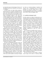
Figure 1. . ATR-FTIR spectra of untreated polymer (a) PE (i) and chitosan (ii), PE after UV-ozone treatment (b), after chitosan coating PE
membranes (c) with 0.25 wt/vol chitosan (i), 0.75 wt/vol chitosan (ii), and 2 wt/vol chitosan (iii). Magnified spectra of represent the expanded
spectra of PE membranes coated with 2 wt/vol chitosan (d) with peaks assigned.
and that of 1151 cm−1 arising from asymmetric stretching of the
C−O−C bridge is well evident. The peaks at 1070 and 1031
cm−1 further confirm the saccharine structure of chitosan.31
Figure 1b shows the FTIR of the UV treated PE, and this
showed peak of carboxyl group (CO) at 1730 cm−1, shoulder
at 1410 cm−1, and hydroxyl group (OH) group broad 3000−
3700 cm−1. This confirms that the UV-ozone treatment led to
the carboxyl acid group in PE membranes. Figure 1c and d
shows the FTIR spectra of chitosan immobilized PE which
showed distinct characteristic peaks, corresponding to primary
and secondary amine of chitosan at 1650 and 1550 cm−1
respectively. A new peak at 1737 cm−1 is evident which
corresponds to CO stretch of the ester group.32 Hence, from
FTIR, it is evident that coated membranes contain both
characteristic peaks of PE and chitosan, with the formation of
new ester linkage. Thus, from FTIR, we can conclude that
chitosan was successfully coated onto PE token membranes.
Further complete etching of PEO phase was ensured by
taking the IR spectra of PE membranes as shown in Figure S1.
From Figure S1 it is evident that no peaks of PEO were
observed.
washing with phosphate buffered saline (PBS). Thus, obtained pellets
were resuspended in PBS (of pH 7.4) for required final concentration
of cells of ∼107−108 CFU/mL. The three replicates were performed
before reporting the antibacterial activity. The membranes of specific
dimensions were immersed in the PBS culture. The suspended
membranes were incubated at 37 °C for 4 h. After 4 h, the supernatant
of 100 μL was used for plating on the nutrient agar after suitable
dilution. After 12 h of incubation colonies formed were counted.
■
RESULTS AND DISCUSSION
Characterizing Chitosan Immobilized PE Membranes.
FITR. The surface grafting of chitosan upon UV-ozone
treatment was characterized by FTIR. Figure 1a shows the
FTIR spectra of untreated PE and chitosan. Untreated PE
membranes showed absorption peaks at 2920 and 2850 cm−1
corresponding to C−H stretching of methylene. The peaks at
1464 and 720 cm−1 can be attributed to C−H bending and C−
H rocking of methylene group, respectively. The untreated
chitosan exhibited a distinct absorption peak at 1650 cm−1
which is attributed to N−H bending of amide I and the peak at
1585 cm−1 due to C−C stretching (in-ring) of the aromatic
ring. The peak corresponding to 1381 cm−1 is due to amide III
864
DOI: 10.1021/acssuschemeng.5b00912
ACS Sustainable Chem. Eng. 2016, 4, 862−870
Research Article
ACS Sustainable Chemistry & Engineering
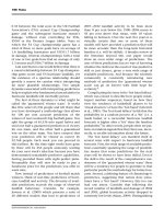
XPS. Further evidence of chitosan immobilization onto PE is
based on XPS, as shown in Figure 2a, which reveals no traces of
Scheme 1. Proposed Mechanism of UV-Ozone Treatment,
Possible Products of UV-Ozone Treatment (a) and Possible
Chitosan Coating on PE Membranes via Ester Linkage (b)
et al.16 for plasma treated PE films. Further, neutralization of
these chitosan coated PE membrane can lead to regeneration of
NH2 group which is very important from antibacterial point of
view and will be discussed in detail later.
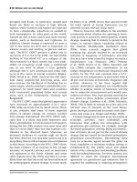
Kjeldahl Nitrogen Analysis. To obtain a quantitative picture,
the concentration of nitrogen present on PE was assessed using
Kjeldahl nitrogen analysis. Both, the UV-ozone treated and
untreated PE membranes were dipped in chitosan solution of
varying concentration followed by repeated washing and
sonication to remove the unbound chitosan. Subsequently,
the membranes were neutralized. For the present study,
chitosan of 0.25, 0.75, and 2.0% wt/vol chitosan was dissolved
in acetic acid and used for nitrogen analysis. The untreated PE
membranes were free of chitosan whereas, for the UV-ozone
treated samples the concentration of nitrogen scaled with
increasing concentration of chitosan (see Figure 3). For
instance, PE membranes which were subjected to 0.25% wt/
Figure 2. Wide XPS spectra (a) of PE (i) before coating with chitosan
and (ii) after coating with 2 wt/vol chitosan N-1s scan (b) of PE (i)
before coating with chitosan and (ii) after coating with 2 wt/vol
chitosan.
nitrogen on neat PE. However, chitosan coated membranes
exhibited a peak of N-1s (see Figure 2a and b). From XPS, the
mass % and the atomic % of nitrogen was estimated to be 1.90
and 1.64% respectively, for 2% wt/vol chitosan coated
membranes. Further N-1s peak exhibited a binding energy of
402.8 eV and no such peak was observed for untreated PE
membranes. This suggests that elemental nitrogen is present
only on treated PE membranes, which is arising from chitosan
present on the surface.
A possible mechanism of grafting chitosan on PE can be
explained, as shown in Scheme 1. When subjected to UV-ozone
treatment, the PE chains undergo chain scission and rearrangement to form carboxylic group, as shown in Scheme 1a. This
carboxylic group can react with the ester linkage of chitosan.
Both NH2 and OH groups of chitosan have equal probabilities
to react, however, due to acidic media the carboxylic group is
protonated. Thus, the protonated carboxyl moieties will react
with the hydroxyl group of chitosan to form ester linkage. It is
envisaged that formation of amide linkage is hindered in the
presence of acidic media. Thus, the proposed mechanism is in
line with FTIR wherein the peak at 1737 cm−1 arising due to
CO stretch of ester group confirms the grafting of chitosan
onto PE. Similar observations have been reported by Theapsak
Figure 3. Amount of chitosan coated on PE membranes obtained
using Kjeldahl nitrogen analysis of treated and untreated PE.
865
DOI: 10.1021/acssuschemeng.5b00912
ACS Sustainable Chem. Eng. 2016, 4, 862−870
Research Article
ACS Sustainable Chemistry & Engineering
vol of chitosan coating exhibited weight of 13 ± 0.5 μg·mm−2 of
chitosan, whereas the 2.0% wt/vol chitosan coated membranes
exhibited weight of 32.7 ± 0.7 μg·mm−2 of chitosan. This
clearly suggests that UV-ozone treatment enhances the
interaction between the membranes and chitosan.
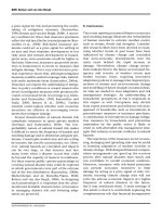
Staining of Chitosan. In order to understand the
distribution and chitosan coverage on the porous PE
membranes, the samples were stained with amide black. It is
envisaged that amide black interacts with the amine groups of
chitosan and gets adsorbed on the surface. Figure 4 shows the
The process of grafting chitosan on PE surface is carried out
in a liquid media. As soon as the media is separated, chitosan
tend to phase separate in air as it tries to undergo shape
relaxation resulting in a spherical shape in air. It is important to
note that the dispersion of chitosan is strongly contingent on
the availability of functional moieties on the PE surface and also
on the chitosan concentration. The solution with 2.0% wt/vol
chitosan exhibits slightly higher viscosity due to higher
molecular weight of chitosan. The higher molecular weight of
chitosan tends to form aggregated structures resulting in a
nonuniform distribution of droplets over the surface.
Surface Optical Profiles. Figure 5 shows the surface optical
profiles acquired using an optical profilometer that reflects the
surface roughness of untreated and 2.0% wt/vol chitosan
coated PE membranes. The untreated PE membranes exhibited
a roughness (Ra) of ca. 16.0 ± 3.8 μm and the membrane with
2 wt/vol chitosan exhibited a Ra of ca. 23.1 ± 3.2 μm. This
indicates an increase in surface roughness upon immobilizing
chitosan. These results clearly hint at the fact that the presence
of chitosan is mainly on the PE surface and not in the pores.
This will help in retaining the pure water flux even after
chitosan immobilization and will be discussed subsequently.
Designer Porous Membranes through Selective
Etching of PEO from PE/PEO Blends: Morphology. Figure
6 shows the SEM micrographs of the surface and cross-section
of PE membranes before and after chitosan coating. PE/PEO
blends are immiscible,2,5,6,33 wherein PEO is dispersed in the
PE matrix as observed from the SEM micrographs. The average
droplet size of PEO in the blends is ca. 1.13 μm with a
polydispersity index of 1.10.5,6 Interestingly, after chitosan
immobilization, the pore diameter was similar. Thus, it can be
concluded that chitosan is only coated on the membrane
surface and this is also supported by surface staining
experiments and optical profilometry as well. In order to
validate the hypothesis proposed earlier in context to the
distribution of chitosan on the surface, EDAX mapping was
carried out.
Figure 7 shows the EDAX one to one surface mapping of the
membrane coated with 2.0% wt/vol chitosan. Figure 7a
represents the surface morphology of the PE membrane, and
Figure 7b and c represents the one to one mapping of C-1s
(carbon) and N-1s (nitrogen), respectively. It is evident that
Figure 4. Optical microscopic images of untreated PE (a), 0.25% wt/
vol chitosan coated PE (b), 0.75% wt/vol chitosan coated PE (c), and
2.0% wt/vol chitosan coated PE (d).
optical microscopic images of untreated PE membranes and
membranes coated with chitosan. From Figure 4a, it is evident
that untreated PE membrane does not exhibit any color.
However, membranes with chitosan immobilized on the surface
show dark patches due to adsorption of amide black on the
surface. This phenomenon is consistent with increasing
chitosan concentration. This analysis clearly shows that
chitosan is well distributed on the surface of the membrane
which also suggests that UV-ozone treatment actually assists in
generating free radicals on the surface that facilitate in chitosan
immobilization. Similar observations have been reported in
literature.16
Figure 5. Surface and roughness profile of untreated PE (a) and 2.0% wt/vol chitosan (b) obtained by optical profilometer (Ra indicates the
roughness value of whole scanned surface).
866
DOI: 10.1021/acssuschemeng.5b00912
ACS Sustainable Chem. Eng. 2016, 4, 862−870
Research Article
ACS Sustainable Chemistry & Engineering
Scheme 2. Typical Cross Flow Test Cell for Estimating the
Transmembrane Flux As a Function of Pressure
Figure 6. SEM micrograph of surface topology of untreated PE
membrane (a) and across the surface (b) and PE membranes after
2.0% wt/vol chitosan on the surface (c) and across the surface (d).
from nitrogen mapping, which is attributed to the presence of
amine, that the chitosan is present on the surface of the
membrane. Further, the distribution of N-1s suggests uniform
coating of chitosan on the surface and this observation supports
the staining experiments as well. EDAX spectra also revealed
the atomic concentration of C and N to be 71.77 and 28.23%,
respectively (and weight % of C and N to be 68.55 and 31.45%,
respectively). Further traces of N-1s are absent in EDAX
spectra of untreated PE membranes, as shown in Figure S2
confirming the absence of nitrogen on the surface. Thus, the
presence of nitrogen on the chitosan coated surface arises
would arise from chitosan present on the surface. Hence, the
presence of chitosan on the PE surface can be confirmed.
Membrane Performance. The membrane performance was
evaluated by estimating the trans-membrane flux as a function
of pressure using an indigenously developed cross-flow setup as
shown in Scheme 2. Figure 8 illustrates the flux at various
pressures for membranes with varying chitosan coating. It is
Figure 8. Flux measurement at various trans membrane pressure for
PE membranes.
observed that as pressure increases, flux increases for
membranes. From Figure 8, it is evident that the flux did not
vary significantly with the coating of chitosan on PE
membranes. However, a decreased flux was noted for the
highest concentrations of chitosan i.e. at 2% wt/vol, which
presumably could be attributed to the resistance offered by
excess of chitosan. Interestingly, in 0.75% wt/vol chitosan
coated PE membranes, the flux increased from 4062 ± 266 to
4749 ± 252 L m−2 h−1. This increase in flux with respect to
Figure 7. SEM micrograph of 2 wt/vol chitosan coated PE membrane (a), C-1s mapping on the surface (b), N-1s mapping on the surface (c), and
EDAX spectra of the surface (d).
867
DOI: 10.1021/acssuschemeng.5b00912
ACS Sustainable Chem. Eng. 2016, 4, 862−870
Research Article
ACS Sustainable Chemistry & Engineering
unmodified PE membrane is ascribed to the hydrophilic
chitosan coating on a rather hydrophobic PE membrane.
Chanachai et al.34 reported a similar effect where an increase in
hydrophilicity of the surface led to a decrease in repulsive forces
between hydrophobic membrane and water. Further, hydrophilicity facilitates in enhanced diffusion and thus increases the
flux.35 But higher concentration of chitosan decreased the flux
due to a thicker layer of chitosan which presumably blocked the
pores and offered additional resistance to flow.
Antibacterial Studies. The antibacterial activity of the
chitosan coated PE membranes was studied using E. coli as
Gram-negative and S. aureus as Gram-positive bacteria (see
Figures 9 and 10). The antibacterial activity is expressed here in
Figure 10. Total agar plate counts of E. coli (i) and S. aureus (ii)
colonies after 12 h of inoculation of negative control (a) untreated PE
(b), 0.25% wt/vol chitosan (c), 0.75% wt/vol chitosan (d), and 2.0%
wt/vol chitosan (e).
Figure 10i and ii, it is evident that the number of colonies
decreases with increasing chitosan concentration which
indicates that chitosan eventually inhibits the bacterial growth.
The decrease in colonies clearly indicates the antibacterial
nature of the membranes. The mechanism of bacterial
population reduction upon chitosan coating could be due to
the interaction of free NH2 group (of chitosan) with the
phospholipids36−39 present in the bacterial cell membrane.
Second, it can be envisioned that protonated NH3+ groups of
the chitosan can form a complex with the phosphate groups in
phospholipid bilayer of bacterial cell membrane. This complex
might as well result in the disruption of osmotic balance further
resulting in the release of intracellular electrolytes such as
potassium ions, glucose, nucleic acid, etc., thus resulting in cell
death. Further, at pH 6.0 similar reductions in bacterial
population were noted for E. coli. It was observed with increase
in chitosan concentration, antibacterial property was found to
increase as shown in Figure 9ii.
■
SUMMARY
In this study, we were able to immobilize chitosan on UVozone treated PE membranes which was further confirmed by
FTIR, XPS, EDAX, Kjeldhal analysis, and amide black staining
experiments. Thus, the hydrophobic polyolefin was converted
to hydrophilic and in addition, rendered an antibacterial
surface. The concentration of chitosan immobilization was
optimized by varying the concentration of chitosan solution
and through the flux measurements. For instance, 32 μg mm−2
of chitosan was estimated when the PE membranes were
treated with 2% wt/vol chitosan solution, however, the flux was
reduced. But, PE membranes with 0.75% wt/vol chitosan
coating exhibited increase in flux from 4062 ± 266 to 4749 ±
252 L m−2 h−1 with respect to untreated PE membranes.
Intriguingly, with 2 wt/vol of chitosan coating on the surface,
the bacterial reduction efficiency of 90 and 94% for E. coli and
S. aureus respectively was observed. Thus, this study clearly
demonstrates that chitosan coated sustainable antibacterial
membranes can be derived by etching one of the phases from
binary polyolefinic blends and can further be explored for water
purification.
Figure 9. Dependence of E. coli and S. aureus (CFU mL−1) on the
chitosan coating on the membranes after 4 h of inoculation at pH 7.4
(i). Dependence of E. coli at pH 6.0 (ii) (of negative control (cells
without membranes) (a) untreated PE membranes (positive control)
(b), 0.25% wt/vol chitosan (c), 0.75% wt/vol chitosan (d), and 2.0%
wt/vol chitosan (e)).
terms of colony forming units per milliliter (CFU mL−1).
Figure 9 shows the colony count after 4 h of inoculation. From
Figure 9i, it is evident that untreated PE membranes exhibited a
colony count of 2.8 × 10⁷ per mL whereas 2% wt/vol chitosan
coated PE membranes showed a colony of 6.0 × 10⁶ i.e., about
90% reduction in E. coli is observed with respect to initial count.
Similarly a reduction from 4.7 × 10⁷ to 1.7 × 10⁷ i.e., about 94%
reduction in S. aureus is observed with respect to initial count.
Figure 10 exhibits agar plate counts of bacterial colonies after
12 h of inoculation of untreated and chitosan coated PE. From
■
ASSOCIATED CONTENT
S Supporting Information
*
The Supporting Information is available free of charge on the
ACS Publications website at DOI: 10.1021/acssuschemeng.5b00912.
868
DOI: 10.1021/acssuschemeng.5b00912
ACS Sustainable Chem. Eng. 2016, 4, 862−870
Research Article
ACS Sustainable Chemistry & Engineering
■
(14) Ulbricht, M.; Matuschewski, H.; Oechel, A.; Hicke, H.-G.
Photo-Induced Graft Polymerization Surface Modifications for the
Preparation of Hydrophilic and Low-Proten-Adsorbing Ultrafiltration
Membranes. J. Membr. Sci. 1996, 115, 31−47.
(15) Revanur, R.; McCloskey, B.; Breitenkamp, K.; Freeman, B. D.;
Emrick, T. Reactive Amphiphilic Graft Copolymer Coatings Applied
to Poly (Vinylidene Fluoride) Ultrafiltration Membranes. Macromolecules 2007, 40, 3624−3630.
(16) Theapsak, S.; Watthanaphanit, A.; Rujiravanit, R. Preparation of
Chitosan-Coated Polyethylene Packaging Films by Dbd Plasma
Treatment. ACS Appl. Mater. Interfaces 2012, 4, 2474−2482.
(17) Deng, J.; Wang, L.; Liu, L.; Yang, W. Developments and New
Applications of Uv-Induced Surface Graft Polymerizations. Prog.
Polym. Sci. 2009, 34, 156−193.
(18) Wolska, J.; Smolinska-Kempisty, K.; Bryjak, M.; Kujawski, W.
Polypropylene Membranes with the Double Sensitivity Effect. J. Appl.
Polym. Sci. 2015, 132, 1097−4628.
(19) Yu, H.-Y.; Xie, Y.-J.; Hu, M.-X.; Wang, J.-L.; Wang, S.-Y.; Xu, Z.K. Surface Modification of Polypropylene Microporous Membrane to
Improve Its Antifouling Property in Mbr: Co 2 Plasma Treatment. J.
Membr. Sci. 2005, 254, 219−227.
(20) Shim, J. K.; Na, H. S.; Lee, Y. M.; Huh, H.; Nho, Y. C. Surface
Modification of Polypropylene Membranes by Γ-Ray Induced Graft
Copolymerization and Their Solute Permeation Characteristics. J.
Membr. Sci. 2001, 190, 215−226.
(21) Jiang, J.; Zhu, L.; Zhu, L.; Zhang, H.; Zhu, B.; Xu, Y. Antifouling
and Antimicrobial Polymer Membranes Based on Bioinspired
Polydopamine and Strong Hydrogen-Bonded Poly (N-Vinyl Pyrrolidone). ACS Appl. Mater. Interfaces 2013, 5, 12895−12904.
(22) Yu, H.-Y.; Xie, Y.-J.; Hu, M.-X.; Wang, J.-L.; Wang, S.-Y.; Xu, Z.K. Surface Modification of Polypropylene Microporous Membrane to
Improve Its Antifouling Property in Mbr: Co2 Plasma Treatment. J.
Membr. Sci. 2005, 254, 219−227.
(23) Morra, M.; Occhiello, E.; Garbassi, F. Adhesion Improvement
by Uv Grafting onto Polyolefin Surfaces. J. Adhes. 1994, 46, 39−47.
(24) Jiang, J.-H.; Zhu, L.-P.; Li, X.-L.; Xu, Y.-Y.; Zhu, B.-K. Surface
Modification of Pe Porous Membranes Based on the Strong Adhesion
of Polydopamine and Covalent Immobilization of Heparin. J. Membr.
Sci. 2010, 364, 194−202.
(25) Musale, D. A.; Kumar, A.; Pleizier, G. Formation and
Characterization of Poly(Acrylonitrile)/Chitosan Composite Ultrafiltration Membranes. J. Membr. Sci. 1999, 154, 163−173.
(26) Aravind, U. K.; Mathew, J.; Aravindakumar, C. T. Transport
Studies of Bsa, Lysozyme and Ovalbumin through Chitosan/
Polystyrene Sulfonate Multilayer Membrane. J. Membr. Sci. 2007,
299, 146−155.
(27) Boributh, S.; Chanachai, A.; Jiraratananon, R. Modification of
Pvdf Membrane by Chitosan Solution for Reducing Protein Fouling. J.
Membr. Sci. 2009, 342, 97−104.
(28) Sarkar, K.; Banerjee, S.; Kundu, P. Removal of Anionic Dye in
Acid Solution by Self Crosslinked Insoluble Dendronized Chitosan.
Hydrol.: Curr. Res. 2012, 3, 133.
(29) Carlson, R. P.; Taffs, R.; Davison, W. M.; Stewart, P. S. AntiBiofilm Properties of Chitosan-Coated Surfaces. J. Biomater. Sci.,
Polym. Ed. 2008, 19, 1035−46.
(30) Desai, S. M.; Singh, R., Surface Modification of Polyethylene. In
Long Term Properties of Polyolefins; Springer, 2004; pp 231−294.
(31) Peniche, C.; Argüelles-Monal, W.; Davidenko, N.; Sastre, R.;
Gallardo, A.; San Román, J. Self-Curing Membranes of Chitosan/PAA
IPNS Obtained by Radical Polymerization: Preparation, Characterization and Interpolymer Complexation. Biomaterials 1999, 20, 1869−
1878.
(32) Gonzalez, E.; Hicks, R. F. Surface Analysis of Polymers Treated
by Remote Atmospheric Pressure Plasma. Langmuir 2010, 26, 3710−
3719.
(33) Mural, P. K. S.; Madras, G.; Bose, S. Positive Temperature
Coefficient and Structural Relaxations in Selectively Localized Mwnts
in PE/PEO Blends. RSC Adv. 2014, 4, 4943−4954.
Figure S1: FTIR spectra of PE membrane after etching
PEO phase. Figure S2: SEM micrograph and EDAX
mapping of untreated PE membrane (PDF)
AUTHOR INFORMATION
Corresponding Author
*E-mail address:
Notes
The authors declare no competing financial interest.
■
ACKNOWLEDGMENTS
The authors would like to acknowledge the Department of
Science and Technology and INSA (India) for the financial
support and CeNSE, IISc, for various characterization facilities.
In addition, the authors are grateful to Prof. Jayant Modak for
extending his facilities, IISc, for extending their help in
antibacterial studies.
■
REFERENCES
(1) Pendergast, M. M.; Hoek, E. M. V. A Review of Water Treatment
Membrane Nanotechnologies. Energy Environ. Sci. 2011, 4, 1946−
1971.
(2) Trifkovic, M.; Hedegaard, A.; Huston, K.; Sheikhzadeh, M.;
Macosko, C. W. Porous Films Via PE/PEO Cocontinuous Blends.
Macromolecules 2012, 45, 6036−6044.
(3) Geise, G. M.; Lee, H.-S.; Miller, D. J.; Freeman, B. D.; McGrath,
J. E.; Paul, D. R. Water Purification by Membranes: The Role of
Polymer Science. J. Polym. Sci., Part B: Polym. Phys. 2010, 48, 1685−
1718.
(4) Ulbricht, M. Advanced Functional Polymer Membranes. Polymer
2006, 47, 2217−2262.
(5) Mural, P. K. S.; Banerjee, A.; Rana, M. S.; Shukla, A.;
Padmanabhan, B.; Bhadra, S.; Madras, G.; Bose, S. Polyolefin Based
Antibacterial Membranes Derived from PE/PEO Blends Compatibilized with Amine Terminated Graphene Oxide and Maleated PE. J.
Mater. Chem. A 2014, 2, 17635−17648.
(6) Mural, P. K. S.; Sharma, M.; Shukla, A.; Bhadra, S.;
Padmanabhan, B.; Madras, G.; Bose, S. Porous Membranes Designed
from Bi-Phasic Polymeric Blends Containing Silver Decorated
Reduced Graphene Oxide Synthesized Via a Facile One-Pot Approach.
RSC Adv. 2015, 5, 32441−32451.
(7) Han, M.; Lim, B.; Jung, H.; Hyun, J.; Kim, S.; Kim, W. Reactive
Blends of Poly (Butylene Terephthalate)/Polyamide-6 with Ethylene
Glycidyl Methacrylate. Korea-Australia Rheology Journal 2001, 169−
177.
(8) Utracki, L. A. Polymer Blends Handbook; Kluwer Academic
Publishers: Dordrecht, The Netherlands, 2002; Vol. 1.
(9) Sharma, M.; Madras, G.; Bose, S. Unique Nanoporous
Antibacterial Membranes Derived through Crystallization Induced
Phase Separation in PVDF/PMMA Blends. J. Mater. Chem. A 2015, 3,
5991−6003.
(10) Rubira, A. F.; da Costa, A. C.; Galembeck, F.; Leite Escobar, N.
F.; da Silva, E. C.; Vargas, H. Polyethylene and Polypropylene Surface
Modification by Impregnation with Manganese (Iv) Oxide. Colloids
Surf. 1985, 15, 63−73.
(11) Wavhal, D. S.; Fisher, E. R. Membrane Surface Modification by
Plasma-Induced Polymerization of Acrylamide for Improved Surface
Properties and Reduced Protein Fouling. Langmuir 2003, 19, 79−85.
(12) McCloskey, B. D.; Park, H. B.; Ju, H.; Rowe, B. W.; Miller, D. J.;
Freeman, B. D. A bioinspired fouling-resistant surface modification for
water purification membranes. J. Membr. Sci. 2012, 413-414, 82−90.
(13) Ulbricht, M.; Belfort, G. Surface Modification of Ultrafiltration
Membranes by Low Temperature Plasma Ii. Graft Polymerization
onto Polyacrylonitrile and Polysulfone. J. Membr. Sci. 1996, 111, 193−
215.
869
DOI: 10.1021/acssuschemeng.5b00912
ACS Sustainable Chem. Eng. 2016, 4, 862−870
Research Article
ACS Sustainable Chemistry & Engineering
(34) Chanachai, A.; Meksup, K.; Jiraratananon, R. Coating of
Hydrophobic Hollow Fiber Pvdf Membrane with Chitosan for
Protection against Wetting and Flavor Loss in Osmotic Distillation
Process. Sep. Purif. Technol. 2010, 72, 217−224.
(35) Dickson, J. M.; Childs, R. F.; McCarry, B. E.; Gagnon, D. R.
Development of a Coating Technique for the Internal Structure of
Polypropylene Microfiltration Membranes. J. Membr. Sci. 1998, 148,
25−36.
(36) Palermo, E. F.; Lee, D.-K.; Ramamoorthy, A.; Kuroda, K. Role
of Cationic Group Structure in Membrane Binding and Disruption by
Amphiphilic Copolymers. J. Phys. Chem. B 2011, 115, 366−375.
(37) Singh, S. K.; Singh, M. K.; Kulkarni, P. P.; Sonkar, V. K.; Grácio,
J. J. A.; Dash, D. Amine-Modified Graphene: Thrombo-Protective
Safer Alternative to Graphene Oxide for Biomedical Applications. ACS
Nano 2012, 6, 2731−2740.
(38) Kumar, S.; Raj, S.; Kolanthai, E.; Sood, A. K.; Sampath, S.;
Chatterjee, K. Chemical Functionalization of Graphene to Augment
Stem Cell Osteogenesis and Inhibit Biofilm Formation on Polymer
Composites for Orthopedic Applications. ACS Appl. Mater. Interfaces
2015, 7, 3237−3252.
(39) Kumar, S.; Bose, S.; Chatterjee, K. Amine-Functionalized
Multiwall Carbon Nanotubes Impart Osteoinductive and Bactericidal
Properties in Poly (E-Caprolactone) Composites. RSC Adv. 2014, 4,
19086−19098.
870
DOI: 10.1021/acssuschemeng.5b00912
ACS Sustainable Chem. Eng. 2016, 4, 862−870