human body
Bạn đang xem bản rút gọn của tài liệu. Xem và tải ngay bản đầy đủ của tài liệu tại đây (18 MB, 56 trang )
About the pagination of this eBook
Due to the unique page numbering scheme of this book, the
electronic pagination of the eBook does not match the pagination
of the printed version. To navigate the text, please use the
electronic Table of Contents that appears alongside the eBook or
the Search function.
For citation purposes, use the page numbers that appear in the text.
Encyclopædia Britannica, Inc.
Chicago
■
London
■
New Delhi
■
Paris
■
Seoul
■
Sydney
■
Taipei
■
Tokyo
Britannica Illustrated Science Library
Britannica Illustrated Science Library
HUMAN BODY I
HUMAN BODY I
© 2
008 Editorial Sol 90
All rights reserved.
Idea and Concept of This Work: Editorial Sol 90
Project Management: Fabián Cassan
Photo Credits: Corbis, ESA, Getty Images, Graphic News,
NASA, National Geographic, Science Photo Library
Illustrators: Guido Arroyo, Pablo Aschei, Carlos Francisco
Bulzomi, Gustavo J. Caironi, Hernán Cañellas, Leonardo César,
José Luis Corsetti, Vanina Farías, Manrique Fernández Buente,
Joana Garrido, Celina Hilbert, Inkspot, Jorge Ivanovich, Iván
Longuini, Isidro López, Diego Martín, Jorge Martínez, Marco
Menco, Marcelo Morán, Ala de Mosca, Diego Mourelos, Laura
Mourelos, Pablo Palastro, Eduardo Pérez, Javier Pérez, Ariel
Piroyansky, Fernando Ramallo, Ariel Roldán, Marcel Socías,
Néstor Taylor, Trebol Animation, Juan Venegas, Constanza
Vicco, Coralia Vignau, Gustavo Yamin, 3DN, 3DOM studio
Composition and Pre-press Services: Editorial Sol 90
Translation Services and Index: Publication Services, Inc.
Portions © 2008 Encyclopædia Britannica, Inc.
Encyclopædia Britannica, Britannica, and the thistle logo are
registered trademarks of Encyclopædia Britannica, Inc.
Britannica Illustrated Science Library Staff
Editorial
Michael Levy, Executive Editor, Core Editorial
John Rafferty, Associate Editor, Earth Sciences
William L. Hosch, Associate Editor, Mathematics and
Computers
Kara Rogers, Associate Editor, Life Sciences
Rob Curley, Senior Editor, Science and Technology
David Hayes, Special Projects Editor
Art and Composition
Steven N. Kapusta, Director
Carol A. Gaines, Composition Supervisor
Christine McCabe, Senior Illustrator
Media Acquisition
Kathy Nakamura, Manager
Copy Department
Sylvia Wallace, Director
Julian Ronning, Supervisor
Information Management and Retrieval
Sheila Vasich, Information Architect
Production Control
Marilyn L. Barton
Manufacturing
Kim Gerber, Director
Britannica Illustrated
Science Library
Britannica Illustrated
Science Library
Enc
yclopædia Britannica, Inc.
Jacob E. Safra, Chairman of the Board
Jorge Aguilar-Cauz, President
Michael Ross, Senior Vice President, Corporate Development
Dale H. Hoiberg, Senior Vice President and Editor
Marsha Mackenzie, Director of Production
International Standard Book Number (set):
978-1-59339-797-5
International Standard Book Number (volume):
978-1-59339-809-5
Britannica Illustrated Science Library: Human Body I 2008
Printed in China
w
ww.britannica.com
Human
Body I
Contents
What Are We
Made Of?
Page 6
Bones and
Muscles
Page 18
Internal Systems
and Organs
Page 34
The Senses
and Speech
Page 68
Control
Centers
Page 80
What are cells like, and how do they form
tissue? What is blood, and why are proteins
so important? The heart, usually thought of as
the wellspring of love and the emotions, is
actually the engine of the circulatory system.
It is because of the heart that all the cells of
the body receive a constant supply of
nutrients, oxygen, and other essential
substances. The heart is so powerful that it
pumps about 10 pints (4.7 l) of blood per
minute. The nervous system is the most
intricate of all the body's systems. It works
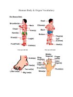
A LIVING STRUCTURE
The skeleton consists of
206 separate bones,
which differ in form, size,
and name. It supports
and shapes the body,
protects the internal
organs, and—in the
bone marrow of certain
bones—manufactures
various types of blood cells.
every second of every day, gathering
information about the organism and its
surroundings and issuing instructions so that
the organism can react. It is this computer
that permits us to think and remember and
that makes us who we are.
T
he nervous system is a complex network
of sensory cells, originating in the brain
and spinal cord, that transmits signals
throughout the body, employing a caravan of
chemical messengers to make sense of this
marvelous complex that we catalogue as
touch, taste, smell, hearing, and vision. In fact,
at this precise moment, because of an
extraordinary relationship between our eyes
and our brain, we are able to see and
understand what we are reading. Modern
cameras are designed on the same basic
principles as our eye, but they have never been
able to equal the visual power of the eye. The
focus and the automatic aperture of the human
eye are perfect. Our ears share a similar
complexity and allow us to have excellent
hearing. The external ear operates by receiving
sound waves in the air. Sound waves travel
through the auditory canal and are transmitted
by the bones of the intermediate ear toward
the cochlea, which contains liquid and is
spiraled like the shell of a small sea snail. The
cochlea converts waves of air into vibrations of
liquid, which are detected by special filaments
in the ear that are of many lengths and that
detect sound waves of different lengths. These
filaments then transmit nerve impulses to the
brain and provide us with our ability to
interpret what we hear. This book will also
tell you about the function of our skin, the
largest organ of the body, which serves as an
elastic barrier covering and protecting
everything inside our bodies. Captivating
images will show you how each of our
extraordinary body systems function, and
incredible facts will help you understand why
the human body is so amazing.
H
ow can we understand what we are?
What are we made of? Are we aware
that all that we do—including reading
this book—is the work of a marvelous
machine? We know very little about how we
are able to be conscious of our own actions;
nevertheless, even though we are usually not
very aware of it, this community of organs
that is the body—an integrated system that
includes the brain, heart, lungs, liver, kidneys,
muscles, bones, skin, and endocrine glands—
acts together in exquisitely regulated
harmony. It is interesting that various
mechanisms work together to keep the
temperature of the body at 98.6° F (37° C);
thanks to the dynamic structure of bones
and cartilage, the body is maintained in
perfect balance. The body also has a
fantastic ability to transform the food it
ingests into living tissues, bones, and
teeth, all of which contribute to its growth.
By this same process, we obtain the energy
for working and playing. It is hard to
imagine that not long ago the cells of the
body of the person reading this book were
autonomous and were duplicating
themselves freely within the walls of a
mother's uterus. Certainly no one
reading this book could recognize herself
or himself in those cells. Nevertheless,
each cell carried within it the information
necessary for the development of that
person. Everything that happens inside us is
truly fascinating. Therefore, we invite you to
enjoy this book. It is full of incredible facts
and illustrations that will show you the
complex ways each part of the body works.
A Perfect
Machine
What Are We Made Of ?
T
o understand the truest and
most elementary characteristics
of life, we must begin with the
cell-the tiny organizing
structure of life in all its forms.
Most cells are too small to be
observed with the naked eye, but they
can be distinguished easily through an
ordinary microscope. Human body
tissues are groups of cells whose size
and shape depend on the specific
tissue to which they belong. Did you
know that an embryo is a mass of
rapidly dividing cells that continue to
develop during infancy? We invite you
to turn the page and discover many
surprising things in this fascinating
and complex world.
UNDIVIDED ATTENTION 8-9
WATER AND LIQUIDS 10-11
THE CELL 12-13
MITOSIS 14-15
SYSTEMS OF THE BODY 16-17
MITOSIS
An enlarged view that shows
the process of mitosis, the
most common form of
cellular division
HUMAN BODY I
98
WHAT ARE WE MADE OF?
Undivided
Attention
From birth the infant's brain
cells develop rapidly,
making connections that can
shape all of life's
experiences. The first three
years are crucial. When
neurons receive visual,
auditory, or gustatory stimuli,
they send messages that
generate new physical
connections with
neighboring cells. The signals
are sent through a gap called
a synapse by means of a
complex electrochemical
process. What determines
the formation of a person's
synapses and neural
networks? One key factor is
believed to be the undivided
attention and mental effort
exerted by the person.
THE SENSE OF TOUCH
It is predominant in the fingers and hands. The
information is transmitted through neurotransmitters,
nerves that carry these impulses to the brain and
that serve to detect sensations such as cold, heat,
pressure, and pain.
SKIN
The skin is one of the
most important organs of the
body. It contains approximately
five million tiny nerve endings
that transmit sensations.
Learning
Each child has his or her own intellectual filter; the
quality of the filter depends on undivided attention and
on how the child responds to a broad variety of stimuli.
Brain
At birth the infant brain contains 100
billion neurons. That is about as many
nerve cells as there are stars in the
entire Milky Way Galaxy! Then as the
infant receives messages from the
senses, the cerebral cortex begins its
dynamic development.
Respiration
Respiration is usually an involuntary,
automatic action that allows us to take in
the oxygen we need from the air and exhale
carbon dioxide. These gases are exchanged
in the pulmonary alveoli.
Neurons
Each neuron in the brain can be
connected with several thousand other
neurons and is capable of receiving
100,000 signals per second. The signals
travel through the nervous
system at a speed of 225 miles per hour
(360 km/h). Thanks to this complex
communication network, the brain is
capable of remembering, calculating,
deciding, and thinking.
A WORLD OF SENSATIONS
The tongue recognizes four tastes (sweet,
salty, sour, and bitter), and the nasal fossas
contain cells that have more than 200 million
filaments, called cilia, which are capable of
detecting thousands of odors.
DENDRITES
They are the branches
through which a neuron
receives and sends messages.
With this system each neuron
can be stimulated by
thousands of other neurons,
which in turn can stimulate
other neurons, and so forth.
3 pounds
(1.4 kg)
IS THE WEIGHT OF
A HUMAN BRAIN.
225
(360 km/h)
THE VELOCITY OF THE NERVOUS
SYSTEM'S SIGNALS
miles
per hour
HUMAN BODY I
11
10
WHAT ARE WE MADE OF?
Water and Fluids
W
ater is of such great importance that it makes up almost
two thirds of the human body by weight. Water is present in all
the tissues of the body. It plays a fundamental role in digestion and
absorption and in the elimination of indigestible metabolic waste. Water also
serves as the basis of the circulatory system, which uses blood to distribute
nutrients to the entire body. Moreover, water helps maintain body temperature
by expelling excess heat through the skin via perspiration and evaporation.
Perspiration and evaporation of water account for most of the weight a person
loses while exercising.
N
3% NITROGEN
Present in proteins
and nucleic acids
Water Balance and Food
In its continuous process of taking in and
eliminating water, one of the most
important functions of the body is to maintain a
continuous equilibrium between the water that
enters and the water that leaves the body.
Because the body does not have an organ or
other place for storing water, quantities that are
lost must be continuously replenished. The
human body can survive for several weeks
without taking in food, but going without water
for the same length of time would have tragic
consequences. The human being takes in about
2.5 to 3 quarts (2.5-3 l) of water per day. About
half is taken in by drinking, and the rest comes
from eating solid food. Some foods, such as fruits
and vegetables, consist of 95 percent water.
Eggs are 90 percent water, and red meat and
fish are 60 to 70 percent water.
HOW THIRST IS CONTROLLED
Thirst is the sensation through
which the nervous system informs
its major organ, the brain, that the
body needs water. The control
center is the hypothalamus. If the
concentration of plasma in the blood
increases, it means the body is losing
water. Dry mouth and a lack of
saliva are also indications that the
body needs water.
HOW WATER IS ABSORBED
Water for the body is obtained
primarily by drinking and ingesting
food and through internal chemical
reactions.
HOW WATER IS
ELIMINATED
Water is expelled not only with
urine but also with sweat, through
the elimination of feces, and through
evaporation from the lungs and skin.
50%
of the water
comes from
ingesting
fluids.
35%
of the water
is obtained
from food.
15%
comes from
metabolic
activities.
60%
is eliminated
with urine.
18%
is eliminated by
sweating and through
evaporation from
the skin.
14%
is eliminated during
exhalation by the
lungs.
8%
is eliminated
in excrement.
C
18% CARBON
Present in all
organic molecules
O
65% OXYGEN
Present in water and in
almost all organic molecules
H
10% HYDROGEN
Present in water,
nutrients, and
organic molecules
Chemical Elements
The body contains many chemical elements. The most common are oxygen, hydrogen,
carbon, and nitrogen, which are found mainly in proteins. Nine chemical elements are
present in moderate amounts, and the rest (such as zinc) are present only in very small
amounts, so they are called trace elements.
0.004% IRON
Fluids and tissues, bones,
proteins. An iron deficiency
causes anemia, whose
symptoms include fatigue
and paleness. Iron is
essential for the formation
of hemoglobin in the blood.
THE PERCENTAGE OF A PERSON'S
WEIGHT THAT IS DUE TO WATER. IN
GENERAL, A 10 PERCENT LOSS OF WATER
LEADS TO SERIOUS DISORDERS, AND A
LOSS OF 20 PERCENT RESULTS IN DEATH.
60%
SULFUR 0.3%
Contained in numerous
proteins, especially in the
contractile proteins
S
POTASSIUM 0.3%
Nerves and muscles;
inside the cell
K
SODIUM 0.15%
Fluids and tissues, in
the form of salt
Na
MAGNESIUM 0.05%
Lungs, kidneys, liver,
thyroid, brain, muscles,
heart
Mg
PHOSPHORUS 1%
Urine, bones
P
CHLORINE 0.2%
maintains the
equilibrium of water
in the blood.
Cl
CALCIUM 1.5%
Bones, lungs, kidneys,
liver, thyroid, brain,
muscles, heart
Ca
0.0004% IODINE
Urine, bones. When
consumed, iodine passes
into the blood and from
there into the thyroid gland.
Among its other functions,
iodine is used by the thyroid
to produce growth
hormones for most of the
organs and for brain
development.
Fe
I
Proteins
Proteins are formed through
the combination of the four
most common chemical
elements found in the body.
Proteins include insulin, which
is secreted by the pancreas to
regulate the amount of
sugar in the blood.
12
WHAT ARE WE MADE OF? HUMAN BODY I
13
The Cell
I
t is the smallest unit of the human body—and of all
living organisms—able to function autonomously. It is
so small that it can be seen only with a microscope.
Its essential parts are the nucleus and cytoplasm,
which are surrounded by a membrane. Each cell
reproduces independently through a process called
mitosis. The animal kingdom does have single-
celled organisms, but in a body such as that of
a human being millions of cells are organized
into tissues and organs. The word “cell”
comes from Latin; it is the diminutive of
cella, which means “hollow.” The science
of studying cells is called cytology.
MATHIAS
SCHLEIDEN
NUCLEUS
ROUGH
ENDOPLASMIC
RETICULUM
MITOCHONDRIA
THEODOR
SCHWANN
Cell Theory
Before the invention of the
microscope, it was impossible to
see cells. Some biological theories were
therefore based on logical speculations
rather than on observation. People believed
in “spontaneous generation” because it was
inconceivable that cells would regenerate.
The development of the microscope,
including that of an electronic version in
the 20th century, made detailed
observation of the internal structure of the
cell possible. Robert Hooke was the first to
see dead cells in 1665. In 1838 Mathias
Schleiden observed living cells, and in 1839,
in collaboration with Theodor Schwann, he
developed the first theory of cells: that all
living organisms consist of cells.
Mitochondria
The mitochondria provide large amounts of
energy to the cell. They contain a variety of
enzymes that, together with oxygen, degrade
products derived from glycolysis and carry out
cellular respiration. The amount of energy
obtained in this process is almost 20
times as great as that released by
glycolysis in the cytoplasm.
Mitochondria are very different
from other organelles because
they have a unique structure: an
external membrane enclosing an
internal membrane with a great
number of folds that delimit
the internal area, or
mitochondrial matrix. In
addition, the mitochondria
have a circular chromosome
similar to that of bacteria
that allows the mitochondria
to replicate. Cells that need a
relatively large amount of
energy have many
mitochondria because the
cells reproduce frequently.
TRANSPORT MECHANISMS
The cell membrane is a semipermeable barrier. The cell
exchanges nutrients and waste between its cytoplasm
and the extracellular medium via passive and active
transport mechanisms.
DIFFUSION It is a passive
transport mechanism in which
the cell does not use energy. The
particles that cross the cell
membrane do so because of a
concentration gradient. For example,
water, oxygen, and carbon dioxide
circulate by diffusion.
FACILITATED DIFFUSION
Passive transport in which
substances, typically ions (electrically
charged particles), that because
of their size could not otherwise
penetrate the cell's bilayer can do so
through a pore consisting of proteins.
Glucose enters the cell in this way.
ACTIVE TRANSPORT
It occurs
by means of proteins and requires
energy consumption by the cell
because the direction of ion transport
is against the concentration gradient.
In some cells, such as neurons, the
Na+/K+ pump uses active transport
to move ions into or out of the cell.
UNDER
THE MICROSCOPE
This cell has been magnified
4,000 times with an electron
microscope. The nucleus is
clearly visible, along with some
typical organelles in the green-
colored cytoplasm.
CENTRIOLES
They are cylindrical,
hollow structures that
are part of the
cytoskeleton.
NUCLEUS
The nucleus consists
of chromatin and
regulates cell
metabolism, growth,
and reproduction.
PORE
A discontinuity in
the nuclear
membrane
formed by
proteins
SMOOTH ENDOPLASMIC
RETICULUM
Various membranes, whose
functions include transport
and synthesis. They are
tube-shaped and do not
have ribosomes.
VESICLE
A closed
compartment.
It transports
or digests cell
products and
residues.
NUCLEOLE
The nucleole can
be single or
multiple. The
nucleole consists
of ribonucleic
acid and proteins.
CELLULAR MEMBRANE
The covering of the cell
surrounding the cytoplasm.
It is also known as the
plasma membrane.
VACUOLE
Transports and
stores ingested
materials, waste,
and water
DNA
It is organized
into chromosomes
within the nucleus.
DNA is genetic material
that contains information
for the synthesis and
replication of proteins.
GOLGI APPARATUS
This structure processes
proteins produced by the
rough endoplasmatic
reticulum and places them
in sacs called vesicles.
CYTOPLASM
The region located
between the plasma
membrane and the
nucleus. It contains
organelles.
MITOCHONDRIA
An organelle of the
eukaryotic cell responsible
for cellular respiration
LYSOSOME
This is the “stomach”
of the cell because it
breaks down waste
molecules with its
enzymes.
RIBOSOME
This organelle is
where the last
stages of protein
synthesis take
place.
CYTOSKELETON
Composed of fibers,
the cytoskeleton is
responsible for cell
motion, or
cytokinesis.
ROUGH
ENDOPLASMATIC
RETICULUM
A labyrinthine assembly of
canals and membranous
spaces that transport
proteins and are involved
in the synthesis of
substances.
PEROXISOME
Organelles present
in eukaryotes that
function to metabolize
and eliminate toxic
substances
from cells
100 billion
THE AVERAGE NUMBER OF CELLS IN THE
BODY OF AN ADULT. ONE CELL ALONE CAN
DIVIDE UP TO 50 TIMES BEFORE DYING.
14
WHAT ARE WE MADE OF? HUMAN BODY I
15
Mitosis
I
t is the cell-division process that results in the formation of
cells that are genetically identical to the original (or mother)
cell and to each other. The copies arise through replication
and division of the chromosomes, or genetic material, in such a
way that each of the daughter cells receives a similar inheritance
of chromosomes. Mitosis is characteristic of eukaryotic cells. It
ensures that the genetic information of the species and the
individual is conserved. It also permits the multiplication of cells,
which is necessary for the development, growth, and regeneration of
the organism. The word “mitosis” comes from the Greek mit os,
which means “thread,” or “weave.”
THE ESTIMATED NUMBER OF CELLS
REPLACED EVERY SECOND IN THE HUMAN
BODY THROUGH CELLULAR DIVISION
50,000
50 MITOSES MARK THE
LIFETIME OF A CELL AND
ARE KNOWN AS THE
“HAYFLICK LIMIT.” THIS
IDEA IS NAMED AFTER
LEONARD HAYFLICK, WHO IN
1961 DISCOVERED THAT THE
SECTION OF DNA CALLED
THE TELOMERE INFLUENCES
CELL LIFE SPAN.
Limit
The Ever-Changing Skin
Mitosis, or cellular division, occurs intensely within the
skin, a fundamental organ of the sense of touch. The dead
cells on the surface are continuously being replaced by new cells,
which are produced by mitosis in the lowest, or basal, layer. From
there the cells move upward until they reach the epidermis, the
outer layer of the skin. A person typically sheds 30,000 dead
skin cells every minute.
Antioxidants
Antioxidants are various types of substances (vitamins,
enzymes, minerals, etc.) that combat the pernicious effects of
free radicals—molecules that are highly reactive and form as a result
of oxidation (when an atom loses an electron), which is often caused
by coming into contact with oxygen. A consequence of this oxidative
action is the aging of the body. One action of antioxidants is the
regulation of mitosis. Preventive geriatrics has focused on using
antioxidants to prevent disease and to slow aging, in part because
properly regulated mitosis is fundamental to these processes.
CENTROMERE
SUPERFICIAL
CELLS
GRANULAR
CELLS
SPINOUS
CELLS
BASAL
CELLS
SPINDLE
FILAMENT
CELLULAR
MEMBRANE
CENTRIOLE
CYTOPLASM
CHROMATIN
NUCLEUS
NUCLEUS
ORGANELLES
SISTER
CHROMOSOMES
CHROMATID
CHROMOSOME
SHEDDING SUPERFICIAL CELLS LAYERS OF THE SKIN
1.
INTERPHASE
An independent stage
that precedes mitosis.
The chromatin
consists of DNA.
2.
PROPHASE
In prophase the chromatin
condenses to form chromosomes.
The karyotheca (nuclear
envelope) begins to disappear.
Chromosomes are formed by two
chromatids that are joined
together by a centromere.
3.
METAPHASE
It is characterized by the
appearance of the spindle. The
centromere—the “center” of
each chromosome—and the
chromatids are joined together
and align at the center of the
spindle complex. The nuclear
membrane disappears.
4.
ANAPHASE
In this crucial stage the copies of
genetic information separate: the
chromatids move apart and form
sister chromosomes that migrate to
opposite poles of the cell.
5.
TELOPHASE
The spindle disappears, and a
new nuclear membrane begins
to form around each new set of
chromosomes. The membrane
divides, resulting in two new
cells that are identical
daughters of the original cell.
NUCLEUS
16
WHAT ARE WE MADE OF? HUMAN BODY I
17
Systems of the Body
T
he body has various systems with different functions. These
functions range from reproducing a cell to developing a new
human being, from circulating the blood to capturing
oxygen from the air, and from processing food through
grinding and chemical transformations to absorbing nutrients
and discarding waste. These functions act in harmony, and
their interaction is surprisingly efficient.
Muscular
System
Its function is to define the shape of the organism and
protect it. The muscular system is essential for
producing movement. It consists of muscles, organs
made of fleshy tissue, and contractile cells. There are
two types of muscles: striated and smooth. Striated
muscles are attached to the bones and govern
voluntary movement. Smooth muscles also obey the
brain, but their movement is not under voluntary
control. The myocardium, the muscle tissue of the
heart, is unique and is in a class by itself. See page 30.
Respiratory
System
Air from the external world enters the body
through the upper airways. The central
organs, the lungs, absorb oxygen and expel
carbon dioxide. The lungs send oxygenated
blood to all the cells via the circulatory
system and in turn receive blood that requires
purification. See page 46.
Endocrine
System
The endocrine system is formed by glands that
are distributed throughout the body. Its
primary function is to produce approximately
50 hormones, the body's chemical messengers.
The endocrine system secretes the hormones
into the bloodstream so that they can reach
the organs they are designed to influence,
excite, or stimulate for such activities as
growth and metabolism. See page 62.
MALE
The various male organs contribute
one of the two cells needed to
create a new human being. Two
testicles (or gonads) and a penis are
the principal organs of the system.
The system is continuously active,
producing millions of tiny cells called
spermatozoa. See page 64.
Reproductive
System
FEMALE
A woman's internal organs are the vagina, the
uterus, the ovaries, and the fallopian tubes.
The basic functions of these organs are the
production of ova and the facilitation of
fertilization of an ovum by a spermatozoon (a
mature male sperm cell). When fertilization
occurs, it sets a group of processes in motion
that result in pregnancy. See page 66.
Skeletal System
The skeleton, or skeletal system, is a solid structure
consisting of bones that are supported by ligaments
and cartilage. The main functions of the system
are to give the body form and to support it, to
cover and protect the internal organs, and to
allow motion to occur. The skeleton also
generates red blood cells (called
erythrocytes). See page 20.
Circulatory System
This system carries blood to and from the heart and reaches the
organs and cells in every part of the body. The supreme pump—the
heart—drives the vital fluid—blood—through the arteries and
collects it by means of the veins, with a continuous driving impulse
that makes the heart the central engine of the body. See page 36.
Nervous
System
The central nervous system consists of the
brain, which is the principal organ of the
body, along with the spinal cord. The
peripheral nervous system consists of the
cranial and spinal nerves. Together they
send external and internal sensations to the
brain, where the sensations are processed
and responded to whether the person is
asleep or awake. See page 82.
Lymphatic System
Its basic functions are twofold. One is to defend the body
against foreign organisms, such as bacteria or viruses. The
other is to transport interstitial fluid and substances from the
digestive system into the bloodstream via the lymphatic
drainage system. See page 42.
Digestive System
This system is a large tract that changes form and
function as it goes from the mouth to the rectum and
anus, passing through the pharynx, the esophagus, the
stomach, and the small and large intestines. The liver and
pancreas help process ingested food to extract its
chemical components. Some of these components are
welcome nutrients that are absorbed by the system, but
others are useless substances that are discarded and
eliminated. See page 50.
Urinary System
This system is a key system for homeostasis—that is,
the equilibrium of the body's internal conditions. Its
specific function is to regulate the amount of water
and other substances in the body, discarding any that
are toxic or that form an unnecessary surplus. The
kidneys and the bladder are the urinary system's
principal organs. The ureters transport the urine from
the kidneys to the bladder, and the urethra carries
the urine out of the body. See page 58.
JOINTS 28-29
MUSCULAR SYSTEM 30-31
MUSCLE FIBER 32-33
Bones and Muscles
T
he musculoskeletal system
consists of the skeletal system
of bones, attached to each other
by ligaments to form joints, and
the skeletal muscles, which use
tendons to attach muscles to bone. The
skeleton gives resistance and stability
to the body and serves as a support
structure for the muscles to work and
produce movement. The bones also
serve as a shield to protect the internal
organs. In this chapter you will see in
detail—even down to the inside of a
muscle fiber—how each part works.
Did you know that bones are constantly
being regenerated and that, besides
supporting the body, they are charged
with producing red blood cells? In this
chapter you will find incredible images,
curiosities, and other information.
SKELETON 20-21
BONE TISSUE 22-23
CRANIUM AND FACE 24-25
THE GREAT AXIS OF THE BODY 26-27
MUSCLES OF THE THORAX
They play an important role in
breathing by facilitating the
contraction and expansion of
the thoracic cavity.
Skeleton
20
BONES AND MUSCLES
HUMAN BODY I
21
T
he skeleton, or the skeletal system, is a strong,
resistant structure made up of bones and their
supporting ligaments and cartilage. The skeleton
gives the body form and structure, covers and
protects the internal organs, and makes movement
possible. The bones store minerals and produce blood
cells in the bone marrow.
CRANIUM
Holds and protects
the brain
INFERIOR MAXILLARY
The only movable bone
of the head, it forms the
mandible (or jaw).
SHOULDER
BLADE
Joins to the
humerus
Sexual Differences
Bone structure is basically the same for
both sexes. In women, though, the center
opening of the pelvis is larger in order for an
infant's head to pass through it during childbirth.
The pelvic girdle is formed by two coxal, or hip,
bones, which are joined in the rear with the
sacral bone and are fused together in the front
in the pubis. The pelvic girdle is involved in the
joining of the hips, where it connects to the
femur (thigh bone), serving the function of
transmitting weight downward from the upper
part of the body. The pelvic girdle and sacrum
form the pelvis, which contains the organs of
the digestive, reproductive, and urinary systems.
Types of Bones
Depending on their characteristics, such as
size or shape, the bones of the human body
are generally classified as follows:
SHORT BONES: have a spherical or conical shape.
The heel bone is a short bone.
LONG BONES: have a central section that lies between
two end points, or epiphyses. The femur is a long bone.
FLAT BONES: form thin bony plates. Most bones of
the cranium are flat bones.
IRREGULAR BONES: take various shapes. The
sphenoids (“wedgelike” bones) in the skull are
irregular bones.
SESAMOID BONES: are small and round. The
patella and the bones between tendons and
in the joints of the hands and feet are
sesamoid bones.
Well-Defined Form
The structure of the skeleton can be described as a
vertical column of chained vertebrae with a pair of
limbs at each end and topped off by the cranium. The upper
limbs, or arms, are connected to the shoulder blades and
clavicles in what is called the scapular belt, and the lower
limbs, or legs, are connected at the hips, or pelvic belt. The
joints reach such a level of perfection that modern engineering
often uses them as a model in the study of levers when
designing such objects as cranes or desk lamps. Although the
bones that make up the skeleton are solid, they have a flexible
structure and to a large degree consist of spongy tissue.
Nevertheless, a small bone is capable of supporting up to
9 tons without breaking. A comparable weight would
crush a block of concrete. For a long time
anatomists thought that bones themselves were
not alive and that their strength merely
provided support for the other organs.
Modern medicine recognizes that bones
are actively living, furnished with
nerves and supplied with blood.
The body has 80 of these bones, which belong to
the part of the skeleton formed by the spinal
column, the ribs, and the cranium.
Axial Bones
THESE COMPRISE THE OTHER 126 BONES: THOSE OF THE ARMS, SHOULDERS,
HIPS, AND LEGS. THESE BONES PERMIT A GREAT RANGE OF MOTION.
Appendicular Bones
THE LENGTH OF THE SHORTEST
BONE OF THE BODY. IT IS THE
STIRRUP, A BONE IN THE EAR.
0.12 inches
(3 mm)
THE SIZE OF THE LARGEST
BONE OF THE BODY, THE FEMUR
17inches
(43 cm)
In the Renaissance, the cradle of
modernity, Leonardo da Vinci was one
of the first to make precise drawings of
human bones. Such drawings were
needed for studying anatomy since
there were no photographs or X-rays.
Leonardo
The total number of bones in the body is
between 206 and 208, depending on the
individual. The variation occurs with the
supernumerary bones (bones of the skull) and
the sesamoids (bones found in the joints of the
hands and feet or embedded within tendons).
208 bones
OCCIPITAL BONE
Forms part of the
back of the
cranium
CARPALS
The bones of the wrist
METACARPALS
The bones of
the palm of the
hand
COCCYX (TAILBONE)
CLAVICLE
Connects the shoulder
blade with the sternum
SPINAL COLUMN
The core of the body's
structure
STERNUM
Connected
to the ribs
by bands of
cartilage
PELVIS
Contains and
supports the
abdominal
organs
HUMERUS
The bone of the
upper part of the
arm, extending
from the shoulder
to the elbow
SACRUM
ILIUM
Forms the
posterior, or back,
part of the pelvis
RIBS
Surround and
protect the
heart and the
lungs
CUBITUM
The inside bone
of the forearm
RADIUS
The shorter bone
of the forearm
CALCANUM
Heel bone, the
largest bone of
the foot
PHALANGES
The bones of
the fingers
KNEECAP
The knee bone, or
patella, which is
enveloped by tendons
FIBULA
The thin outside
bone of the lower
part of the leg
FEMUR
The thigh bone, the
largest bone in the
body. It extends from
the hip to the knee.
TIBIA
The bone that
supports most of the
weight of the lower
part of the leg
TARSALS
Ankle bones
METATARSALS
Five small bones
between the ankle
and the toes
PHALANGES
Bones of
the toes
SACRUM
COXALS
SACROILIAC
The joint that transmits
the weight of the body
from the spinal column
to the pelvis
TWO TYPES OF BONE CELLS
22
BONES AND MUSCLES HUMAN BODY I
23
Bony Tissue
T
he primary mission of the bones is to protect the organs of the body. Bones are solid and
resilient, which allows them to endure blows and prevent damage to the internal organs.
The hard exterior is balanced by the internal spongy part. Over a person's lifetime bones
are continuously regenerated; this process continues even after a person reaches maturity.
Besides supporting the body and enabling movement, the bones are charged with producing
red globules: thousands of millions of new cells are produced daily in the bone marrow, in
a never-ending process of replacing old cells.
BLOOD VESSELS
carry blood to and
from the bones to the
rest of the body.
PERIOSTEUM
A thin membrane
that covers the
exterior surface
of the bone
OSTEOBLAST
produces
osseous, or
bone, tissue,
which
maintains the
strength of
the bone.
Bone Marrow
A soft, fatty substance that
fills the central cavities and
produces red blood cells. Over
time bone marrow in the large
bones loses its ability to
produce red blood cells.
OSTEOCLAST
breaks down
the tissue so
that it can be
replaced with
newer tissue.
IN AN INFANT
In a newborn infant the ends of the long
bone (epiphyses) are made of cartilage.
Between the bone shaft and an epiphysis,
an area called a “growth plate” produces
cartilage to lengthen the bone.
EPIPHYSIS
Secondary
ossification centers,
to aid in long-term
bone growth and to
shape the bones
GROWTH PLATE
Continues to act,
depositing bone on
the diaphysis face
of the growth plate
GROWTH PLATE
consists of cartilage. It
deposits new bone on the
diaphysis face of the
growth plate so the bone
will grow.
EPIPHYSIS
The end of a long bone,
which at birth consists
of cartilage
Canals
The structure of compact
bone, showing concentric
rings, or laminae, and canals
called Havers conduits.
COMPACT BONE
Exterior covering,
dense and heavy.
It is one of the
hardest materials
in the body.
FUSION
Epiphysis,
growth plates,
and diaphysis
are transformed
into continuous
bone.
DIAPHYSIS
Water is
deposited in the
new bone.
DIAPHYSIS
Also called
“bone shaft”
Calcium and Marrow
All the hard parts that form the skeleton in vertebrates, such as
the human being, are called bones. They may be hard, but they are
nevertheless formed by a structure of living cells, nerves, and blood
vessels, and they are capable of withstanding pressure of up to 1,000
pounds (450 kg). Because of their constitution and characteristics,
they can mend themselves when fractured. A resistant exterior layer
called the periosteum covers the outside of the compact bone. The
endosteum, a thin layer of connective tissue lining the interior cavity of
bone, contains the trabecular, or spongy mass, which is characterized
by innumerable pores. The bone marrow, located in the center of the
large bones, acts as a virtual red blood-cell factory and is also known as
the medulla ossea. Minerals such as calcium go into making the bones. The
fact that calcium is found in foods such as milk explains why
healthy bones are usually associated with drinking a lot
of milk. Calcium and phosphorous, among other
chemical substances, give bones strength and
rigidity. Proteins such as
collagen provide flexibility
and elasticity.
Evolution of Bone
Bone development is completed at about 18 or
20 years of age in a process that begins with an
infant's bones, which are largely cartilage, and
continues with the ongoing generation of bone in the
person as an adult. Calcium is an indispensable element
for the healthy development of bones through this
process. Until the age of six months, an intake of 0.007
ounce (210 mg) of calcium per day is recommended.
Spongy Bone
Internal layer of the bone. It is a
network in the form of a honeycomb
consisting of struts or rigid partitions
called trabeculae, with spaces or
cavities between them.
The osseous tissue consists of two types of cells,
osteoblasts and osteoclasts. Both are produced by
the bone marrow, and their interaction and
equilibrium ensure the integrity and continuous
renewal of the bone. An osteoclast reabsorbs bone
tissue, leaving empty spaces, and an osteoblast fills
them. The function of the osteocytes, a variant of the
osteoblasts, is to maintain the shape of the bone.
WHY FRACTURES HEAL
Bone has great regenerative capacity. Bone tissue
has an extraordinary ability to repair itself after a
fracture through processes that include the
relatively rapid generation of cells. Medicine can
guide these processes to cure other lesions,
deformities, etc.
A
A fracture occurs, and
the blood cells
coagulate to seal the
broken blood vessels.
1
IN A CHILD
In a child ossification
continues to completion
during epiphysis,
generating long-term
bone growth.
2
IN AN ADULT
The process is complete when a
person reaches about 18 years of
age. The epiphysis, growth plates,
and bone shaft fuse and become
ossified into a continuous bone.
3
B
Over a few days a
fibrous mesh forms,
which closes the ends of
the bone and replaces
the coagulate.
C
Within one to two weeks
new spongy bone
develops on a base of
fibrous tissue. The spaces
created by the fracture
are filled, and, finally, the
ends are fused.
D
Within two to three
months, new blood
vessels have developed.
Compact bone forms on
the bony callous.
VEIN
ARTERY
DIAPHYSIS
contains the bone
marrow, which
produces red
blood cells and
has a network of
blood vessels.
24
BONES AND MUSCLES HUMAN BODY I
25
Cranium and Face
T
he cranium surrounds and protects the brain, cerebellum, and cerebral trunk (sometimes
called the encephalus). In an adult the cranium consists of eight bones that form the skull and
the base of the cranium. The face is the anterior part of the skull. It consists of 14 bones, all
of which are fixed except the lower maxillary, which makes up the mandible. The total number of
bones in the head as a whole exceeds the total of the face and cranium (22) because it includes the
little bones of the middle ear.
Cranial Sinuses
The sinuses are air-filled cavities whose principal known
function is to humidify and heat the air that enters the
respiratory tract via the nose. The sinuses reduce the
weight of the head, and they also act as resonance
cavities, giving the voice its timbre. The sinuses are
covered by a moist membrane and are connected via
small openings with the interior of the nasal cavity.
When the sinuses become inflamed or filled with mucus,
there is a risk of infection.
Vibration
When a person speaks, the bones
of the cranium vibrate. In Japan
a technology was developed
based on this vibration. In 2006
the firefighters of the Madrid
municipality in Spain adopted
this technology. A helmet,
furnished with a cranial contact
microphone, amplifies the
vibrations produced in the bones
of the cranium during speech
and sends them to radio
equipment.
FRONTAL SINUS
ETHMOID SINUS
SPHENOID SINUS
MAXILLARY SINUS
Foramen Magnum
In Latin this term means “big hole.” It is a circular
opening, also called the occipital orifice, which is
located at the base of the cranium. The foramen
magnum allows for the passage of the spinal
column, the medulla oblongata, the vertebral
arteries, and the spinal nerve. The placement of the
foramen magnum toward the bottom of the skull is
associated with more highly evolved species.
The cranium can be compared
to a sphere, which consists of
separate bones at birth and closes
completely at maturity. The
narrow separations between the
bones, which appear as lines in
the fetus for the first months of
its life, are called sutures.
Spaces called fontanels form
where the sutures meet. Their
separation has the functional
purpose of allowing the brain
to grow. Therefore, when brain
growth is complete, the sphere
closes tightly, because its
function is to protect the brain.
Cranial Bones (8)
PARIETAL (2)
The superior and lateral parts of the cranium
OCCIPITAL (1)
Together with the temporals,
it forms the base of the cranium.
FRONTAL (1)
It makes up the forehead.
TEMPORAL (2)
The lateral part of the cranium
SPHENOID (1)
The front part of the base of the cranium
and part of the orbital bone (eye socket)
ETHMOID (1)
Upper part of the nasal cavity
Facial Bones (14)
ZYGOMATIC (2)
The cheekbones
PALATINES (2)
Internal bones that form
the roof of the mouth
LACHRYMAL BONES (2)
form the eye socket.
SUPERIOR MAXILLARIES (2)
The upper mandible
NASAL CONCHAS (2)
Independent of
the ethmoid conchas
VOMER (1)
divides the nasal cavity
into two halves.
NASAL BONE (2)
forms the bridge of the nose
(the rest of the nose is cartilage).
INFERIOR MAXILLARY (1)
constitutes the mandible
and is the only facial bone
that can move freely.
FORAMEN
MAGNUM
22
THE TOTAL
NUMBER OF BONES
IN THE CRANIUM
83
(1,360 cu cm)
THE TYPICAL VOLUME
OF THE CRANIUM
Sutures
and Fontanels
cubic
inches
9
THE WEIGHT OF AN
ADULT HUMAN HEAD
pounds
(4 kg)
26
BONES AND MUSCLES
HUMAN BODY I
27
T
he vertebral, or spinal, column is the flexible axis that lends support
to the body. It consists of a series of bones jointed together in a
line, or chain, called the vertebrae. The spinal column forms a
protective inner channel through which the spinal cord runs. The ribs
perform a similar function, wrapping and shielding the vital internal
organs, which include the heart and lungs.
Downwards
All the vertebrae except the cervical
axis and atlas have a cylindrical body,
which gives them a particular
characteristic: as they approach the
pelvis they tend to be longer and
stronger.
CARPALS (8)
1. LUNATE
2. PISIFORM
3. TRIQUETRUM
4. TRAPEZIUM
5. TRAPEZOID
6. CAPITATE
7. SCAPHOID
8. HAMATE
TARSUS (7)
1. MEDIAL CUNEIFORM
2. INTERMEDIATE
CUNEIFORM
3. LATERAL CUNEIFORM
4. TALUS
5. TARSAL SCAPHOIDS
6. CALCANEUS
7. CUBOIDS
METACARPALS (5)
CARPALS (8)
METATARSALS (5)
PHALANGES (14)
PHALANGES (14)
Bones of the Hands and Feet
Each hand (see the drawing below) has 27 bones, and each
foot (see above) has 26. The hand has great mobility, and each
of its fingers (five in all) has three phalanges (distal, medial,
and proximal), except for the thumb, which has two. The
complex of carpal bones makes up the wrist and is connected
to the forearm. The metacarpal bone sustains the medial part.
The feet function in a similar manner; the toes have first,
second, and third phalanges, except for the big toe.
Stability and Motion
The vertebrae have a centrum that allows
them to support the body's weight, each
vertebra upon the next, as well as the weight of the
rest of the body. The vertebrae also have extensions
that allow them to articulate with other vertebrae or
act as supports for the ligaments and the muscles.
This system gives the axis of the body both strength
and flexibility. In addition, most of the nerves of the
peripheral system (that is, those responsible for
voluntary movement, for pain, and for the sense of
touch) are connected to the spinal cord inside the
spinal column. In the centrum the vertebrae are
separated from each other by intervertebral
disks that are made of cartilage and have a
gelatinous interior. When an intervertebral
disk is damaged, some of this material can
escape and pinch a nerve. This condition,
called a herniated disk, can be very painful.
1
1
2
3
4
7
8
5
6
2
3
7
6
4
5
SACRUM
This bone is
formed by five
fused vertebrae.
COCCYX
This bone is
composed of four
fused vertebrae.
The Ribs and the Rib Cage
The 12 pairs of ribs, which also extend from the
spinal column, protect the heart, lungs, major
arteries, and liver. These bones are flat and
curved. The seven upper pairs are called “true
ribs,” and they are connected to the sternum (a
flat bone consisting of fused segments) by
cartilage. The next two or three pairs (called
“false ribs”) are connected indirectly. The
remaining pairs (“floating ribs”) are not
attached to the sternum. The rib cage,
formed by the ribs and its muscles, is flexible:
it expands and contracts during breathing.
SACRAL
CANAL
Nerves pass
through the
sacral canal.
BLADE
LUMBAR VERTEBRAE
There are five of them, and
they bear the weight of the
upper part of the body.
The Three Curves
The three types of natural
curvature in the spinal
column include cervical
lordosis (forward, or inward,
bending in the cervical
region of the spine),
kyphosis (outward bending
of the thoracic region of the
spine), and lumbar lordosis
(forward bending of the
lower back). Shown here
is the right side of the
spinal column.
AXIS
The second cervical
vertebra. Together with
the atlas, it permits the
movement of the head.
CERVICAL
These seven
vertebrae (including
the atlas and the
axis) support the
head and the neck.
THORACIC, OR DORSAL,
VERTEBRAE
There are 12, and they are
joined to the ribs.
ATLAS
This bone is the first of
the seven cervical bones;
it unites the spinal
column with the head.
PARTS OF THE VERTEBRAE
1. SPINAL APOPHYSIS
2. TRANSVERSE
APOPHYSIS (2)
3. ARTICULAR
APOPHYSIS (4)
(2 SUPERIOR AND
2 INFERIOR)
4. LAMINAE (2)
5. PEDICULAE (2)
6. FORAMEN MAGNUM
7. BODY
1
2
3
7
6
4
5
STERNUM
LUNG
LIVER
DIAPHRAGM
HEART
SPLEEN
STOMACH
RIB
CARTILAGE
33 bones
OR VERTEBRAE, MAKE UP THE
SPINAL COLUMN.
DEPENDING ON THE
INDIVIDUAL, SOMETIMES
THERE ARE 34. THEY ARE
CONNECTED BY DISKS OF
CARTILAGE THAT ACT AS
SHOCK ABSORBERS. THE
SACRUM AND THE COCCYX ARE
A RUDIMENTARY TAIL LOST
DURING EVOLUTION.
The Great Axis
of the Body
HUMAN BODY I
29
28
BONES AND MUSCLES
The Knee
The knee is the biggest
joint of the body. It
maintains its stability because it is
constrained by four ligaments: the
anterior and posterior cruciate and
the internal and external lateral. The
ligaments link the femur (the thigh
bone) with the tibia (a bone of the
leg). The knee is protected by the
kneecap, a bony disk covered with
cartilage that encases the anterior
and superior part of the knee
joint. Like the majority of the
joints, it is synovial.
FEMUR
The thigh bone,
which is the
upper region of
the lower limb
MUSCLE
MUSCLE
TIBIA
The larger of
the two bones
of the lower leg
KNEECAP
Protective
bony disk
covered with
cartilage
SYNOVIAL
MEMBRANE
produces the
synovial liquid.
PATELLAR
LIGAMENT
This ligament
crosses over
the kneecap
and encases it.
MENISCUS
Fibrous
cartilage that
helps the
weight-
supporting
bones to
absorb a blow
EXTERNAL
LIGAMENTS
Stabilize the joint
during movement.
The knee also has
internal ligaments.
ARTERY
The femoral artery
(artery of the femur)
changes into the
popliteal artery at the
posterior face of the
knee. Like all arteries
it carries oxygenated
blood from the heart.
Joints
T
hey are the structures where two or more bones come together, either directly or by
means of strong fibrous cords called ligaments. The skeleton has movement thanks to
its joints. Most joints, like the knee, are synovial joints. They are characterized by
mobility, versatility, and lubrication. The muscles that surround them contract to cause
movement. When they work as a whole, the bones, muscles, and joints—together with the
tendons, ligaments, and cartilage—constitute a grand system that governs the motor
activity of the body and allows us to carry out our daily physical activities.
Flexion
Extension
Circumduction
MOVEMENTS
The complex of joints,
together with the muscles
and bones, allows the
body to perform
numerous actions, with
movements that include
turns and twists.
Hypermobility
The versatility of the joints refers
to their characteristic range of
motion. Just as there are mobile,
semimobile, and fixed joints, there is also
a group of joints that are hypermobile.
Such joints are less common but are
easily recognizable, especially in children
and adults who have not lost the
flexibility of their joints. The elbows,
wrists, fingers, and knees can at an early
age and in certain individuals have a
greater-than-normal range of motion.
For people with hypermobile joints this
extra range of motion can be
accomplished without difficulty or risk
of dislocation.
Rotation
Abduction
Dorsiflexion
Plantar
Flexion
Adduction
FIBULA
The smallest bone
of the lower leg
A CHARACTERISTIC OF THE JOINTS
IS THAT THEY CAN MAKE A SOUND,
SUCH AS THAT MADE WHEN
SOMEONE CRACKS HER OR HIS
KNUCKLES. THIS IS BECAUSE THERE
IS AN EXPLOSIVE RELEASE OF GAS
THAT PERMITS A SHOCK-ABSORBING
FLUID TO FLOW IN THE JOINT.
Noise
IN THIS YEAR PROFESSOR
KENJI TAKAGI OF JAPAN
USED A CYSTOSCOPE FOR
THE FIRST INTERNAL
OBSERVATION OF THE KNEE.
Technological advances now
permit arthroscopy to make
precise observations for diagnosis.
1918
IN THE FORM OF A PIVOT
The joint of the upper bones of the neck.
One bone is nested within the other and
turns within it. This is the case of the atlas
and the axis, in the upper part of the neck,
which allow the head to turn from side to
side. This is a limited movement.
SPHEROID
Articulation of the shoulder.
A bone that has a spherical end
that can be inserted into another
bone. The motion is extremely
varied, such as that of the
shoulders.
HINGE
Articulation of the knee. One bone
with a cylindrical end is inserted into
the patellar groove of the other. There
is flexion and extension, as in the knee.
PLANE
Articulation of the foot. Two
surfaces that slide, one on top of the
other, forward, backward, and sideways,
as in some joints of the foot and wrist.
ELLIPSOID
The joint between
the humerus and the
radius. A bone with an
oval end is inserted into
the cavity of another
bone. The motion is
varied, but there is
minimal rotation, as is
the case for the wrists.
BASAL JOINT
The joint at the base
of the thumb. The ends
of the two bones come
together at a right
angle. This allows them
to turn, and they move
backward and forward,
as occurs with the
thumbs.
Mobile
These are also called diarthroses; they
are the joints with the greatest range of
motion. The ends of the bones linked
together are structured in various ways
that facilitate their movement relative to
each other, while ensuring the stability of
the joint. Most joints in the body are of
this type.
Semimobile
Also known as amphiarthroses. The
surfaces of the bone that make contact
have cartilaginous tissue. One example
is the vertebral joints: they have little
individual movement, but as a whole
they have ample flexion, extension, and
rotation.
Fixed
Also known as synarthroses. Most fixed
joints are found in the cranium and have
no need for motion because their primary
function is to protect internal organs.
They are connected by bone growth or
fibrous cartilage and are extremely rigid
and very tough.
Where the
patellar tendon
connects to
the bone
Muscular System
30 BONES AND MUSCLES
HUMAN BODY I
31
T
he muscles are organs formed by fleshy tissue consisting of contractile
cells. They are divided into striated, smooth, and, in a unique case,
cardiac (the myocardium is the muscular tissue of the heart). Muscles
shape and protect the organism. The muscles of the skeleton are attached
to the bones to permit voluntary movement, which is consciously directed
by the brain. The smooth muscles are also directed by the brain, but their
motion is not voluntary, as in the case of digestion. These muscles get
most of their energy from alimentary carbohydrates, which can be stored
in the liver and muscles in the form of glycogen and can later pass into the
blood and be used as glucose. When a person makes a physical effort,
there is an increased demand for both oxygen and glucose, as well as an
increase in blood circulation. A lack of glucose leads to fatigue.
FRONTAL MUSCLE
wrinkles the forehead.
ORBICULAR MUSCLE
allows blinking.
STERNOCLEIDOMASTOID
allows the head to turn and
move forward.
PECTORALIS MAJOR
stretches the arm forward. It turns it
and brings it close to the body.
BRACHIAL BICEP
bends the arm at the elbow.
EXTERNAL OBLIQUE
turns the trunk and bends it to both sides.
RECTUS ABDOMINIS
bends the trunk forward.
SPLENIUS
keeps the head erect.
TRAPEZIUM
turns the head and the
shoulders forward. It
stabilizes the shoulders.
OCCIPITAL
pulls the scalp backward.
ANTERIOR TIBIA
lifts the foot and is
connected to the metatarsal
bones of the foot.
EXTENSOR DIGITORUM
LONGUS
Called the “pedis,” it
connects to the dorsal part
of the foot.
FEMORAL QUADRICEPS
A powerful muscular complex
that stretches the knee when
a person runs and kicks. The
quadriceps include four
muscles, with their upper
extremes connected to the
femur and the pelvis and their
lower extremes anchored in
the tibia. When the muscles
contract, the lower part of
the leg is thrust forward.
STRIATED
They are also called “skeletal” (because they
cover the skeleton) and “voluntary.” They
are composed of cells and fibers that
contract rapidly.
CARDIAC
Composed of small interconnected fibers,
which maintain the rhythmic and
continuous pumping of the heart.
SMOOTH
Perform unconscious actions such as
digestion. Their fibers contract slowly over
an extended period of time.
ACHILLES TENDON
connects the gastrocnemius to
the calcaneus bone (talus bone).
GASTROCNEMIUS
Also called “twins.”
There are two, and they
extend from the femur
to the calcaneus. They
bend the leg.
DELTOID
A triangular muscle surrounding
the shoulder. It lifts the arm to
the side and causes it to swing
when walking.
FEMORAL BICEP
bends the leg at
the knee.
Clearly, a lot fewer
muscles are needed to
smile than to frown.
FOREHEAD
WRINKLE THE
EYEBROWS
UPPER LIP
ELEVATOR
ZYGOMATIC
MINOR
MUSCLES FOR
FROWNING
MUSCLES
FOR SMILING
GLUTEUS MAXIMUS
extends from the hip
to the thigh.
BRACHIAL TRICEP
stretches the arm at the elbow.
When the Skeleton Moves
The great number of muscles of
voluntary action available to the human
body makes possible thousands of distinct
movements. Actions from the simple blink of
an eyelid to the twisting of a belt are
accomplished by muscular action. The eye
muscles involve the most activity because
they carry out 100,000 movements per day.
Some 30 muscles control all the movements
of the face and define an infinite possible
combination of facial expressions. It is
calculated that to pronounce one word, the
organs for speech and respiration move some
70 muscles. The stirrup muscle, which
controls the stirrup of the ear, is one of the
smallest in the body. It measures
approximately 0.05 inch (1.2 mm). There are
other muscles that are very large, including
the latissimus dorsi of the shoulder. The foot
has 40 muscles and more than 200
ligaments. Because the muscles are
connected by a great number of nerves, a
lesion or blow causes the brain to react,
producing pain. Approximately 40 percent of
the total weight of the body consists of the
muscular system. When the organism
reduces the quantity of calories it normally
ingests (for example, when a person goes on
a diet), the first thing the body loses is
water, which is reflected in a rapid weight
loss. Then the metabolism adapts to the diet,
and the body resorts to using up muscle
tissue before drawing on the fats stored for
burning calories. For this reason, when the
diet begins this second phase, the
consequences can be lack of vigor and loss of
muscle tone, which is recovered when the
diet returns to normal.
OR VOLUNTARY MUSCLES ARE IN THE
TYPICAL HUMAN BODY.
650
skeletal
muscles
RISORIUS
ZYGOMATIC
MAJOR
OCULAR ORBIT
NASAL
LOWER LIP
DEPRESSOR
MENTALIS
MUSCLE
PLATYSMA
THE THREE TYPES OF MUSCLES
HUMAN BODY I
33
32
BONES AND MUSCLES
Muscular Fiber
A
fiber is the long, thin cell that, when organized
by the hundreds into groups called fascicles,
constitutes the muscles. It is shaped like an
elongated cylinder. The amount of fiber present
varies according to the function accomplished by
each muscle. Fibers are classified as white, which
contract readily for actions that require force and
power, and red, which perform slow contractions in
movements of force and sustained traction. Each
muscle fiber contains in its structure numerous
filaments called myofibers. Myofibers, in
turn, have two classes of protein
filaments: myosin, also called thick
filaments, and actin, or thin filaments.
Both kinds of fibers are arranged in
tiny matrices called sarcomeres.
MUSCLE
Composed of
hundreds of fiber
bundles
MUSCLE
FIBER
MYOFIBRIL
A filament that usually has a
sticklike form and that is found
inside a muscle fiber
PERINEURIUM
The sheath of connective
tissue that surrounds
each fascicle
AXON
The extension of the
nerve cell, whose end
makes contact with the
muscle and other cells
SARCOMERE
Each small
internal cylinder
of the myofibril,
consisting of
actin and myosin
CONNECTED
FILAMENTS
Actin and myosin are
linked through these
filaments.
MYOSIN AND
ACTIN FILAMENTS
The actin and myosin
filaments overlap
each other to cause
muscular contraction.
Z BAND
marks the
boundary
between
sarcomeres.
THE HEAD OF A MOLECULE
The head of a myosin molecule
extends. It makes contact
with the actin, and the myocin
and actin overlap each other,
producing a muscular
contraction.
THICK
MYOFILAMENT (MYOSIN)
The principal protein in the
thick muscles, which
enables the reaction that
leads to contraction
THIN
MYOFILAMENT
(ACTIN)
determines muscular
contraction when
linked with myosin.
The order to contract given by the
nervous system ceases, and the
muscle fibers return to a position
of rest. This happens to all muscles,
regardless of the duration of
contraction.
Relaxation
The nervous system orders the muscle
fibers, no matter which type, to
shorten. In order to create muscle
contraction, calcium is released within
the muscle cell, which allows the actin
and the myosin to come together and
overlap each other.
Contraction
THE LENGTH A
MUSCLE FIBER CAN
REACH
12 inches
(30 cm)
THE POTENTIAL
CONTRACTION OF A
MUSCLE FIBER IN TERMS
OF THE FIBER'S LENGTH
70%
Marathon runners may have
as much as 90 percent red,
or slow, fibers in their twin
muscles. Champions in the
100-meter dash have only
20 to 25 percent.
Running
Specialization
The quantity of muscle fiber varies according to the size
and function of the muscle. Also, the same muscle can
combine white fibers (rapid contracters) and red fibers (slow
contracters). Even though their percentages differ from one
person to the next, the composition of the muscles of the upper
limbs tends to be the same as that of the lower in the same
person. In other words, the relation between motor neurons and
muscle fibers is inscribed in a person's genes. Depending on the
type of neuron that stimulates them, the fibers are differentiated
into slow fibers (when the neuron or motor neuron innervates
between five and 180 fibers) and rapid fibers (when the neuron
innervates between 200 and 800 fibers). The neurons and the
fiber constitute what is called a motor unit.
A Bone Lever
In a lever system a force is applied to one end of a
bar that is placed on a fixed point of support (the
fulcrum) to move a weight at the other end. In the
body the bones are the bars, and the joints act
like a fulcrum. The force is proportional to the
muscular contraction.
FASCICLE
Each of the hundreds
of fiber bundles that
make up one muscle
CAPILLARIES
These bring blood to
the muscle fibers.
FIRST CLASS LEVER
The joint is located between the
muscular contraction and the body
part that is moved. Examples are
the muscles that pull the cranium
to move the head backward.
1
SECOND CLASS LEVER
The body part that is moved is
located between the joint and the
muscular contraction. Examples
are the muscles of the calf that lift
the heel.
2
THIRD CLASS LEVER
The most common type in the body,
where the muscular contraction is
applied between the joint and the
body part moved. Examples are the
muscles that bend the elbow.
3
Opposites
The muscles contract or relax according to the movement
to be accomplished. To make the brain's directive take
effect, the muscles involved carry out opposing actions.
EXTENDED
ARM
FLEXED ARM
Relaxed
Biceps
Contracted
Triceps
Contracted
Biceps
Relaxed
Triceps
Force
Fulcrum
Weight
Force
Fulcrum
Weight
Force
Fulcrum
Weight
KIDNEYS 60-61
ENDOCRINE SYSTEM 62-63
MALE REPRODUCTIVE SYSTEM 64-65
FEMALE REPRODUCTIVE SYSTEM 66-67
Internal Systems
and Organs
I
t is difficult to explain that the
sexual attraction between a man and
woman—something that appears to
be so natural and intimate—is a
chemical phenomenon. What is
certain is that when a couple feels they
are in love, it is because hormones have
gone into action. Without them, amorous
thoughts and sexual fantasies would be
drab and dull. We invite you to find out to
what extent hormones determine many
of our actions and also to investigate in
detail, one by one, how the body's
systems function. You will learn to
understand how various organs of the
body work as a team. Although each
organ accomplishes specific tasks on
its own, they all communicate with
each other, and together they form a
complete human being.
LUNGS 48-49
DIGESTIVE SYSTEM 50-51
STOMACH 52-53
LIVER, PANCREAS, BILE 54-55
LARGE AND SMALL INTESTINE 56-57
URINARY SYSTEM 58-59
CIRCULATORY SYSTEM 36-37
ALL ABOUT THE HEART 38-39
COMPONENTS OF THE BLOOD 40-41
LYMPHATIC SYSTEM 42-43
GANGLIA 44-45
RESPIRATORY SYSTEM 46-47
THE CHEMISTRY OF LOVE
Even a light kiss results in
the release of adrenaline,
causing a sensation of
euphoria and joy.
Circulatory System
36
INTERNAL SYSTEMS AND ORGANS
HUMAN BODY I
37
I
ts function is to carry blood to and from all the organs of the body. To
drive the constant movement of the blood, the system uses the pumping
of the heart, the organ that acts as the system's engine. The arteries
bring oxygen-rich blood to all the cells, and the veins retrieve the blood so
that it can be oxygenated once again and so that wastes can be removed.
Veins
The veins are the conduits that transport
deoxygenated blood back toward the heart after
it has traveled to different parts of the body. The
veins have thin walls with less muscular fiber and
less elasticity than the arteries. The principal
veins have valves to prevent the reflux of blood,
forcing it to travel in only one direction.
Capillaries
These are branchings of the arterioles, small
vessels into which the arteries are subdivided.
The capillaries are tiny, and they come together
to form small veins, which combine to form larger
veins. The capillaries are crucial in the exchange
of oxygen, nutrients, and waste, and they form a
network to carry out this activity. Ten capillaries
together are as thick as a human hair.
THE EXTERNAL DIAMETER
OF THE AORTA (THE
LARGEST ARTERY) AND
THE VENA CAVA (THE
LARGEST VEIN)
1inch
(2.5 cm)
TEMPORAL ARTERY
runs along the side of
the head.
SUPERIOR VENA CAVA
brings the blood from
the upper part of the
body for purification.
The superior vena cava
and the inferior vena
cava together form the
largest vein.
INFERIOR VENA CAVA
takes blood arriving
from the area below
the diaphragm and
brings it up to the
heart.
LEFT PRIMITIVE
ILIAC VEIN
This is the primary
vein of the hip area.
JUGULAR VEINS
There are two on
each side of the neck:
the internal and the
external.
LEFT CAROTID ARTERY
runs along the neck and
supplies blood to the
head.
AORTIC ARTERY (AORTA)
The body's principal artery
HEART
The great
engine
HUMERAL ARTERY
(Axillary) The right one arises
from the brachiocephalic
trunk and the left from the
aortic arch.
TUNICA
ADVENTITIA
ELASTIC
MEMBRANET
TUNICA
MEDIA
OUTSIDE
OF TUNICA
INTIMA
INSIDE
OF TUNICA
INTIMA
PULMONARY ARTERY
carries blood to the lungs.
PALMAR
VENOUS ARCH
channels the hand's
venal blood flow.
FEMORAL ARTERY
carries oxygenated
blood along the thigh.
TIBIAL ARTERY
irrigates the leg.
BLOOD
DISTRIBUTION
DURING
CIRCULATION
SUBCLAVIAN VEIN
connects the axillary with
the superior vena cava.
A System That Goes Around
The center of the system is the heart, which, together with a
network of vessels, forms the cardiovascular machinery. This vital
engine beats more than 30 million times a year-approximately 2 billion
times in a person's lifetime. With each beat it pumps about 5 cubic
inches (82 ml) of blood. This means that an adult heart could fill a
2,000-gallon (8,000-l) tank in just one day. Beginning at the heart, the
circulatory system completes two circuits: the main, or systemic,
circulation via the aortic artery and the minor, or pulmonary,
circulation. The main circulation brings oxygenated blood to the
capillary system, where the veins are formed; the minor circulation
brings oxygen-poor blood through the pulmonary artery to be enriched
with oxygen and to have carbon dioxide removed from it, a process
called hematosis. Other secondary circuits are the hepatic portal
system and the hypophyseal portal system.
TRUNCUS OF THE
PORTAL VEIN
It terminates in the
sinusoids of the liver.
RENAL VEIN
Blood exits the kidneys
through this vein.
TEMPORAL VEIN
runs along the side
of the head.
RADIAL ARTERY
runs along the radial
side of the forearm.
FEMORAL VEIN
runs along the thigh,
channeling the
deoxygenated blood
toward the heart.
TIBIAL VEIN
LEFT PRIMITIVE
ILIAC ARTERY
provides blood to the
pelvis and the legs.
THE TOTAL LENGTH OF THE
BLOOD VESSELS. NINETY-
EIGHT PERCENT OF THEM
ARE CAPILLARIES.
60,000 miles
(100,000 km)
THE RANGE IN DIAMETER OF
CAPILLARIES THE AVERAGE
LENGTH IS 0.04 INCH (1 MM).
0.00001
to 0.1 inch
(0.001 to 0.2 mm)
67%
VEINS
17%
ARTERIES
11%
HEART
5%
CAPILLARIES
EXTERNAL MEMBRANE
INTERNAL COVERING
VALVE
MUSCULAR MEMBRANE
CAPILLARY WALL
NUCLEUS
Arteries
Muscular elastic blood vessels. Their
function is to bring oxygenated blood
from the heart (from the primary
artery, the aorta) to all the cells of the
body. Arteries have thick walls,
allowing them to withstand the high
pressure of the blood.
38
INTERNAL SYSTEMS AND ORGANS HUMAN BODY I
39
T
he heart is the engine of the circulatory apparatus: it supplies 10 pints (4.7 l) of
blood per minute. Its rhythmic pumping ensures that blood arrives in every part
of the body. The heart beats between 60 and 100 times per minute in a person
at rest and up to 200 times per minute during activity. The heart is a hollow organ,
the size of a fist; it is enclosed in the thoracic cavity in the center of the chest above
the diaphragm. The name of the stomach's entrance, or cardias, comes from the Greek
word for heart, kar dia. Histologically, one can distinguish three layers of tissue in the
heart, starting from the inside out: the endocardium, the myocardium, and the pericardium.
DIASTOLIC
The atria and the ventricles
are relaxed. The blood,
supercharged with carbon
dioxide, flows from all the
corners of the body and
enters the right atrium, while
the blood that was
oxygenated through the
work of the lungs returns to
the left part of the heart.
THE SEQUENCE OF THE HEARTBEAT
SUPERIOR
VENA CAVA
brings the
blood to be
oxygenated
from the lower
part of the
body.
RIGHT
ATRIUM
It sends the
blood through
the tricuspid
valve to the
right ventricle.
LEFT
VENTRICLE
receives the
oxygenated
blood via the
mitral valve.
LEFT ATRIUM
receives the
oxygenated blood
from the lungs
TRICUSPID
VALVE
opens so that
blood can pass
from the atrium to
the ventricle and
then closes to
prevent it from
going back.
PAPILLARY
MUSCLES
MITRAL VALVE
This valve, also known
as the bicuspid valve,
opens the path for the
blood from the left
auricle toward the
ventricle and then
prevents it from
returning.
SEPTUM
The interventricular
wall that separates
the two inferior
cavities
PULMONARY
VALVE
Through this valve
blood to be
oxygenated passes
from the right
ventricle toward the
pulmonary artery.
AORTA
The principal
artery of the
body.
Oxygenated
blood exits
through this
artery.
AORTIC VALVE
regulates the passage of
the oxygenated blood
toward the aorta.
VALVES
The valves control the blood
flow between the atria and the
ventricles. In the graphic above
(right) the pressure of the
blood pumped by the heart
forces the valve open. The
graphic below shows that once
the blood has entered, its own
weight leads to a pressure
reversal that causes the valve
to close.
IS THE AVERAGE WEIGHT OF
AN ADULT HEART (RANGE: 7 TO
14 OUNCES [200 TO 400 G]).
The Return Flow of Blood
These cells are phantom cells,
because all they contain is a large
amount of hemoglobin, a protein that
has a great affinity for combining with
oxygen. The red blood cells, which
circulate in the blood, bring oxygen to
the cells that need it, and they also
remove a small part of the carbon
dioxide that the cells are discarding
as waste. Because they cannot
reproduce themselves, they must
be replaced by new red blood
cells that are
produced by the
bone marrow.
AORTA
PULMONARY
VEIN
PORTAL
VEIN
PULMONARY
ARTERY
Network of
vessels in the
upper part of
the body
Network of
vessels in the
lower part of
the body
Network of vessels
in the liver
Network of
vessels in the
right lung
Network of
vessels in
the left lung
Network of
vessels in the
digestive
apparatus
SUPERIOR
VENA CAVA
INFERIOR
VENA CAVA
IS THE APPROXIMATE NUMBER OF
TIMES THAT THE HEART BEATS PER
MINUTE. IT PUMPS 2,000 GALLONS
(8,000 L) OF BLOOD PER DAY.
70
A RED BLOOD CELL
TRAVERSES THE
BODY IN 20 SECONDS.
THEREFORE, THE
DISTANCE THAT IT
TRAVELS AMOUNTS
TO 12,000 MILES
(19,000 KM).
1
ATRIAL SYSTOLE
The atria contract to push
the blood down toward the
ventricles. The right
ventricle receives the blood
that will have to be sent to
the lungs to be oxygenated.
The left ventricle receives
blood coming from the
lungs, which is already
oxygenated and must be
pumped toward the aorta.
2
VENTRICULAR SYSTOLE
The ventricles contract
after a brief pause. The
systole, or contraction, of
the right ventricle sends
impure blood to the lungs.
The contraction of the left
ventricle pumps the
already oxygenated blood
toward the aorta; it is
ready for distribution
throughout the body.
3
RIGHT
VENTRICLE
receives the
blood from its
atrium and
pumps it to the
pulmonary
valve.
TENDINOUS
CORDS
These are the small
fibrous threads
whosefunction is to fasten
the ends of the tricuspid
valve to the heart wall.
LEFT
RIGHT
VALVE
TENDINOUS
CORDS
All About the Heart
10
ounces
(300 g)
20 seconds
40
INTERNAL SYSTEMS AND ORGANS
Components of the Blood
T
he blood is a liquid tissue composed of water, dissolved substances, and blood cells. The
blood circulates inside the blood vessels thanks to the impulse it receives from the
contraction of the heart. A principal function of the blood is to distribute nutrients to all the
cells of the body. For example, the red blood cells (erythrocytes) carry oxygen, which associates
with the hemoglobin, a substance in the cell responsible for the blood's red color. The blood also
contains white blood cells and platelets that protect the body in various ways.
THE APPROXIMATE VOLUME OF
BLOOD PRESENT IN A HUMAN ADULT
5 quarts (4.7 l)
Blood Components
The blood is a tissue, and as
such it is characterized by the
same type of cells and intercellular
substance as tissue. It is
distinguished from the rest of the
tissues in the human body by an
abundance of intercellular material,
which consists primarily of water.
The intercellular material, called
plasma, is yellow, and it contains
abundant nutrients and other
substances, such as hormones and
antibodies, that take part in various
physiological processes.
White Blood Cells,
or Leukocytes
This is what a leukocyte, or white
blood cell, looks like swimming in
blood plasma. They are called white
because that is their color when
viewed under a microscope.
Platelets
are cell fragments that
have separated from the
megakaryocytes, cells
located in the bone
marrow. They have a role
in blood coagulation. Next
to the red blood cells, the
platelets are the most
abundant component of
the blood.
Red Blood Cells
These cells are phantom cells, because all they contain
is a large amount of hemoglobin, a protein that has a
great affinity for combining with oxygen. The red
blood cells, which circulate in the blood, bring oxygen
to the cells that need it, and they also remove a small
part of the carbon dioxide that the cells are discarding
as waste. Because they cannot reproduce themselves,
they must be replaced by new red blood cells that are
produced by the bone marrow.
Plasma
Red and white blood cells and platelets
(which contribute to coagulation)
make up 45 percent of the blood. The
remaining 55 percent is plasma, a fluid
that is 90 percent water and the rest
various nutrients.
Red Blood Cells 4 to 6 million
White Blood Cells 4,500 to 11,000
Platelets 150,000 to 400,000
Normal pH 7. 4 0
COMPONENTS OF THE
BLOOD PER 0.00006 cubic
inch (1 cu ml)
DAILY PRODUCTION
IN MILLIONS
200,000
10,000
400,000
Red Blood
Cells
White
Blood Cells
Platelets
COMPOSITION
GRANULOCYTES Neutrophils
Eosinophils
Basophils
AGRANULOCYTES Lymphocytes
Monocytes
0.0003 INCH (0.008 MM)
90% Water
8% Protein
2% other
(salts, nutrients,
glucose, amino acid
fats, and waste)
0.0003 INCH (0.008 MM)
0.0003 INCH (0.008 MM)
HUMAN BODY I
41
Each person belongs to a blood
group. Within the ABO system the
groups are A, B, AB, and O. Each
group is also identified with an
antigen, or Rh factor, that is
present in the red blood cells of 85
percent of the population. It is of
vital importance to know what
blood group a person belongs to so
as to give only the right type during
a blood transfusion. The immune
system, via antibodies and antigens,
will accept the body's own blood
type but will reject the wrong type.
GROUP A
An individual with red blood cells
with antigen A in its membranes
belongs to blood group A, and that
person's plasma has antibodies
against type B. These antibodies
recognize red blood cells with antigen
B in their membranes as foreign.
FLEXIBILITY
Red blood cells are
flexible and take on a
bell shape in order to
pass through the
thinnest blood vessels.
COMPATIBILITY
Donors of group O can give blood to any group,
but group AB donors can give only to others
with AB blood. The possibility of blood donation
depends on the antibodies of the recipient.
AB AB0
AB AB0
ANTI-B ANTIBODY
ANTIGEN A
ANTIGEN B
ANTI-A
ANTIBODY
BICONCAVE FORM BELL-SHAPED
GROUP B
Members of this group have
antigen B in the membrane of
their red blood cells and anti-A
antibodies in their blood plasma.
GROUP AB
Members of this group have
antigen A and B in the
membrane of their red blood
cells and no antibodies in their
blood plasma.
GROUP O
Members of this group have no
antigens in the membranes of their
erythrocytes and anti-A and anti-B
antibodies in their blood plasma
1
2
3
4
The Blood Groups
ANTIGEN A
ANTIGEN B
ANTI-B
ANTIBODY
ANTI-A
ANTIBODY
THE BLOOD MAINTAINS
THE BODY AT THIS
AVERAGE TEMPERATURE.
98.6º F
(37º C)
IS THE PORTION
OF BODY WEIGHT
REPRESENTED BY
THE BLOOD.
7%