Applied Radiological Anatomy for Medical Students Applied - part 8 pdf
Bạn đang xem bản rút gọn của tài liệu. Xem và tải ngay bản đầy đủ của tài liệu tại đây (912.8 KB, 18 trang )
interclavicular ligament, which lies within the suprasternal notch,
and focal thickening of the joint capsule known as the anterior and
posterior sternoclavicular ligaments. Each joint contains a fibrocarti-
lagenous disk dividing the joint into medial and lateral synovial
compartments.
The joint is capable of small movements, which are associated with
movement at the acromioclavicular joint and which act to increase
the range of movement of the whole upper limb. Movements at the
sternoclavicular joint include elevation and depression, horizontal
forward and backward movement, circumduction, and axial rotation.
The acromioclavicular joint
The acromioclavicular joint is a complex synovial joint between the
lateral border of the clavicle and the medial aspect of the acromion of
the scapula. The joint contains an incomplete fibrocartilaginous disk
and is surrounded by a weak synovial joint capsule. Accessory liga-
ments comprise the aromioclavicular ligament, a fibrous band that
overlies the superior surface of the joint, and the coracoclavicular liga-
ment that extends from the inferior surface of the clavicle to the supe-
rior surface of the coracoid process, providing a strong attachment of
the clavicle to the scapula and lending stability to the joint.
Disruption of the ligaments or the joint capsule itself will result in
widening of the joint space, and the clavicle will override the
acromion.
The supraspinatus tendon runs immediately below the acromioclav-
icular joint. Any degenerative disease in the joint may cause irregular-
ity of the under surface of the joint, which in turn causes wear and
tear of the tendon, and loss of the normal tendon thickness. When
assessing plain radiographs of the shoulder, observe the soft tissues
inferior to the acromioclavicular joint for narrowing of the distance
between it and the humeral head and for calcification within the
supraspinatus tendon. Ultrasound examination of the shoulder pro-
vides useful “real-time” imaging of the rotator cuff (Fig. 12.3). Changes
in the reflectivity of the tendons and the surface of the bony contours
are suggestive of inflammatory or degenerative change. Dynamic
information can also be gained by imaging the shoulder in different
positions and during movement.
The humerus
The hemispherical head of the humerus articulates with the glenoid
fossa of the scapula. The anatomical neck of the humerus is formed by
the boundary of the joint capsule. The surgical neck is the term used
for the slightly narrowed junction between the head of the humerus
and its shaft, because of the tendency of the humerus to fracture at
this point. The lateral aspect of the humeral head forms two promi-
nent tubercles, known as the greater and lesser tuberosities or tuber-
cles, which are separated by the intertubercular or bicipital groove.
The greater tuberosity lies posterior to the lesser tuberosity. Many of
the tendons of the rotator cuff insert onto the humeral tubercles:
supraspinatus, infraspinatus, and teres minor attach to the greater
tuberosity and subscapularis to the lesser tuberosity. The long head of
biceps lies within a vertical channel known as the bicipital groove.
A spiral groove along the posterior aspect of the shaft of the
humerus accommodates the radial nerve. Deltoid inserts onto a small
protrusion on the lateral aspect of the shaft known as the deltoid
tuberosity, triceps attaches posteriorly and brachialis anteriorly. The
neurovascular bundle of the median nerve, brachial artery, and basilic
vein lies more superficially, medial to the humerus.
At the elbow, the humerus expands and flattens to form the medial
and lateral supracondylar ridges and the medial and lateral epi-
condyles, from which the common flexor and extensor origins, respec-
tively, arise. The lateral rounded capitellum and the medial trochlea
form the articular surfaces of the humerus at the elbow. The fat-filled
olecranon fossa posteriorly accommodates the olecranon process of
the ulna during elbow flexion, and a similar fossa anteriorly accom-
modates the head of the radius.
The glenohumeral joint
The glenohumeral or shoulder joint is a synovial ball and socket joint.
The shallow glenoid fossa is deepened by the glenoid labrum, a cir-
cumferential outer fibrocartilaginous ring (Fig. 12.4). Even with the
labrum present, the articular surface of the glenoid remains less than
one-third of the surface area of the humeral head. The joint capsule
attaches to the glenoid labrum and inserts into the articular margin of
the humeral head, except inferiorly where it extends on to the medial
aspect of the humeral neck. The anterior portion of the joint capsule
is strengthened by the three glenohumeral ligaments surrounding the
shoulder joint. The capsule is lax inferiorly, as demonstrated by
arthrography (Fig. 12.5). The tendon of the long head of biceps runs
through the joint capsule, enclosed by the synovial membrane of the
capsule, and can therefore be involved in diseases of the joint. The
transverse humeral ligament is an accessory ligament of the shoulder
joint; it bridges the intertubercular groove between the greater and
lesser tuberosities, holding the long tendon of biceps in place.
The movements of the shoulder joint are:
• Flexion: clavicular head of pectoralis major, anterior fibers of deltoid,
coracobrachialis
• Extension: posterior fibers of deltoid, reinforced in the flexed posi-
tion by latissimus dorsi, pectoralis major, teres major
• Abduction: initiated by supraspinatus, continued by deltoid
• Adduction: pectoralis major, latissimus dorsi, subscapularis, teres
major
• Medial rotation: pectoralis major, anterior fibres of deltoid, latissimus
dorsi, teres major, subscapularis
• Lateral rotation: posterior fibres of deltoid, teres minor, infra-
spinatus.
The upper limb alex m. barnacle and adam w. m. mitchell
115
Lateral Medial
Deltoid
Echo
reflective
border of
supraspinatus
tendon
TendonBony margin of the
head of the humerus
Fig. 12.3. Ultrasound image of the shoulder, showing the hyperechoic superior
border of the supraspinatus tendon. The contour of the bony surface of the
humeral head remains smooth.
As mentioned above, movement of the shoulder girdle increases the
range of movement of the shoulder. Note that flexion/extension at the
shoulder joint does not occur in a true anteroposterior plane; in
flexion, the upper arm moves anteriorly and medially, so that anatom-
ical flexion of the shoulder involves a degree of abduction.
Musculature of the shoulder
Pectoralis major arises from the anterior chest wall structures, which
comprise the sternum, the upper six costal cartilages, the anterior
surface of the clavicle, and the aponeurosis of external oblique. It
inserts on to the lateral lip of the humeral intertubercular groove.
Pectoralis minor lies deep to pectoralis major, arising medially from
the anterior surfaces of the third, fourth, and fifth ribs and inserting
onto the coracoid process of the scapula.
Serratus anterior arises from the lateral aspects of the upper eight
ribs, forming the medial wall of the axilla. It attaches to the costal
surface of medial border of the scapula.
Trapezius is a broad, flat, superficial muscle arising from the nuchal
line of the occiput, the ligamentum nuchae, the thoracic vertebral
spines, and the supraspinous ligaments. It inserts onto the lateral
aspect of the clavicle, the acromion, and the scapula spine. Latissimus
dorsi has an extensive origin, including the spines and supraspinous
ligaments of the lower six thoracic vertebrae, the thoracolumbar
fascia of the back, the posterior part of the iliac crest, and the lower
four ribs. It forms a strap-like tendon that inserts on to the floor of the
intertubercular groove of the humerus.
Levator scapulae and the major and minor rhomboids lie deep to
trapezius, running from the thoracic vertebrae to the medial border
of the scapula.
Deltoid arises from the lateral third of the clavicle, the acromion,
and the scapular spine, inserting on to the deltoid tuberosity of the
body of the humerus.
Teres major forms part of the posterior axillary wall, arising from
the lateral border and angle of the scapula and inserting onto the
medial lip of the intertubercular groove of the humerus.
The muscles of the rotator cuff have been covered in the scapula
section.
Bursae of the shoulder
A bursa is a sac lined with a synovial membrane, which secretes lubri-
cating synovial fluid. Bursas usually occur around joints and serve to
reduce friction at sites where tendons or ligaments rub across bony
structures.
The glenohumeral joint is surrounded by several bursae. The most
clinically significant of these is the large subacromial–subdeltoid
bursa, which lies between the supraspinatus and the inferior surface
of the coracoacromial arch. This bursa does not communicate with
the joint capsule unless the supraspinatus tendon is ruptured. Spill of
contrast medium into the bursa during joint arthrography therefore
implies disruption of the supraspinatus muscle or tendon.
Imaging of the shoulder
The standard plain radiographic views of the shoulder are the antero-
posterior (Fig. 12.1) and axial projections (Fig. 12.6). The axial view
allows assessment of the congruity of the glenohumeral joint. In sus-
pected shoulder dislocation, the trans-scapular view provides informa-
tion on the relationship of the humeral head to the glenoid fossa,
which is projected behind the humeral head (Fig. 12.7). The Striker’s
view is acquired with the beam angled through the axilla to provide
anatomical detail of the posterior aspect of the humeral head, which
is obscured on the axial view and may be damaged in cases of recur-
rent dislocation (Fig. 12.8).
The fibrocartilaginous components of the shoulder joint and its sur-
rounding tendons are well demonstrated on MR. Information regard-
ing the joint capsule, the bony configuration of the humeral head, and
the integrity of the labrum can be acquired by instilling arthrographic
contrast medium into the joint capsule. Arthrography can be per-
formed using air or iodinated contrast medium, and then acquiring
The upper limb alex m. barnacle and adam w. m. mitchell
116
Fig. 12.4. T2 weighted axial MR image at the level of the head of the humerus,
showing the low signal labrum projecting from the margins of the glenoid and
a sliver of high signal synovial fluid within the joint.
Fig. 12.5. Conventional arthrogram of the shoulder. Iodinated contrast medium
has been instilled into the joint through a butterfly cannula, which is seen
overlying the image (arrow). Contrast fills the joint capsule and outlines the
tendon of the long head of biceps.
radiographs to demonstrate the extent of the joint capsule (see
Fig. 12.5). Alternatively, MR contrast agents such as gadolinium can
be instilled prior to MR examination of the shoulder, allowing very
detailed imaging of the labrum and the articular surface (Fig. 12.9).
The axilla
The axilla lies between the lateral chest wall and the upper arm. The
fat-filled pyramidal space contains the axillary artery and vein, cords
and terminal branches of the brachial plexus, the coracobracialis and
biceps muscles, and the axillary lymph nodes. The apex of the space is
formed by the first rib and the middle third of the clavicle. The medial
wall of the axilla is made up of the lateral aspects of the upper four
ribs and their accompanying intercostal muscles and fascia, and serra-
tus anterior. The anterior wall is bounded by pectoralis major and
minor, the posterior wall by subscapularis, latissimus dorsi and teres
major, and the lateral wall by the intertubercular groove of the
humerus onto which the muscles of the anterior and posterior walls
insert. The base of the axilla is formed by skin and superficial fascia.
This allows an excellent window for ultrasound examination of the
axilla, which is useful in the assessment of soft tissue pathology such
as lymphadenopathy. The structures of the axilla are also well demon-
strated on MRI.
The musculature of the arm
The musculature of the upper arm is divided into two compartments
by the medial and lateral intermuscular septa, which extend from the
humerus to fuse with the deep fascia of the arm. The anterior compo-
nent contains the flexor muscles: the biceps, coracobrachialis and
The upper limb alex m. barnacle and adam w. m. mitchell
117
Lesser tubercle
Greater tubercle
Intertubercular
groove
Clavicle
Glenoid cavity
Glenoid cavity
Coracoid process
Spine of
scapula
Coracoid
process
Rib
Humeral
head
Acromion
Glenoid
cavity
Acromio-
clavicular
joint
Clavicle
Fig. 12.6. Axial radiograph of the shoulder.
Fig. 12.7. Trans-scapular radiograph of the shoulder. The body of the scapula is
projected behind the shaft of the humerus and the glenoid fossa is seen en
face.
Fig. 12.8. Striker’s view of the shoulder. This view clearly demonstrates the
posterior aspect of the humeral head.
The upper limb alex m. barnacle and adam w. m. mitchell
118
Shaft of radius
Radial head
Radial
tuberosity
Shaft of ulna
Head of ulna
Radial and
ulna styloid
processes
Carpus
(a)
brachialis. The posterior compartment contains the extensor muscle
group: the medial, lateral, and long heads of the triceps.
The biceps has short and long heads, which unite in the distal third
of the arm; the short head arises from the coracoid process and the
long head from the supraglenoid tubercle. The tendon crosses the
elbow joint, inserting onto the radial tuberosity and fusing via the flat
tendon of biceps, known as the bicipital aponeurosis, with the deep
fascia of the medial aspect of the forearm.
The coracobrachialis arises from the tip of the coracoid process and
inserts onto the medial aspect of the shaft of the humerus.
The brachialis arises from the anterior surface of the humerus and
inserts on to the anterior surface of the coronoid process of the ulna.
The triceps has three heads; the long head arises from the infragle-
noid tubercle of the scapula, and the medial and lateral heads arise
from the posterior aspect of the shaft of the humerus. The heads of
triceps combine to form a single strong tendon that inserts onto the
olecranon of the ulna.
The forearm
The radius
The narrow proximal radius has a small, cupped head, which articu-
lates with the capitellum of the humerus and the radial notch of the
ulnar at the elbow joint (Fig. 12.10). The radial tuberosity, onto which
biceps inserts, projects from the anteromedial surface of the radius,
just beyond the radial head. Supinator and pronator quadratus have
broad insertions onto the proximal and distal radius, respectively. The
distal radius is expanded to accommodate the insertions of the flexor
and extensor muscle groups of the wrist and hand. The distal radius is
angled medially. The lateral margin of the radius forms the styloid
process and the medial surface is grooved to accommodate the ulna at
the distal radioulnar joint.
The ulna
The expanded proximal ulnar has a deep-cupped anterior surface,
known as the trochlear notch, which articulates with the trochlea of
the humerus. The olecranon is formed by the most proximal aspect of
the ulna and fills the olecranon fossa of the humerus on elbow exten-
sion. It gives insertion to the triceps. Anteriorly, the coronoid process
of the ulna projects from the border of the trochlear notch and gives
attachment to brachialis. The annular ligament, which holds the
radial head in articulation with the ulna at the proximal radioulnar
Fig. 12.9. T1 weighted fat-suppressed (“fat-sat”) coronal MR arthrogram of the
shoulder joint. Gadolinium within the joint space is of high signal intensity,
highlighting the joint capsule and outlining the superior aspect of the glenoid
labrum. The articular cartilage is of intermediate signal intensity. No contrast
spills into the subacromial-subdeltoid bursa, confirming that the supraspinatus
tendon is intact.
Fig. 12.10. Radiographs of
the radius and ulna:
(a) anteroposterior view,
(b) lateral view.
The upper limb alex m. barnacle and adam w. m. mitchell
119
joint, attaches to the margins of the radial notch of the lateral aspect
of the ulna. Like the radius, the shaft of the ulna gives origin to some
of the flexor and extensor muscle groups of the forearm. The distal
ulna gives rise to a medial styloid process and a small rounded head.
The radius and ulna are closely related by a strong interosseous
membrane, which divides the forearm into the anterior flexor and
posterior extensor compartments. The ulna stabilizes the forearm and
allows the radius to rotate about its axis. The proximal and distal
radioulnar joints are both synovial pivot joints. The capsule of the
proximal joint is continuous with the synovial capsule of the elbow
joint. The capsule of the distal radioulnar joint does not usually com-
municate with the capsule of the wrist joint. Because of the close rela-
tionship between the radius and ulna, disruption and angulation of
one bone are often accompanied by a fracture or dislocation of the
second. In trauma cases involving a fracture of one of the components
of the forearm, imaging of the remainder of the forearm should be
performed, to include the elbow and wrist, so that further injuries
are not missed.
The ossification centers of the elbow should be considered as one
unit. The pattern of ossification follows the mnemonic CRITOL; the
secondary ossification centre for the Capitulum appears at 1 year of
age, the Radial head and Internal (medial) epicondyle at 5 years of age,
the Trochlea at 11 years, the Olecranon at 12 years and the lateral
Epicondyle at 13 years (Fig. 12.11). Fusion of the epiphyses with the
humerus should be complete by 17 years of age.
Radial head
Radial
tuberosity
Radius
Ulna
Ulna
Distal radius
(b) Fig. 12.10. Continued
Humerus
Capitulum
RadiusUlna
Humerus
Capitulum
RadiusUlna
Oblique view
Oblique view
Humerus
Capitulum
Radius
Ulna
Humerus
Capitulum
Radius
Ulna
Lateral view
Lateral view
Fig. 12.11. Radiographs of the secondary ossification centers of the elbow.
(a) 2 years, (b) 5 years, (c) 5 years, (d) 10–11 years, (e) 12 years.
(a)i
(a)ii
(b)i
Humerus
Capitulum
Radial
epiphysis
Radius
Ulna
Anteroposterior view
The upper limb alex m. barnacle and adam w. m. mitchell
120
Humerus
Medial
epicondyle
Capitulum
Ulna
Radial headRadius
Lateral view
Humerus
Capitulum
Olecranon
Radius and head
Lateral view
Humerus
Capitulum
Ulna
Radial head
Radius
Lateral view
Humerus
Capitulum
Radial head
RadiusUlna
Medial
epicondyle
Anteroposterior view
Humerus
Lateral
epicondyle
Capitulum
Radius and
radial head
Ulna
Trochlea
Medial
epicondyle
Anteroposterior view
Humerus
Capitulum
Olecranon
Ulna
Radius
Lateral view
Humerus
Capitulum
Radial
head
RadiusUlna
Trochlea
Anteroposterior view
Fig. 12.11. Continued
(b)ii (c)i
(c)ii
(d)i
(d)ii
(e)i
(e)ii
The upper limb alex m. barnacle and adam w. m. mitchell
121
The elbow joint
The distal humerus forms the capitulum and the trochlea, which artic-
ulate with the head of the radius and the trochlear notch of the ulna,
respectively, forming a synovial hinge joint (Fig. 12.12). Capsular thick-
enings known as the radial and ulnar collateral ligaments strengthen
the joint capsule. The joint contains two fat pads. The anterior fat pad
is visualized in approximately 15% of normal joints. The posterior fat
pad is only seen when a joint effusion fills the joint space and dis-
places or elevates the fat pads.
The movements of the elbow joint are:
• Flexion: brachialis, biceps, assisted by brachioradialis, pronator
teres
• Extension: triceps.
The movements of the radioulnar joints are:
• Supination: biceps, supinator
• Pronation: pronator teres, pronator quadratus.
Imaging of the elbow joint
Standard radiographic views of the elbow comprise lateral and antero-
posterior projections. The radial head view isolates the radial head so
that the conspicuity of radial head fractures, which are often occult, is
Radial head Coronoid
process
Radius
UlnaOlecranon process
of the ulna
Distal humerus
Lateral view
Groove for
olecranon
fossa
Medial epicondyle
Trochlea
Coranoid
process
Ulna
Capitulum
radial head
Anteroposterior view
increased. A raised anterior fat pad should be interpreted as a fracture
involving the elbow joint, even in the absence of a discernible fracture
line.
Careful attention should be paid to the presence and position of the
epiphyses following injuries to the elbow in children, so that disloca-
tions or fractures involving the growth plates are not missed.
Alignment of the epiphyses must also be carefully assessed on plain
radiographs. On the lateral view, a line parallel to the anterior cortical
line of the humerus should pass through the middle third of the
capitulum (see Fig. 12.12). In a young patient with unfused epiphyses,
a supracondylar fracture through the unossified growth plate can only
be detected by the abnormal alignment of the humeral shaft with the
capitulum.
Musculature of the forearm
The anterior compartment of the forearm contains several muscle
groups, including pronator quadratus and pronator teres, the wrist
flexors, and the long flexors of the fingers and thumb, many of which
arise from the common flexor origin on the anterior aspect of the
medial epicondyle of the humerus. The posterior compartment
includes brachioradialis and the long extensors of the wrist and hand,
some of which arise from the common extensor origin on the anterior
aspect of the lateral epicondyle of the humerus.
Pronator teres arises from the common flexor origin and the coro-
noid process of the ulna, and inserts onto the lateral surface of the
shaft of the radius.
Flexor carpi radialis arises from the common flexor origin and
inserts onto the base of the second and third metacarpals.
Palmaris longus extends from the common flexor origin to the
flexor retinaculum. It is a vestigial muscle and is often absent.
Flexor digitorum superficialis arises from the common flexor origin,
the ulna collateral ligament, the coronoid process, and the radial
head, and passes deep to the flexor retinaculum. Its tendons decussate
to insert onto the sides of the middle phalanx of each digit.
Flexor carpi ulnaris arises from the common flexor origin and the
posterior border of the ulna, inserting onto the pisiform, the hamate
and the medial aspect of the base of the fifth metacarpal.
Flexor pollicis longus arises from the anterior surface of the radius,
passes deep to the flexor retinaculum, and inserts onto the base of the
distal phalanx of the thumb.
Flexor digitorum profundus arises from the anterior and medial
aspects of the ulna. Its four tendons pass deep to the flexor retinacu-
lum, traverse the decussation of the flexor digitorum superficialis and
insert onto the base of the terminal phalanx of each finger.
Pronator quadratus is a broad, flat muscle deep in the forearm,
running between the anterior surfaces of the radius and ulna.
Brachioradialis arises from the lateral supracondylar ridge of the
humerus and inserts onto the lateral aspect of the distal radius.
Extensor carpi radialis longus arises from the lateral supracondylar
ridge of the humerus and inserts onto the base of the second
metacarpal.
Extensor carpi radialis brevis arise from the common extensor
origin and inserts onto the dorsal aspect of the base of the third
metacarpal.
Extensor digitorum arises from the common extensor origin and
forms four tendons distally in the forearm, which pass deep to the
flexor retinaculum in a single synovial sheath. The tendons attach to
the bases of the middle and distal phalanges of the fingers.
Fig. 12.12. Standard anteroposterior and lateral radiographic views of the elbow.
Extensor digiti minimi passes from the common extensor origin to
the dorsal aspect of the little finger.
Extensor carpi ulnaris arises from the common extensor origin and
the posterior aspect of the ulna and attaches to the ulnar side of the
base of the fifth metacarpal.
The supinator arises from the common extensor origin and the pos-
terior aspect of the ulna. The muscle passes laterally, wrapping around
the upper end of the radius to attach to its anterior surface, forming
part of the floor of the antecubital fossa.
The abductor pollicis longus arises from the posterior aspect of the
radius and ulna, and passes laterally, to attach to the radial side of the
base of the first metacarpal.
The extensor pollicis brevis also arises from the posterior aspect of
the radius and ulna, and accompanies abductor pollicis longus to
attach to the base of the proximal phalanx of the thumb.
The extensor pollicis longus arises from the posterior aspect of the
ulna, passes deep to the extensor retinaculum and attaches to the base
of the distal phalanx of the thumb. Extensor indicis arises from the
posterior aspect of the ulna and attaches to the dorsal aspect of the
index finger.
The wrist and hand
The hand
The proximal portion of the hand is made up of the bones of the
carpus, part of which articulates with the bases of the metacarpals.
There are eight carpal bones arranged in two rows (Fig. 12.13). The
proximal row contains three carpal bones: the scaphoid, lunate, and
triquetral (lateral to medial). The distal row comprises four bones: the
trapezium, trapezoid, capitate, and hamate (lateral to medial). The
pisiform is a sesamoid bone, which overlies and articulates with the
triquetral bone in the proximal carpal row. The palmer surfaces of
pisiform and hamate give attachment to flexor carpi ulnaris; several
small muscles of the hand take their origins from both the dorsal and
palmer surfaces of the carpal bones.
The configuration of the carpal bones creates a palmer concavity
or tunnel, bridged by a fibrous strap or retinaculum, which
attaches medially to the pisiform and hook of hamate, and laterally
to the scaphoid tubercle and trapezium. This carpal tunnel contains
several of the flexor tendons and the median nerve. MRI and, less
commonly, ultrasound, are used to assess the soft tissues of this
region (Fig. 12.14).
The bases of the five metacarpals articulate with the distal carpal
row and with each other via synovial joints. The synovial capsules of
the carpometacarpal joints are thickened to form the deep transverse
ligaments of the palm. The heads of the metacarpals articulate with
the proximal phalanges. There are two phalanges in the thumb and
three in each of the fingers. For the sake of clarity, the digits are best
labeled as thumb, index finger, middle finger, ring finger, and little
finger. The interphalangeal joints all form synovial hinge joints. The
shafts of the metacarpals give attachment to the small interossei
muscles of the hand, opponens pollicis, and adductor pollicis; the
phalanges give attachment to the long flexors and extensors of the
digits.
The first metacarpal is rotated on its long axis and has a saddle-like
configuration to the articular surfaces of the carpometacarpal joint.
Flexion and extension therefore occur at right angles to the move-
ments of the other digits. Specifically, this also allows opposition of
the thumb and index finger.
The upper limb alex m. barnacle and adam w. m. mitchell
122
Metacarpals
Hook of hamate
Hamate
Lines of congruence
Triquetral
Pisiform
Ulna
Lunate
Radius
Scaphoid
Capitate
Trapezoid
Trapezium
Fig. 12.13. Standard anteroposterior radiograph of the wrist.
Fig. 12.14. Gradient echo MR image through the wrist, demonstrating the carpal
tunnel and the tendons within it.
As in the elbow, the bones of the carpus ossify at different times
and knowledge of the sequence is important both in wrist injuries
in children and in the assessment of bone age (see later). The timing
of ossification of the carpal bones is relatively predictable. The
capitate and hamate ossify in the first year of life, the triquetral in
the second year, the lunate in the third, the scaphoid, trapezium
and trapezoid in the sixth year, and the pisiform by the twelfth year
(Fig. 12.15).
Bone age
A child’s skeletal maturity can be assessed and monitored by estimat-
ing the patient’s bone age from the epiphyses of the hand. This can
be critical in patients with endocrine disturbances or limb length
discrepancies. An estimate of bone age is made by comparing the epi-
physes of the left hand against radiographic standards from normal
Western populations found in atlases such as Greulich and Pyle (1959).
Of note, bone age is more difficult to estimate in the young child
The upper limb alex m. barnacle and adam w. m. mitchell
123
Capitate
Hamate
Radius
Ulna
Triquetral
Capitate
Hamate
Hamate
Triquetral
Capitate
Scaphoid
Capitate
Hamate
Triquetral
Lunate
Trapezoid
Trapezium
Scaphoid
Capitate
Hamate
Triquetral
Lunate
Trapezoid
Trapezium
Scaphoid
Pisiform
Fig. 12.15. Radiographs
of the carpal bones in
the growing child,
demonstrating
ossification of the carpal
bones during the first
12 years of life:
(a) 1 year, (b) 3 years,
(c) 5 years, (d) 7 years,
(e) 12 years.
(a)
(b)
(c)
(d)
(e)
The upper limb alex m. barnacle and adam w. m. mitchell
124
Fig. 12.16. T1 weighted coronal MR image of the wrist, showing the bones of the
carpus and the low signal triangular fibrocartilage between the carpus and
the ulna.
Fig. 12.17. Contrast has been introduced into the radiocarpal and the mid carpal
joint (these joints do not normally communicate). Contrast does not spill into
the distal radioulnar joint, confirming the triangular fibrocartilage is intact.
due to non-ossification of the carpal bones at that age; in such cases,
radiographs and standard tables of the knee are used to calculate
bone age.
The wrist
The wrist forms a complex synovial joint. On plain radiographs, the
distal ulna appears shorter than the adjacent radius; a fibrocartilagi-
nous disc, known as the triangular fibrocartilage, fills the space
(Fig. 12.16). The distal ulna articulates with the triangular fibrocarti-
lage, which in turn articulates with the triquetral and lunate. The
distal radius articulates directly with the scaphoid and lunate. In
extreme ulnar deviation of the wrist, the radius has some articulation
with the triquetral. Medial and lateral collateral ligaments thicken the
joint capsule. The triangular fibrocartilage is entirely intracapsular.
The presence of the fibrocartilagenous disk and its osseous attach-
ments means that the wrist joint should not communicate with the
distal radioulnar joint.
The composite synovial joint formed between the proximal and
distal carpal rows is known as the midcarpal joint and it is here that
much of the flexion and extension of the wrist occurs. Interosseous
ligaments separate the two rows of carpal bones, so that, in the
majority of people, the radiocarpal and midcarpal joints do not com-
municate (Fig. 12.17).
The movements of the wrist take place at the radiocarpal, mid-
carpal, and carpometacarpal joints together. They are:
• Flexion: flexor carpi ulnaris, flexor carpi radialis
• Extension: extensor carpi radialis longus and brevis, extensor carpi
ulnaris
• Abduction: extensor carpi radialis longus and brevis, flexor carpi
radialis
• Adduction: extensor carpi ulnaris, flexor carpi ulnaris
• Circumduction.
Imaging of the wrist and hand
The alignment of the carpus can be disrupted by trauma and should
be carefully assessed on both anteroposterior and lateral radiographs.
The lateral view of the wrist is critical as disruption of the carpal
alignment is easily missed on the anteroposterior view. On the lateral
radiograph, the lunate should be cupped snugly in the hollow formed
by the distal radial articular surface, and the capitate should be con-
gruent with the concave distal surface of the lunate (Fig. 12.18). When
a fracture of the scaphoid is suspected, multiple supplementary views
may help to avoid missing a subtle fracture line (Fig. 12.19). Missed
scaphoid fractures may have significant consequences, due to the risk
of avascular necrosis of the bone if the blood supply is disrupted. Any
ongoing concerns regarding this diagnosis should be addressed by an
isotope bone scan, which elegantly demonstrates the local blood
supply to the scaphoid bone.
Ultrasound of the wrist and hand is valuable in assessing the
superficial tendons sheaths and tendons. MRI plays an increasingly
central role in detailed examination of this region.
Vascular supply of the upper limb
Arterial supply
The upper limb is supplied by the subclavian artery, which becomes
the axillary artery where it crosses the lateral border of the first rib.
At the lower border of teres major, which marks the inferior bound-
ary of the axilla posteriorly, the axillary artery becomes the brachial
artery (Fig. 12.20). The axillary artery gives off several branches
before becoming the brachial artery; the branches are best remem-
bered by dividing the axillary artery into three segments. The first
segment gives off one branch, the superior thoracic artery; the
second segment gives off two branches, the acromiothoracic artery
and the lateral thoracic artery; the third segment has three branches,
the subscapular artery, and the anterior and posterior circumflex
humeral arteries, which supply the shoulder and form an anastamo-
sis around the surgical neck of the humerus. The brachial artery con-
tinues to the elbow, lying antero-medially within the anterior flexor
compartment of the upper arm, medial to biceps, and its tendon,
giving off small perforators to the surrounding musculature, the
elbow joint, and humerus.
Within the antecubital fossa, the brachial artery divides into the
radial and ulnar arteries. The radial artery lies deep to brachioradialis
within the antero-lateral aspect of the forearm, accompanying a
superficial branch of the radial artery throughout its course. It gives
supply to the musculature of the forearm, the elbow, and the wrist
joints. At the wrist it crosses onto the dorsal aspect of the hand, where
it overlies scaphoid, passing through the first dorsal interosseus
muscle to form the deep palmer arch of the hand. A small branch of
the radial artery contributes to the superficial palmer arch. The deep
palmer arch gives rise to the palmer metacarpal arteries,
The ulnar artery lies within the medial aspect of the anterior com-
partment of the forearm, accompanying the ulnar artery. It too sup-
plies forearm muscles, the elbow, and wrist. Its largest branch is the
interosseous artery, which arises just beyond the origin of the ulnar
artery; its anterior and posterior divisions are intimately related to the
interosseous membrane within the forearm. Distally, the ulnar artery
crosses the anterior aspect of the wrist superficial to the flexor reti-
naculum and terminates adjacent to the pisiform bone of the carpus,
forming the superficial palmer arch of the hand. In a similar pattern
to the radial artery, it has a small supply to the deep palmer arch. The
superficial palmer arch gives rise to digital arteries, which bifurcate to
supply the medial and lateral aspects of adjacent digits (Fig. 12.21).
Imaging of the upper limb vessels may be critical in cases of major
trauma to the upper limb, particularly at the shoulder where the axil-
lary artery is closely applied to the surgical neck of the humerus.
Although contrast-enhanced CT examinations reconstructed to give
images that are termed CT angiograms may delineate the major vascu-
lar structures of the limb, traditional catheter angiography in an inter-
ventional radiology suite allows far greater detail and provides an
opportunity for endovascular treatment of vessel trauma by the inter-
ventional radiologist at the time of the examination.
Venous drainage
The upper limb is drained by deep and superficial veins. The veins of
the hand form an intricate superficial dorsal venous network and a
deeper palmar network. These drain into the superficial veins of the
forearm, the largest of which are the basilic and cephalic veins, lying
medially and laterally, respectively. Deep perforator veins from the
muscles of the forearm anastamose with these forearm veins. At the
elbow, the smaller superficial vessels drain into the cephalic and
basilic veins. The basilic vein lies medial to the brachial artery and to
biceps within the upper arm, and pierces the deep fascia to join the
deep brachial vein within the axilla. The smaller cephalic vein, which
lies superficially within the anterolateral aspect of the upper arm,
becomes more anterior in position at the shoulder. The vessel follows
the anterior border of deltoid and inserts into the axillary vein after
piercing the clavipectoral fascia in the groove between the bellies of
pectoralis major and deltoid, the deltopectoral groove (Fig. 12.22).
The deep veins of the forearm form the brachial vein above the
elbow, which accompanies the brachial artery within the anterior
compartment of the upper arm and becomes the axillary vein at the
lower border of teres major.
These veins are increasingly commonly used for placement of non-
tunnelled long-term venous access catheters (peripherally inserted
central venous catheters, PICCs) and are amenable to ultrasound
guided puncture.
Nerve supply of the upper limb
The upper limb receives its innervation from the anterior divisions or
rami of the lower four cervical and the first thoracic nerves. These
The upper limb alex m. barnacle and adam w. m. mitchell
125
Metacarpals
Capitate
Lunate
Radius and ulna
1st meatcarpal
Trapezium
Scaphoid
Fig. 12.18. Standard
lateral radiograph of
the wrist. The lines
represent the
orientation of the lunate
with the distal radius
and the capitate.
form the five roots of the brachial plexus, which lies between the
anterior and middle scalene muscles of the neck. The roots of the
brachial plexus undergo further complex divisions and fusions to
finally form three distinct cords: the lateral, posterior, and medial
cords. These are named according to their intimate relationship to the
axillary artery as they exit the axilla.
The posterior cord gives innervation to many of the muscles of the
rotator cuff, to the larger muscles of the shoulder and to the shoulder
joint, and then divides to form the radial and axillary nerves.
Radial nerve
The radial nerve lies posteriorly within the axilla. It descends the arm
in the radial groove of the humerus, deep to the medial and lateral
heads of triceps, supplying the extensor muscles of the upper arm and
overlying skin. At the elbow, the radial nerve lies antero-laterally, and
descends the arm deep to the brachioradialis and lateral to the radial
artery. Its largest branch is the posterior interosseous nerve, which
pierces the supinator to supply the extensor compartment of the
forearm. At the wrist, the radial nerve accompanies the radial artery
onto the dorsal aspect of the hand, to give its terminal digital divi-
sions. The radial nerve supplies the extensor muscles of the upper
limb, the elbow, wrist, and intercarpal joints, and the lateral aspect of
the dorsum of the hand, including the dorsal surfaces of the lateral
two digits and the middle finger.
Musculocutaneous nerve
The musculocutaneous nerve arises from the lateral cord of the
brachial plexus and lies anteriorly within the upper arm, between
The upper limb alex m. barnacle and adam w. m. mitchell
126
Fig. 12.19. Standard radiographic projections used to demonstrate the scaphoid bone and ensure that it is clearly visualized in all planes.
biceps and brachialis, before becoming the lateral cutaneous nerve of
the forearm.
Median nerve
The median nerve arises from both lateral and medial cords of the
brachial plexus and accompanies the brachial artery in the medial
portion of the anterior compartment of the upper arm. It passes
medial to the brachial artery in the antecubital fossa and continues in
the anterior compartment of the forearm, giving off its first branch,
the anterior interosseous nerve, which descends on the anterior
surface of the interosseous membrane. The median nerve enters the
wrist deep to the flexor retinaculum and ends by dividing into digital
branches overlying the carpus. The median nerve and its branches
supply most of the forearm flexors and the small muscles of the
thenar eminence. It supplies the skin of the radial aspect of the hand,
including the palmer surfaces of the lateral three digits and the lateral
surface of the index finger.
Ulnar nerve
The ulnar nerve arises from the medial cord of the brachial plexus. It
initially follows the course of the median nerve and brachial artery.
It then pierces the intermuscular septum of the upper arm to exit
the anterior compartment and continue, without branching, adjacent
to the medial head of triceps, passing posterior to the medial epi-
condyles of the humerus at the elbow. Distal to this, the ulnar nerve
lies within the medial aspect of the flexor compartment of the
forearm, deep to the flexor carpi ulnaris. It passes superficial and
The upper limb alex m. barnacle and adam w. m. mitchell
127
Fig. 12.20. Catheter angiogram images of the right axillary artery: (a) the bony structures of the shoulder have been superimposed on the image to aid orientation,
(b) the bony detail of the image has been digitally subtracted to allow clearer visualization of the vascular anatomy. The catheter tip lies just beyond the origin of
the axillary artery. Note that the patient has a right-sided central venous catheter in situ.
Fig. 12.21. Digitally subtracted catheter angiogram of the superficial palmer arch
of the hand. Contrast medium has been injected into the ulnar artery and
opacifies the digital vessels. Some contrast has refluxed into the radial artery.
(a)
(b)
The deep lymph node group, draining the deep muscle and bone,
run along the deep veins of the upper limb and drain into the lateral
group of axillary nodes.
Nuclear medicine studies can be performed to assess the lymphatic
drainage of a limb, by intradermal injection of a radioisotope; radioac-
tivity is then traced through the lymphatic chain, using a gamma
camera. This technique is fast becoming obsolete, given the exquisite
detail of the soft tissues and lymph nodes now available on MR and US
imaging.
Further reading
1 Greulich, W.W. and Pyle, S. (1959) Radiographic atlas of skeletal development of the
hand and wrist, 2nd edn. Stanford University Press, 1959.
medial to the flexor retinaculum at the wrist, dividing into small mus-
cular and cutaneous branches in the hand. The ulnar nerve supplies
some of the flexors of the forearm, most of the small muscles of the
hand, the medial half of the dorsum of the hand, and the medial two
digits and part of the middle finger.
Lymphatic drainage of the upper limb
The major superficial lymph node groups in the upper limb are the
cubital nodes, which are closely related to the medial aspect of the
basilic vein, the axillary nodes, which surround the axillary vein, and
the deltopectoral node, which lies adjacent to the cephalic vein ante-
rior to the shoulder. The deltopectoral node drains into the infraclavic-
ular nodes and then to the axillary nodes.
The upper limb alex m. barnacle and adam w. m. mitchell
128
Fig. 12.22. Digitally subtracted venogram images of the left shoulder. Iodinated
contrast medium has been injected into a distal superficial vein: (a) contrast
outlines the cephalic vein and refluxes into the origin of the left internal jugular
vein. Note the bulging contour of the brachial vein at the sites of the venous
valves. Incidental left superior vena cava noted; (b) in this patient, the brachial
vein is occluded. Contrast injected via the distended cephalic vein refluxes into
the occluded brachial vein. Information such as this is useful in planning the
placement of peripheral venous catheters.
(a) (b)
Imaging methods
The bony pelvis and lower limb are increasingly examined using the
full armoury of imaging modalities as these become more widely
available.
Plain radiography
Plain radiography remains as important as ever, and its more detailed
applications will be discussed further in the relevant anatomical sub-
sections.
Computed tomography (CT)
CT is especially useful in complex skeletal trauma, using three-
dimensional reconstructions to contribute valuable additional
information.
Magnetic resonance imaging (MRI)
MRI has revolutionized the investigation of bone, joint, and soft tissue
abnormalities. Multiplanar imaging capability and high contrast reso-
lution mean that the presence and extent of pathology can be defined
far more accurately.
Ultrasound
Ultrasound is commonly used to investigate the musculoskeletal
system. High frequency (7.5–10 mHz) probes can obtain excellent reso-
lution of the internal architecture of tendons, ligaments, and muscles.
Other applications include the detection of fluid collections around
joints and the initial assessment of soft tissue masses and cysts.
Nuclear medicine
99 m Technetium methylene diphosphonate is the commonest isotope
in routine use and is administered intravenously. The bone scan is
very sensitive to the presence of any pathology but is relatively non-
specific. Areas of increased uptake (“hot spots”) are due to both
increased blood supply and increased osteoblast activity and may be
seen in fractures, malignancy, soft tissue, and bony infection, and
joint disease. Labeled white cells can also be used to assess infections
of the bones and soft tissues.
Angiography
Catheter angiography is still used extensively to treat abnormalities of
the arterial system, but for purely diagnostic purposes is being super-
seded by CT or MR angiography. Ascending venography, commonly
used in the past for diagnosis of deep vein thrombosis has been
almost completely replaced by Colour Doppler US.
The bony pelvis and hip joint
The bony pelvis consists of a ring formed by the paired innominate
bones, the sacrum and the coccyx, (Fig. 13.1). The ring is completed
by the paired sacroiliac joints posteriorly and the pubic symphysis
anteriorly.
The innominate bones are composed of three parts: the ilium,
ischium, and pubis. These meet at the triradiate cartilage, visible in
the immature skeleton as a Y-shaped irregular lucency at the
acetabulum.
The ilium is a curved, flat bone with the iliac crest superiorly. At
either end of the crest are the anterior and posterior superior iliac
spines. Below these lie the anterior and posterior inferior iliac spines,
respectively.
The ischium has a body with a tuberosity inferiorly. From this, the
ischial ramus runs anteriorly to join the inferior pubic ramus at a syn-
chondrosis. Posteriorly, the ischial spine divides the greater sciatic
notch above from the lesser sciatic notch below.
The pubis consists of a body and superior and inferior rami.
The sacrum is formed by the fusion of the five sacral vertebrae. Its
concave anterior surface forms a hollowed posterior wall to the true
pelvis. Its broad base lies superiorly supporting the spinal column.
Inferiorly, its apex articulates with the coccyx, a triangular bone
formed from the fusion of the four coccygeal vertebrae (occasionally
three or five).
Laterally the roughened auricular (“ear-shaped”) surfaces of the
sacrum articulate with the iliac bones. The sacral canal is a continua-
tion of the spinal canal and transmits the lower spinal nerve roots.
The ventral rami exit via four paired anterior foraminae, the dorsal
rami via the dorsal foraminae. The pelvis is divided into the true and
false pelvis by the pelvic brim or inlet, which consists of the sacral
Section 5 The limbs
Chapter 13 The lower limb
A. NEWMAN SANDERS
129
Applied Radiological Anatomy for Medical Students. Paul Butler, Adam Mitchell, and Harold Ellis (eds.) Published by Cambridge University Press. © P. Butler,
A. Mitchell, and H. Ellis 2007.
promontory, ilio-pectineal lines, and symphysis pubis. The pelvic
outlet is bounded by the coccyx posteriorly, the ischial tuberosities
laterally, and the inferior pubic arch anteriorly.
The sacroiliac joints
These are synovial joints between the auricular surfaces of the sacrum
and iliac bones. The iliac surface is covered with hyaline cartilage,
the sacral surface by fibrocartilage. A small amount of rotatory move-
ment occurs at the joint, which is increased in pregnancy and child-
bearing. The joint is strengthened by the ventral and dorsal sacroiliac
ligaments and particularly by the interosseous sacroiliac ligament
which occupies the area immediately above and behind the joint.
Three accessory ligaments also contribute to the stability of the
posterior part of the pelvic ring.
The symphysis pubis
This is a secondary cartilaginous joint, consisting of both hyaline and
fibrocartilage. (Primary cartilaginous joints contain only hyaline carti-
lage.) Each articular surface is covered with a layer of hyaline cartilage
enclosing a fibrocartilaginous disk. The whole joint is covered by
dense ligaments. Virtually no movement is possible at the joint.
There are some gender differences in the appearance of the pelvis
visible on plain radiographs (Fig. 13.2).
Ossification of the pelvis is demonstrated in Fig. 13.3.
The lower limb a. newman sanders
130
Anterior superior iliac spine
Iliopectineal
line
Anterior
inferior
iliac spine
Phleboliths
Head of
femur
Lesser
trochanter
Ischial
ramus
Ischial
tuberosity
Inferior
pubic
ramus
Symphysis
pubis
Superior
pubic
ramus
Obturator
foramen
Bladder
Neck of
femur
Greater
trochanter
Hip joint
Calcification
in vestigeal
intervertebral
disc
Anterior
sacral
foramen
Iliac crest
Sacral crest
Ischial
spine
Sacroiliac
joint
Fig. 13.1. Frontal
radiograph of an adult
female pelvis.
Prominent
muscle
attachments
Narrower
pelvis
inlet
Narrower
pubic arch
Longer sacrum
with relatively
narrow inlet
More prominent
ischial spines
More prominent
sacral promontory
Fig. 13.2. Frontal radiograph of an adult male pelvis. Note the differences
between this radiograph and that of the female pelvis illustrated in Fig. 13.1.
Ilium
8 weeks
Pubis
18–22
weeks
Iliac crest
Acetabular
cup
Ischial
tuberosity
Ischium
18–22
weeks
Fig. 13.3. Ossification of
the bones of the pelvis.
The secondary centres
(hatched) start to ossify
at puberty and fuse at
20–25 years.
The hip joint
This is a synovial articulation of the “ball and socket” type between
the head of the femur and the acetabulum. The articular cartilage is
thickest and broadest superiorly where the weight is borne. The fovea
capitis, where the ligament of the head (ligamentum teres) is
attached, is not covered in cartilage. The articular surface of the
acetabulum is deficient inferiorly over the acetabular notch and cen-
trally where the floor of the acetabulum is filled with a fiberofatty
pad. The fibrocartilaginous acetabular labrum serves to deepen the
articular cup. It bridges the acetabular notch as the transverse acetab-
ular ligament. The fibrous capsule is attached around the rim of the
acetabulum and inferiorly to the transverse acetabular ligament. It is
reinforced by three ligaments (Fig. 13.4). Its femoral attachments are
to the base of the neck and to the inter-trochanteric line.
The capsular retinaculum is made up of fibers that are reflected
proximally along the neck. It carries an important part of the blood
supply to the femoral head and neck.
The synovium arises from the margins of the articular cartilage of
the femoral neck and covers the intracapsular femoral neck, the inner
surface of the capsule, the acetabular labrum, the fibrofatty pad filling
in the floor of the acetabulum and is reflected as a tube sheathing the
ligamentum teres. It may communicate with a bursa beneath the
tendon of psoas major through a deficiency in the fibrous capsule and
iliofemoral ligament.
The movements of the hip joint are
• flexion: iliacus, psoas major, pectineus, rectus femoris, and sartorius;
• extension: gluteus maximus and the hamstrings;
• abduction: gluteus medius and minimus, tensor fascia lata, and
sartorius;
• adduction: adductor longus, brevis and magnus, pectineus, and
gracilis;
• medial rotation: anterior fibers of gluteus medius and minimus,
tensor fascia lata;
• lateral rotation: obturator muscles, gemelli, quadratus femoris,
piriformis, gluteus maximus, and sartorius.
A brief outline of the attachment of the most important muscles of
the lower limb is given below to supplement the images and diagrams
of the cross-sectional anatomy (Fig. 13.5).
Gluteus maximus arises from the superior part of the posterior
surface of the ilium including the crest, the side of the sacrum,
coccyx, and sacrotuberous ligament. The majority of the muscle con-
verges as a tendinous sheet to merge with the iliotibial tract. The
deeper fibers attach to the gluteal tuberosity of the femur. Gluteus
medius arises deep to, and below, gluteus maximus and attaches to
the lateral aspect of the greater trochanter. Gluteus minimus arises
below, and deep to, gluteus medius and is completely covered by it. It
is attached to the anterior surface of the greater trochanter.
Piriformis arises from the front of the sacrum and from the gluteal
surface of the ilium. It passes out of the pelvis through the greater
sciatic foramen and inserts on the upper border of the greater
trochanter.
Obturator internus arises from the pelvic surface of the medial part
of the obturator membrane and the surrounding bone and passes
through the lesser sciatic foramen. Its tendon receives the fibers of the
gemelli muscles and inserts at the medial surface of the greater
trochanter.
Obturator externus takes its origin from the outer surface of the
obturator membrane and the surrounding bone and passes below the
hip joint to insert at the base of the medial surface of the greater
trochanter.
Plain radiography relies mainly on the anteroposterior (AP) view
(Fig. 13.6), and several landmarks should be identified. Shenton’s line
is a smooth curve running from the medial aspect of the femoral neck
to the superior border of the obturator foramen. The iliopectineal line
and ilio-ischial lines should also be smooth symmetrical arcs.
The posterior and anterior rims of the acetabulum and the acetabu-
lar “teardrop” are also illustrated. Kohler’s “teardrop distance” should
be less than 11 mm, and there should not be a difference of more than
2 mm between the two sides.
Imaging of the pelvis and hips
A lateral film is often required to rule out subtle fractures of the
femoral neck (Fig. 13.6). A “frog” lateral is sometimes obtained using
The lower limb a. newman sanders
131
Anterior inferior
iliac spine
Iliofemoral
ligament
Intertrochanteric
line
Iliopectineal
eminence
Ischial tuberosity
Lesser trochanter
Pubofemoral
ligament
Fig. 13.4. The ligaments
of the right hip joint,
(a) anterior, (b) posterior.
Ischiofemoral
ligament
Greater
trochanter
Ischial
tuberosity
Region of
loose attachment
of capsule
Lesser
trochanter
Intertrochanteric crest
(a)
(b)
Gluteus maximus Obturator internus
Psoas
Rectus
femoris
Tensor
fascia lata
Sartorius
Femoral head
Common femoral
artery and vein
Prostate
Gluteus
medius
Fig. 13.5. Proton density axial MRI: the hip joints
an AP radiograph with the hip abducted and externally rotated so
that the knee is lying nearly on the table top. The frog lateral is
particularly useful in assessing the femoral capital epiphyses in chil-
dren and comparing one side with the other. Other views occasionally
used include oblique (Judet’s) views of the acetabulum and pelvic inlet
and outlet views in cases of pelvic trauma.
Ultrasound is able to detect small amounts of fluid within the joint;
the anterior surface of the femoral neck within the joint capsule is
accessible to high resolution scanning.
CT is particularly useful in the evaluation of complex bony injuries
of the pelvis, sacro-iliac joints and for identifying bony fragments in
the acetabulum.
MRI is increasingly being used to make the early diagnosis of avas-
cular necrosis of the hip, a condition for which MR has a high sensitiv-
ity and specificity. It is also able to characterize the soft tissues,
ligaments, and the acetabular labrum.
Arthrography is rarely necessary, although it is often helpful if com-
bined with manual or digital subtraction techniques in the assessment
of hip prostheses, especially in the context of possible loosening.
Arthrography also increases the accuracy of MR in detecting labral
tears and small articular cartilage defects.
Pelvimetry
It is occasionally necessary to assess the female pelvis radiologically to
assess the likelihood of difficulties in labor.
MRI should be used if available as it imparts no radiation dose. CT
pelvimetry is more commonly used, especially outside pregnancy, and
is performed using a lateral scout view and measuring the inlet and
outlet diameters in the sagittal plane.
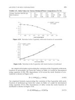
The most important measurement is the AP inlet or conjugate diam-
eter, which is the smallest AP diameter between the posterior
margin of the symphysis pubis and the anterior aspect of the sacrum,
(Fig. 13.7). The normal value varies between 11.0 and 12.5 cm. Values of
less than 10.5 cm indicate increasing likelihood of cephalopelvic dis-
proportion.
The thigh
The femur
The femur (Fig. 13.8), consists of a shaft, a neck, and a head, which
articulates with the acetabulum. The patella is a flattened sesamoid
bone within the quadriceps tendon. Ossification is shown in Fig. 13.9.
The muscles of the thigh (Fig. 13.10)
Anterior femoral muscles
Tensor fascia lata arises from the anterior superior iliac spine (ASIS)
and is inserted into the iliotibial tract, a strong thickened band of the
deep fascia of the lateral aspect of the thigh (fascia lata), which is
attached distally to Gerdy’s tubercle on the antero-lateral condyle of
the tibia.
Sartorius is a narrow strap muscle arising from the ASIS, which
descends diagonally across the front of the thigh to the medial aspect
of the knee, where it inserts on the medial tibial condyle.
Quadriceps femoris is made up of four components. Rectus femoris
arises by a straight head from the anterior inferior iliac spine (AIIS) and
a reflected head from the superior margin of the acetabulum and the
capsule of the hip joint. Its tendon inserts into the superior border of
the patella. Vastus intermedius arises from the anterior surface of the
femoral shaft and inserts into the superior border of the patella deep to
the tendon of rectus femoris. Vastus lateralis arises from the greater
trochanter and the upper part of the linea aspera. Its distal tendon
inserts into the outer border of the patella and blends with the iliotibial
tract. Vastus medialis arises from the lower part of the greater
trochanter and the anterior surface of the femur. Its tendon inserts into
the medial side of the patella. The patellar retinacula are expansions of
the distal tendons of vastus medialis and lateralis.
The lower limb a. newman sanders
132
Anterior
superior
iliac spine
Inferior
superior
iliac spine
Femoral head
Greater
trochanter
Femoral
neck
Obturator
foramen
Ischial
tuberosity
Superior
ramus
Inferior
ramus
of
pubis
Body
Fovea
capitis
Acetabular
teardrop
Lesser
trochanter
Lesser
trochanter
Shenton’s
line
➤
➤
Fig. 13.6. AP and lateral
Radiographs of the right
hip, (a) anteroposterior,
(b) lateral.
Acetabulum
Ischial spine
Vascular
calcification
Greater
trochanter
Lesser
trochanter
Shaft of
femur
Head of femur
Neck of femur
(a)
(b)
C
E
F
A
B
D
Fig. 13.7. Measurements
obtained during CT. AB =
conjugate inlet
diameter; EF = conjugate
outlet diameter
pelvimetry.