Báo cáo sinh học: "Dishevelled and Wnt signaling: is the nucleus the final frontier" potx
Bạn đang xem bản rút gọn của tài liệu. Xem và tải ngay bản đầy đủ của tài liệu tại đây (140.05 KB, 4 trang )
Minireview
Dishevelled and Wnt signaling: is the nucleus the final frontier?
Raymond Habas* and Igor B Dawid
†
Addresses: *Cancer Institute of New Jersey and Department of Biochemistry, UMDNJ-Robert Wood Johnson Medical School, Piscataway, NJ
08854, USA.
†
Laboratory of Molecular Genetics, National Institutes of Child Health and Human Development, Bethesda, MD 20892-2790,
USA.
Correspondence: Igor Dawid. E-mail:
Wnt signaling
Wnt proteins comprise a large family of secreted glycopro-
teins that regulate key developmental processes including
cell-fate determination, proliferation, motility and the
establishment of the primary axis of the body during verte-
brate embryogenesis [1-3]. Defects in Wnt signaling are also
implicated in a host of pathologies including cancer and
neural tube defects. Wnt ligands can transform cells, and
mutations in components of the Wnt signaling pathway,
such as -catenin, have causative roles in colon cancers in
humans, while mutations in Dishevelled (Dsh) are impli-
cated in neural-fold closure disorders [1,4]. To date, 18 Wnt
ligands have been identified in humans [5,6]. This large
number of ligands is paralleled by an equally impressive
number of receptors and co-receptors, which are encoded in
the Frizzled and low-density-related lipoprotein receptor
5/6 (LRP5/6) gene families, which have ten and two
members, respectively, in the human genome [1,6].
Through intensive studies spanning over two decades, a mol-
ecular pathway for Wnt signaling has emerged (Figure 1).
Upon binding of Wnt to its receptor, either Frizzled or a
complex comprising Frizzled and LRP5/6, a signal is trans-
duced to the cytoplasmic phosphoprotein Dsh. There are
three Dsh proteins in mammals (Dsh-1, Dsh-2, and Dsh-3),
and Dsh family members in all organisms are comprised of
three highly conserved domains: an amino-terminal DIX
domain (named for Dsh and Axin), a central PDZ domain
(named for Postsynaptic density-95, Discs-large and Zonula
occludens-1), and a carboxy-terminal DEP domain (for
Dsh, Egl-10 and Pleckstrin) [7]. At the level of Dsh, the Wnt
signal branches into three separate pathways, the so-called
canonical, non-canonical or planar cell polarity (PCP), and
Wnt-Ca
2+
pathways (Figure 1) [1,8,9]. In all three pathways
Dsh is a key transducer of the Wnt signal that operates at the
plasma membrane or in the cytoplasm. But now, a new
study [10] suggests that Dsh also functions within the
nucleus. To put this study in context, we must first review
what is known of the three pathways.
For canonical signaling, which mediates gene induction
events (Figure 1a), Wnt signaling utilizes the DIX and PDZ
Abstract
The phosphoprotein Dishevelled (Dsh) is an essential component of Wnt signaling pathways
and transduces signals into three separate branches, the canonical, non-canonical and Ca
2+
pathways. How Dsh focuses signaling into these branches remains mysterious, but a new
study reveals the importance of nuclear localization of Dsh for pathway-specific activation.
BioMed Central
Journal
of Biology
Journal of Biology 2005, 4:2
Published: 17 February 2005
Journal of Biology 2005, 4:2
The electronic version of this article is the complete one and can be
found online at />© 2005 BioMed Central Ltd
domains of Dsh to induce the stabilization of cytosolic
-catenin; this allows for cytoplasmic accumulation and sub-
sequent translocation of -catenin into the nucleus [1]. Reg-
ulation of -catenin stability is mediated via a complex of
proteins including Axin, glycogen synthase kinase 3 (GSK3),
GSK3-binding protein (GBP) and casein kinase 1 (CK1). In
the absence of Wnt stimulation, -catenin is targeted for
degradation through the proteosomal pathway via the -
transducin repeat containing protein (-TrCP), but -catenin
is stabilized when a Wnt signal is received [1,6,11]. In the
nucleus, -catenin forms complexes with members of the
LEF/TCF family of transcription factors and other factors,
and mediates transcription of Wnt target genes [1].
The non-canonical or PCP pathway mediates cell polarity,
cell movements during gastrulation, and other processes, by
signal transduction through the PDZ and DEP domains of
Dsh, leading to a modification of the actin cytoskeleton
(Figure 1b) [8,12]. At the level of Dsh, two independent and
parallel pathways lead to the activation of the small GTPases
Rho and Rac. Activation of Rho requires the formin-homol-
ogy protein Daam1 that binds to the PDZ domain of Dsh,
leads to the activation of the Rho-associated kinase ROCK,
and mediates cytoskeletal re-organization [8,13,14]. Rac acti-
vation is independent of Daam1, requires the DEP domain
of Dsh, and stimulates Jun kinase (JNK) activity [8,15,16].
Other Dsh-binding molecules that influence the PCP
2.2 Journal of Biology 2005, Volume 4, Article 2 Habas and Dawid />Journal of Biology 2005, 4:2
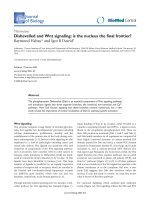
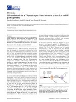
Figure 1
A schematic representation of the Wnt signal transduction cascade. (a) For the canonical pathway, signaling through the Frizzled (Fz) and LRP5/6
receptor complex induces the stabilization of -catenin via the DIX and PDZ domains of Dishevelled (Dsh) and a number of factors including Axin,
glycogen synthase kinase 3 (GSK3) and casein kinase 1 (CK1). -catenin translocates into the nucleus where it complexes with members of the
LEF/TCF family of transcription factors to mediate transcriptional induction of target genes. -catenin is then exported from the nucleus and
degraded via the proteosomal machinery. (b) For non-canonical or planar cell polarity (PCP) signaling, Wnt signaling is transduced through Frizzled
independent of LPR5/6. Utilizing the PDZ and DEP domains of Dsh, this pathway mediates cytoskeletal changes through activation of the small
GTPases Rho and Rac. (c) For the Wnt-Ca
2+
pathway, Wnt signaling via Frizzled mediates activation of heterotrimeric G-proteins, which engage
Dsh, phospholipase C (PLC; not shown), calcium-calmodulin kinase 2 (CamK2) and protein kinase C (PKC). This pathway also uses the PDZ and
DEP domains of Dsh to modulate cell adhesion and motility. Note that for the PCP and Ca
2+
pathways Dsh is proposed to function at the
membrane, whereas for canonical signaling Dsh has been proposed to function in the cytoplasm; a recent study [10] implicates nuclear localization of
Dsh in this pathway. See text for further details.
TCF
GBP
CK1
DIX PDZ DEP
Cell movements
Daam1
Rac
JNK
ROCK
Rho
PKC CamK2
DIX PDZ
Gene induction
Dishevelled
(a) Canonical pathway (b) Non-canonical or planar
cell polarity pathway
(c) Wnt-Ca
2+
pathway
Nucleus
G-protein
DEP
Plasma membrane
DIX PDZ DEP
APC
GSK3
Axin
Axin
LRP5/6
β-TrCP
β-catenin
degradation
β-catenin
accumulation
Fz
Fz
Fz
Wnt
Wnt
Wnt
Multiple functions
pathway include Strabismus and Prickle, but their mecha-
nisms of action remain incompletely understood [8,12,17].
The Wnt-Ca
2+
pathway (Figure 1c) is thought to influence
both the canonical and PCP pathways [9]. Wnt signaling
through Frizzled receptors leads to the release of intra-cellular
Ca
2+
in a process mediated through heterotrimeric G-proteins
and involving numerous other molecules, including phos-
pholipase C (PLC), calcium-calmodulin-dependent kinase 2
(CamK2) and protein kinase C (PKC) [9,18]. The Wnt-Ca
2+
pathway is important for cell adhesion and cell movements
during gastrulation.
Signal specificity
With such a daunting number of Wnt ligands and Frizzled
receptors, two challenging questions that remain unan-
swered are whether (and if so which) Wnt ligands are spe-
cific to particular pathways, and how signals are channeled
to each pathway. Notably, some Wnt ligands are known to
activate both canonical and non-canonical pathways such
as Wnt3a, whereas others such as Wnt5a appear to be spe-
cific to non-canonical signaling. Equally elusive is the
understanding of how the signal is transmitted from the
receptor/coreceptor complex to Dsh, although two recent
studies have revealed a direct interaction between Dsh and
Frizzled [19,20]. Most importantly, the way in which Dsh
couples and distributes Wnt signaling into the three signal-
ing branches remains at best poorly understood.
Dsh occupies a key position at the crossroads of all branches
of the Wnt signaling cascade. It has been proposed that both
the subcellular localization of Dsh and the choice of effector
molecules downstream of Dsh govern the selectivity of spe-
cific pathway activation. Dsh-localization studies in
Drosophila [21] and recently Caenorhabditis elegans [22] have
shown a correlation between localization of Dsh at the mem-
brane and activation of the PCP pathway. Indeed, mutations
in the DEP domain, which is required for PCP signaling,
show impaired membrane localization that is correlated with
impaired PCP signaling [16,21]. These studies have furthered
the hypothesis that the membrane localization of Dsh is
required for at least one output of Wnt signaling. In unstimu-
lated cells, Dsh localizes to punctate vesicular structures in the
cytoplasm [23] by a process that requires the DIX domain; in
response to certain Wnt ligands, Dsh translocates to the
plasma membrane or to the perinuclear/nuclear area, and the
membrane localization in all cases studied requires the DEP
domain [24,25]. The significance of nuclear/perinuclear
localization remained unclear, but it is noteworthy that a
number of components of the canonical signaling pathway,
such as APC, Axin, and GSK3, appear to traffic between the
cytoplasm and the nucleus along with -catenin [26-28].
This multitude of studies forms the background for the
current paper by Sokol and colleagues in Journal of Biology
[10], which identifies two additional domains in Dsh that
modulate both its subcellular distribution and its ability to
activate canonical Wnt signaling. The first newly identified
domain, located carboxy-terminal to the DEP domain,
modulates localization of Dsh through its action as a
nuclear export signal. Dsh protein lacking this domain or
harboring a mutation in a critical lysine residue strongly
accumulates in the nucleus of both Xenopus embryos and
cultured mammalian cells. Surprisingly, however, these
mutant proteins retain their ability to mediate canonical sig-
naling as effectively as wild-type Dsh. Pharmacological
agents impeding nuclear export, and cellular fractionation
studies, further provide evidence that endogenous Dsh
enters the nucleus, supporting the view that Dsh shuttles
between the cytoplasmic and nuclear compartments. The
authors then identified a second domain, located just
carboxy-terminal to the PDZ domain, that is required for
nuclear localization; the sequence of this domain is atypical
for a nuclear localization sequence (NLS). Mutation of this
second domain abolished nuclear accumulation of Dsh in
the presence of nuclear export inhibitors and, remarkably,
impaired the ability of Dsh to induce -catenin stabilization
and to transduce the canonical Wnt signal. Interestingly,
replacement of this atypical NLS with the prototypical NLS
of the T antigen from the simian virus SV40 redirected Dsh
to the nucleus and largely restored Wnt signaling. The
authors further bolster their findings by demonstrating that
stimulation of cultured mammalian cells with Wnt3a results
in the accumulation of a portion of endogenous Dsh (Dvl2
in this case) in/around the nucleus.
Making sense of nuclear localization
So what is the role for Dsh in the nucleus and is the Wnt
field ready to accommodate such a role for Dsh? This new
finding [10] comes as a surprise, because Dsh has been
studied extensively over the past two decades and its nuclear
localization remained unappreciated. To support their con-
clusions the authors showed that Dsh is found in nuclear
fractions, but this approach is not fully conclusive for one
may argue that Dsh exhibits perinuclear localization and co-
fractionates with the outer nuclear envelope. The strongest
evidence for a nuclear role for Dsh comes from experiments
in which nuclear import and export are manipulated,
showing that import is critical for function. Yet, when the
basic conclusion of a nuclear localization and function of
Dsh is accepted, several questions remain. If Dsh function is
required in the nucleus for canonical Wnt signaling, why is
no hyperactivation of the pathway observed by targeting
Dsh to the nucleus? The authors note this point and postu-
late that a ‘steady state’ rather than just localization is
Journal of Biology 2005, Volume 4, Article 2 Habas and Dawid 2.3
Journal of Biology 2005, 4:2
required for function. However, one would at least be com-
pelled to posit that -catenin, which should be stabilized by
such Dsh-targeted approaches, should increase signaling,
and this was not observed.
Perhaps a more salient question is why many studies have
observed Dsh translocation to the plasma membrane in
response to Wnt stimulation or Frizzled expression
[8,12,17,29], but have not detected Dsh in the nucleus. It is
possible that a small but selective pool of Dsh translocates
to the nucleus to mediate canonical signaling while most
Dsh goes to the membrane. Yet, if this is the case, is the
membrane relocalization of the majority of Dsh just a gratu-
itous cellular behavior without meaning? Finally, what is
the function of Dsh in the nucleus? It is possible that
nuclear Dsh acts in transcriptional regulation independent
of -catenin to mediate Wnt signaling, as Sokol and col-
leagues have previously suggested for the Dsh-binding
protein Frodo [30]. These remain important questions that
no doubt will stimulate future studies. Perhaps the nuclear
localization of Dsh will indeed provide clues to elucidate
the final frontier of understanding the diverse mechanisms
of Wnt regulation of gene transcription in the nucleus.
References
1. Logan CY, Nusse R: The Wnt signaling pathway in develop-
ment and disease. Annu Rev Cell Dev Biol 2004, 20:781-810.
2. Wodarz A, Nusse R: Mechanisms of Wnt signaling in devel-
opment. Annu Rev Cell Dev Biol 1998, 14:59-88.
3. Harland R, Gerhart J: Formation and function of Spemann’s
organizer. Annu Rev Cell Dev Biol 1997, 13:611-667.
4. Ueno N, Greene ND: Planar cell polarity genes and neural
tube closure. Birth Defects Res C Embryo Today 2003, 69:318-324.
5. He X: A Wnt-Wnt situation. Dev Cell 2003, 4:791-797.
6. He X, Semenov M, Tamai K, Zeng X: LDL receptor-related
proteins 5 and 6 in Wnt/

-catenin signaling: arrows point
the way. Development 2004, 131:1663-1677.
7. Wharton KA Jr: Runnin’ with the Dvl: proteins that associ-
ate with Dsh/Dvl and their significance to Wnt signal
transduction. Dev Biol 2003, 253:1-17.
8. Veeman MT, Axelrod JD, Moon RT: A second canon. Func-
tions and mechanisms of

-catenin-independent Wnt sig-
naling. Dev Cell 2003, 5:367-377.
9. Miller JR, Hocking AM, Brown JD, Moon RT: Mechanism and
function of signal transduction by the Wnt/

-catenin and
Wnt/Ca2+ pathways. Oncogene 1999, 18:7860-7872.
10. Itoh I, Brott BK, Bae GU, Ratcliffe MJ, Sokol S: Nuclear localiza-
tion is required for Dishevelled function in Wnt/

-catenin
signalling. J Biol 2005, 4:3.
11. Liu C, Kato Y, Zhang Z, Do VM, Yankner BA, He X:

-Trcp
couples

-catenin phosphorylation-degradation and regu-
lates Xenopus axis formation. Proc Natl Acad Sci USA 1999,
96:6273-6278.
12. Wallingford JB, Fraser SE, Harland RM: Convergent extension:
the molecular control of polarized cell movement during
embryonic development. Dev Cell 2002, 2:695-706.
13. Habas R, Kato Y, He X: Wnt/Frizzled activation of Rho regu-
lates vertebrate gastrulation and requires a novel Formin
homology protein Daam1. Cell 2001, 107:843-854.
14. Marlow F, Topczewski J, Sepich D, Solnica-Krezel L: Zebrafish
Rho kinase 2 acts downstream of Wnt11 to mediate cell
polarity and effective convergence and extension move-
ments. Curr Biol 2002, 12:876-884.
15. Habas R, Dawid IB, He X: Coactivation of Rac and Rho by
Wnt/Frizzled signaling is required for vertebrate gastrula-
tion. Genes Dev 2003, 17:295-309.
16. Boutros M, Paricio N, Strutt DI, Mlodzik M: Dishevelled acti-
vates JNK and discriminates between JNK pathways in
planar polarity and wingless signaling. Cell 1998, 94:109-118.
17. Keller R: Shaping the vertebrate body plan by polarized
embryonic cell movements. Science 2002, 298:1950-1954.
18. Kuhl M: Non-canonical Wnt signaling in Xenopus: regula-
tion of axis formation and gastrulation. Semin Cell Dev Biol
2002, 13:243-249.
19. Wong HC, Bourdelas A, Krauss A, Lee HJ, Shao Y, Wu D, Mlodzik
M, Shi DL, Zheng J: Direct binding of the PDZ domain of
Dishevelled to a conserved internal sequence in the C-ter-
minal region of Frizzled. Mol Cell 2003, 12:1251-1260.
20. Cong F, Schweizer L, Varmus H: Wnt signals across the
plasma membrane to activate the

-catenin pathway by
forming oligomers containing its receptors, Frizzled and
LRP. Development 2004, 131:5103-5115.
21. Axelrod JD, Miller JR, Shulman JM, Moon RT, Perrimon N: Differ-
ential recruitment of Dishevelled provides signaling speci-
ficity in the planar cell polarity and Wingless signaling
pathways. Genes Dev 1998, 12:2610-2622.
22. Walston T, Tuskey C, Edgar L, Hawkins N, Ellis G, Bowerman B,
Wood W, Hardin J: Multiple Wnt signaling pathways con-
verge to orient the mitotic spindle in early C. elegans
embryos. Dev Cell 2004, 7:831-841.
23. Capelluto DG, Kutateladze TG, Habas R, Finkielstein CV, He X,
Overduin M: The DIX domain targets Dishevelled to actin
stress fibres and vesicular membranes. Nature 2002,
419:726-729.
24. Torres MA, Nelson WJ: Colocalization and redistribution of
dishevelled and actin during Wnt-induced mesenchymal
morphogenesis. J Cell Biol 2000, 149:1433-1442.
25. Endo Y, Wolf V, Muraiso K, Kamijo K, Soon L, Uren A,
Barshishat-Kupper M, Rubin JS: Wnt-3a-dependent cell motil-
ity involves RhoA activation and is specifically regulated
by Dishevelled-2. J Biol Chem 2004, 280:777-786.
26. Franca-Koh J, Yeo M, Fraser E, Young N, Dale TC: The regula-
tion of glycogen synthase kinase-3 nuclear export by
Frat/GBP. J Biol Chem 2002, 277:43844-43848.
27. Wiechens N, Heinle K, Englmeier L, Schohl A, Fagotto F: Nucleo-
cytoplasmic shuttling of Axin, a negative regulator of the
Wnt--catenin pathway. J Biol Chem 2004, 279:5263-5267.
28. Cong F, Varmus H: Nuclear-cytoplasmic shuttling of Axin
regulates subcellular localization of -catenin. Proc Natl
Acad Sci USA 2004, 101:2882-2887.
29. Rothbacher U, Laurent MN, Deardorff MA, Klein PS, Cho KW,
Fraser SE: Dishevelled phosphorylation, subcellular localiza-
tion and multimerization regulate its role in early
embryogenesis. EMBO J 2000, 19:1010-1022.
30. Hikasa H, Sokol SY: The involvement of Frodo in TCF-
dependent signaling and neural tissue development.
Development 2004, 131:4725-4734.
2.4 Journal of Biology 2005, Volume 4, Article 2 Habas and Dawid />Journal of Biology 2005, 4:2