21st Century Manufacturing Episode 2 Part 10 pot
Bạn đang xem bản rút gọn của tài liệu. Xem và tải ngay bản đầy đủ của tài liệu tại đây (687.26 KB, 20 trang )
37.
Biotechnology Chap.9
Additionally, enzymes can be produced synthetically and are used in everyday
situations-for example in laundry detergents to break down proteins that may have
been spilled on clothing.
9.5.4 The Genome and Chromosomes
The genome is the complete genetic information or total DNA content of an
organism. It is packaged into chromosomes, which are genetic structures consisting
of tightly coiled threads of deoxyribonucleic acid (DNA) and associated proteins.
The genome functions as a blueprint for making an organism. It contains all instruc-
tions for building cell structures and directing life processes throughout the lifetime
of the organism. Different organisms have different numbers of chromosomes per
nucleus. Humans have 46 chromosomes arranged in 22 pairs plus XX or XY -except
in the sperm or the egg, which has 22 chromosomes plus X or Y. The nuclei of most
human cells contain two sets of chromosomes, each set contributed by one parent.
Chromosomes can be observed under a microscope; when stained with certain dyes,
they reveal a distinctive pattern of light and dark bands. In karyotype analysis,
observable differences in size and banding patterns distinguish the chromosomes
from one another. It is also possible to detect major chromosomal abnormalities such
as that of Down's syndrome, in which there is an extra copy of chromosome 21.
9.5.5
DNA l.DeoxyriboNucleic Acid) as the Carrier of Genetic
Information
The science and engineering of deoxyribonucleic acid (DNA) are central to most
biotech industrial processes. Why is DNA so special? The answer is that DNA is the
genetic material for probably all living organisms," (It should be pointed out that
some viruses use RNA rather than DNA as their genetic materiaL) Specifically,
DNA carries the genetic information that is vital for the cells in a person's body to
grow, function, and divide normally. Thus, DNA has a universal role in defining and
regulating life on the planet, from simple bacteria to complex organisms like us. It is
also remarkable for other reasons, including the fact that DNA can replicate itself in
a precise fashion over the lifetime of an organism. DNA directs the production of
proteins necessary for all cellular functions including its own synthesis.
Earlier in the text, the double-helical structure of DNA was compared with a twisted
rope ladder. To further understand the structure of DNA, imagine two strands of "beads"
that have been twisted together. This double-helical structure is made up as follows:
• Each "bead" is a 5-carbon, sugar/phosphate molecule called a nucleotide. Each
nucleotide contains a base shown on the right side of Figure 9.5. There are four
possible bases: two purines (adenine lA] or guanine [G)), or one of two pynm-
idines (thymine [T} or cytosine [Cn (Figures 9.6 and 9.7).
40. Mascarenhas (1999), in his lectures, makes an informal but pedagogically helpful analogy
between the genetic information stored in DNA and the notes and music stored on a standard cassette tape.
Each track of the cassette tape contains the information for one song, just like one length of DNA stores
the information for one gene. Between each track (or gene) there is a break before the next song (or gene)
starts. Abo, with the naked eye, nothing can be seen on the cassette tape-but the music is there ready fa
be expressed. Similarly,in the DNA strand, the genetic information is there, ready to be expressed.
9.5 A Bioscience Review
375
5'end
O·
I
-O~p=O
I
o
HJ ~
~o}f-1
h~H
?H
-o-p=o
6
I
H,C
, 0
HH
H"
o
H
I
-o-p=o
6
H2t.'H_OOH~ ~
Base
~H
o
H
Base
3'end
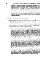
F1gun 9.5 Schematic representation of the nucleotide that
makes up DNA.
RNA only
DNA
llDd
RNA
DNA only
0
H"N/
H
NX"'N/
H
H-<~IH
H-<N
I
NAN/H
I I
H H
I
H
Guanine
Adenine
0
H"N/
H
0
I
NX:N~H
I
HX'w_
H
H3C~ ~ H
I
I
I
I
H N/'O
H
N/'O
H
/'0
I I
I
H
H
H
Uracil Cytosine
Thymine
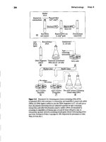
FiJune 9.6
The five nitrogenous bases of DNA, and RNA-see Section 9.7.2
378
Biotechnology Chap. 9
(a)
Adenme
Thymine
H;:Q
i)='
s""H
··H H
H
0
Sugar
• Carbon atom • Nitrogen atom
(b)
H
H~ H-N/ H
S,." HQ-H
/N-H 0 Sugar
H
Guanine Cytosine
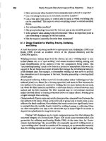
Fipre 'J.7 Base-pairing arrangements in DNA. (From An Introduction to Genefic
Engilluring
by Desmond S.T.Nicholl,
©
1994. Reprinted with the permission of
Cambridge University Press.)
• To create the links within one strand, the nucleotides are attached to each other
by a phosphodieeter bond between the phosphate group at the 5' carbon atom
of one nucleotide and the :\' carbon atom of the next nucleotide. These bonds
are shown on the left of Figure 9.5. This creates a directionality of 5'
?
3' to
the string of nucleotides .
• To create the links between the two strands, the bases on one strand are loosely
attached with hydrogen bonds to the bases on the opposite strand (Figure 9.7).
Base pairing follows a fixed rule:
A
always pairs with T;
G
always pairs with C.
Thus, a sequence of
ATGG
on one strand means the other strand will have
a complementary sequence ofTACC
5' -
ATGGCfACCAAGGTA - 3'
3' -TACCGATGGTICCAT - 5'
The genome can be described in terms of the number of these base pairs. DNA
in a simple bacterial cell such as E. coli is made up of about 4 X 10
6
base pairs: all of
these are shown together in Figure 9.8.By contrast, a human genome is about 3 bil-
lion base pairs.
9.5.6 DNA Replication and Its Relationship to Cell Division
Before a cell in an organism divides, the DNA replicates, as illustrated in Figures
9.9,9.10, and 9.11. As shown in Figure 9.9, the double-stranded DNA molecule
unwinds into two single strands separating the A-T and
CG
base pairs. Next, with
Adenme
Thymine
Carbon atom Nitroeen atom
'Sugar
Sugar'
Sugar
Sugar
Guanine Cytosine
9.5 A Bioscience Review
377
Hgurt' 9.8 Electron micrograph of an
Eschonctn« coli (E. wli)
DNA molecule
the help of certain enzymes, each strand picks up the bases of free nucleotides in
the cell. This is shown schematically in Figure 9.10. Once again, the base-pairing
rules apply in this pickup process. In this way, each new double helix becomes a
duplicate of the original one because, as shown in both Figure 9.10 and Figure 9.11,
the original strands act as the template, specifying which bases are to be added to
the growing strands. Thus when the cell proceeds to divide, each of the new
Figure
9.9 Untwisting of linear DNA strands during
replication. The strands untwist by rotating about the axis of
the unreplicated DNA double helix.
Template strand
Presence of
DNA
polymerase
_\J 5'
Pairs of
DNA
strands
"
Figure
9.10 DNA replication involves the addition (bottom left) offree
nucleotides. The bases are seen in this diagram as the hexagon shapes, and the
sugar/phosphate bonds that become the strands are the attached "bar lines." DNA
polymerase and other proteins catalyze the process.
378
-Position of next
nucleotide added
10growing chain
I'reenucleo6des
9.6 Bioprocesses
379
3'
FllJlll'e9.11 TWoDNA molecules being created from one, leading to two daughter
cells on the upper right and left of the figure.
daughter cells receives a set of DNA molecules identical to that of the original
cell-as indicated in Figure 9.11.
•.6 BIOPROCESSES
9.6.1 Gene Expre ••lon: Connection between Gen •••DNA.
RNA, and Proteins
Genes are the basic unit of heredity. Each gene is a specific sequence of DNA
nuc1eotides that carries the information to direct synthesis of a protein. The process
by which the genetic information encoded on DNA flows into the cell's "protein fac
tories" is a complex set of biochemical reactions (Figure 9.12).
To daughter
ceu z
rodaughter
ceu
i
380
Biotechnology Chap.9
The genetic material
J1gure 9.12 Summary of gt:n", expression. (From
An Introduction to
Genetic
Engineering
by
Desmond S.T. Nicholl,
©
1994.Reprinted with the permission of Cambridge University Press.)
9.6.2
RNA tHiboN-ucleic Acid)
A key substance in gene expression is ribonucleic acid (RNA). The synthesis offunc-
tional proteins out of information encoded in DNA requires several types of RNA.
These include ribosomal RNA (rRNA), messenger RNA (mRNA), and transfer
RNA (tRNA). Gene expression also requires several proteins, for example, RNA
polymerases.
Is arranged as
Arra'nged'
as a ),.
~
""peo"
"~::,~",,
~
Requires In prokaryotes In eukaryctes
Ttanscriptjcn R,'''''''Y
I~~
I"
I
simple P
By
Translation Genes. Geneshave
Whi,ha'''UP'd''
~ Operons I~::::"
~
Which are
~i'h~:::,.:.:::'Y~~<d'"
~ (mRNA) ~
Cosmpo,,"",o,s,p,,,,,,, A::O.'::,
Ribosomes Codons andamino
acids
9.6 Btoproceeees
38'
RNA resembles DNA, but there are some major differences:
• RNA is a chainlike molecule but single stranded.
• RNA also has four bases, but RNA contains the base uracil (U) instead of the
base thymine (T) that is used in DNA.
•The sugar in RNA is ribose, whereas the sugar in DNA is deoxyribose. Both
are 5-carbon sugars (pentoses), but the DNA sugar lacks an oxygen atom at
the 2' carbon atom. The RNA sugar is thus called ribose, while the DNA sugar
is called deoxyribose-that is, a ribose without oxygen at the 2' carbon atom.
The abbreviated terms RNA and DNA are derived from these descriptions of
the sugar molecules.
9.6.3
Transcription
The first step in gene expression is a process known as transcription, in which a DNA
molecule carrying the genetic information isused as a template to synthesize an mRNA
moleculef Ttanscription is similar to DNA replication in that a single strand of DNA is
used as the template for the synthesis of a complementary nucleic acid sequence.
In Figure 9.13a, the shaded ellipse represents the enzyme called
RNA poly-
merase. As implied by the arrow, the polymerase moves along the DNA. Over short
regions, the polymerase temporarily unwinds the two DNA strands as it moves along
the genetic information. RNA polymerase also plays a role in linking the proper
ribonucleotides of mRNA to each other, in order to form the mRNA chain. The
DNA acts as the template to specify which is the correct ribonucleotide to add. This
proceeds according to the base-pairing rules except that uracil (U) in the RNA
replaces thymine (T) in the DNA. Note that the newly formed mRNA strand has the
same sequence as the nontemplate DNA strand. For any given gene, only one of the
DNA strands is used as the template for mRNA synthesis.
9.6.4 Promoters
How does this whole process get triggered? The answer is that in addition to these
base-pairing rules, there are other important regulatory sequences and orderings
specified in the DNA strands (see Nicholl, 1994; Okamura, 1998). Thus "upstream"
from the specific gene being expressed on the top right of Figure 9.13a, there are
DNA sequences to which the RNA polymerase can bind. These sites for starting
transcription-that is,which can temporarily bind the RNA polymerase to the DNA
strands-are known as promoters. These promoter sites can also be seen as clever
switches. Thus, when certain food products enter our system, the switches of the pro-
moter sites are activated, thereby setting off the desirable chain reaction consisting
of (DNA
+
mRNA
+
protein synthesis
+
needs of the cell
+
needs of the body).
Speaking colloquially, this is why one generation of parents should encourage the
next generation to eat a good breakfast. For example, a group of genes known as the
lac operon in the E. coli bacteria in our intestines codes for the enzyme that can
"Transcription also refers to the synthesis of rRNA and tRNA, but it is tbe ItlRNA thai is trans-
latedinloproteins.
382
Biotechnology Chap. 9
(a)
5' ~ATGGCTACCAAOGTA II
3'-TACCGATGGTTCCAT I
5'
RNA polymerase
(b)
NH,
F1pre 9.13
Transcription
in the
upper
sketch and translation in the lower two
sketches. In (a) the DNA strands are temporarily separated by RNA polymerase,
so lhat the mRNA ClUI be formed during gene expression. (From An IntrodU{;tion to
Gtnmc Engineering
by Desmond S.T. Nicholl, C 1994. Reprinted with the
permission of Cambridge University Pr~)
break down lactose. or milk:sugar. However, the "upstream" operator, or promoter,
for this group of genes is a site that can only get triggered in the presence of lactose.
The E. coli bacteria and lactose thus begin a complex chain reaction for breaking
down other food in the intestine.
9.8.5 Translation or Protein Synthesis
'D'anslation is the process by which proteins are synthesized using the information
carried by mRNA. the product of transcription. Recall that proteins are made up of
amino acids. The specific nucleotide sequence of the mRNA determines which
amino acids are to be linked to create a particular protein. A combination of three
nucleotides (e.g.tACO, GOO. CAO) is called a codon and specifies an amino acid.
Figure 9.14 describes the correspondence between the codons and the amino acids
9.6 Bioprocesses
383
F1pft '.14
The "genetic code": nucleotide sequences for amino acids.
A
total of
64 three-letter oodons can be formed from the four letters (A, C. G, and T) of the
DNA
nucleotide bases. However, there exist oniy20 amiDo acids, not 64.This
means that there is more than one codon for most of the amino acids: for example,
Valine (Val) is shown in four entries at the bottom left of the table.1hree of the
COltons(UAA.
VAG,
and UGA) signal "stop" and the end of an mRNA chain.
that they specify.The process by which the mRNA sequence is "translated" into an
amino acid sequence is described in the following paragraph.
Translation requires ribosomes. which are composed of ribosomal RNA
(rRNA) and ribosomal proteins. Ribosomes are the protein factories of cells and are
depicted in Figures 9.13b and 9.13c as the indented elliptical sbapes'' The ribosome
moves along the mRNA chain, and successive amino acids are linked into a growing
protein chain, as shown in Figure 9.13c.1his process requires another type of RNA-
transfer RNA, or tRNA. There is at least one type of tRNA for each amino acid.With
the rules of base pairing coming into play again, the mRNA's codon sequence is rec-
ognized by tRNA molecules that are (1) carrying an anticodon of complementary
ribonucleotides and (2) carrying the amino acid that is specified by the codon. Thus,
as the ribosome moves along the mRNA, tRNAs bring the appropriate amino acids
to be added to the growing protein chain. 'Ibis process continues until the last codon,
the "stop" codon, is reached. As shown in Figure 9.14, this last codon can be UAA,
UAG, or UGA, which do not specify an amino acid but rather signal that the protein
~icholl (1994) describes the ribosome as a "complex structure that essentially acts as a 'iig'-{e.g.,
see Chapter 7,Figures 7.17 to 1.19}-which holds the mRNA in place 50 that the correct amino acid can
be added to the growing chain."
384
Biotechnology Chap. 9
is complete and can be released from the ribosome to carry out its function. Some
proteins remain in the cytoplasm for cell maintenance and operations. Others are
needed in the nucleus; some proteins, such as digestive enzymes, leave the cells for
other functions.
9.6.6
Summary
The following analogy is somewhat exaggerated but:
• The genetic information carried on DNA is analogous to the designer at the
tup of Hgure 4.17in Chapter 4.
• The mRNA and transcription are analogous to the process planning steps that
convert design information to the specific details of manufacturing.
• Ribosomes are the sites where proteins are translated or synthesized, and thus
analogous to fabrication machinery.
• The proteins are the manufactured product. which then do the work of the
cells.
• These proteins can be seen as the "workers," who bring energy back into the
overall system. The created proteins and enzymes subsequently enable other
processes, such as "triggering" the promoters, which then make copies of the
genes. The whole process, summarized here, thus becomes self-replicating.
With the review of both biosciences in Section 9.5 and bioprocesses in Sec-
tion 9.6, some aspects of biotechnology and specifically genetic engineering and
manufacturing can now be considered in more detail.
9.7
GENETIC ENGINEERING I: OVERVIEW
9.7.1 Motivation and Goals
The most common type of genetic engineering in biotech uses recombinant DNA
technologies to transfer genetic information from one organism to another for a
potentially useful purpose. For example, in agriculture, there is now the possibility of
new breeds of plants and animals. In medicine, there is also help for people with
genetic diseases. Genes have now been identified that are involved in Huntington's
disease, cystic fibrosis, sickle-cell anemia, retinoblastoma, and Alzheimer's disease.
Also in medicine, the manufacturing of useful proteins through genetic engi-
neering is a basic goal of many biotech firms. There is growing interest in the manu-
facture of cytokines (such as interferon), which are important for immune system
function.
Recombinant DNA technologies can also be used to produce insulin. Diabetic
people fail to produce sufficient quantities of the protein insulin and hence are
unable to control their sugar metabolism. A daily injection of insulin can regulate
their system without interfering directly with the other bodily chemical reactions.
In
the past, the insulin could be obtained only by expensive extractions from a hog pan-
creas. Now, genetic engineering has enabled bacteria to produce a plentiful supply,
plus help for patients who were allergic to hog insulin. Injecting natural or synthetic
9.7 Genetic Engineering I:Overview
385
proteins (such as insulin) into an organism can induce temporary changes in the
organism's protein content and function. Permanent changes can also be obtained in
an organism,
if
it is possible to manipulate protein synthesis inside its cells.
9.7.2
The Essential Steps
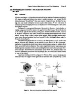
Figure 9.15,by Nicholl (1994),provides a "snapshot" of manufacturing by gene cloning.
• Step 1: The original DNA of an organism, plant, or animal is first cut into frag-
ments.
• Step 2: It is then joined to a carrier, called a vehicle or a vector.
• Step 3: This recombined DNA is then reintroduced into the cell of a host
organism. With the proper manufacturing controls, clones grow.
Gene splicing is one of the most routine and fundamental steps in gene cloning
and is a mainstay of biotech research activities. It is essentially a cut-and-paste
process similar to film splicing, except instead of film stock, pieces of DNA are used,
as shown in Figure 9.16.
9.7.3
Step 1: Cutting DNA Using Restriction Enzymes
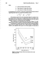
In the first step of Figure 9.15, DNA is cut into fragments at precisely defined
nucleotide sites by restriction enzymes. This process is shown in Figure 9.17.
(1)
t t t
DNAfrllgments
Introduce into host cell
Join to vector
22 s!
Flame 9.15 Schematic diagrams of gene cloning. (From An Introduction to
Genetic
Engineering
by
Desmond S.T. NichoU, C 1994. Reprinted with the
pennistion of Cambridge University Press.)
Grow clones
3.6
Biotechnology Chap. 9
Figure 9.16 Movie-film splicing as an analogy for gene splicing.Only a short
section of
II
gene is represented, and in fact many nucleotide pairs and movie-film
frames would be contained in the two "bracketed" areas on the right of the figure
(from Understllnding DNA and Gene Cloning by Drlica, Copyright © 1992.
Reprinted by permission of John Wiley
&
Sons, Inc.},
9.7.4
Step 2: Joining DNA Using the Enzyme DNA Ligase
Once isolated. a DNA fragment is ready to be joined to another DNA molecule. This
requires the presence of another enzyme called DNA ligase Thisjoining process is a key
aspect of recombinant DNA technology:the DNA ofone organism canbe linked to the DNA
of a completely different organism
Thisisshown in step 2 of Figure9.15and in Figure 9.18.
This second DNA molecule is usually a circular plasmid or a phage-short for
bacteriophage. These are shown in Figures 9.19 and 9.20. The plasmid or phage is
caned a vector.
9.7.5
Step 3: Vectors, Hosts, and Cloning
The plasmid or phage vector is used to carry the recombinant DNA into a host cell
where the genetic material can be propagated. Host cells are most commonly bac-
teria or yeast, single-celled organisms that can exhibit phenomenal growth rates.
They are therefore an important tool in cloning. In a manufacturing context, these
hosts can be viewed as the "transfer line for mass production."
When the host cells with the recombinant DNA divide, they produce a large
number of genetically identical clones, as shown in part 3 of Figure 9.15.The most
common bacterial host is Escherichia coli. E. coli is in our intestines right now, as well
as in those of most animals. (Out of interest, this bacterium gained notoriety in recent
years when several people were killed or sickened by undercooked hamburgers that
had an excess of E. coli. However, this form of E. coli is different from laboratory
strains, which are not pathogenic.) E. coli is the bacteria of choice for gene cloning
because it has been so well studied over the last few decades that it is now charac-
terized and its functions well understood. E. coli cells-at high magnification-are
IGeneticletter
(nucleotide pair)
0<",
,
Scene
I
Frame
Film
DNA
9.7 Genetic Engineering I: Overview
387
Q1
DNA
Flpft
9.17 CUtting ofDNA into short "staggered" pieces.When a restriction enzyme
is added to the DNA, it binds to the DNA and cuts
it
Note there are three cut sitesin
the top diagram.1bis converts the DNA molecule into four shorter moleeules:a, b,c,
and d. Each has "sticky ends" that can subsequently form base pairs with other DNA
in the splicing procedures (from
Undmt4ndillg DNA and Gene Ckming
by Drlica,
Copyright C 1992.Reprinted by permission of John Wiley
&
Sons,Inc.).
shown in Figure 9.21;growing bacterial colonies-more or less at life size-are shown
in Figure 9.22.
9.7.6
Transgenic Plants and Animals
A transgenic plant or animal is one that has been altered to contain a gene from
another organism, usually from another species. Genetic manipulation of plants is
well established as the science of selective breeding. Direct manipulation of plant
genes is a newer but relatively commonplace technique substantially similar to the
gene cloning methods for bacteria and yeast.
Add restriction
endouuclease
c-
to cut DNA
Recognition sequences
388
Biotechnology Chap. 9
DNA
~nCubate fragments to allowjoining
[IjDNAligase
seaIsnicks-
in
DNA
FIpre 9.18 Joining two DNA fragments. The complementary ends of DNA
facilitate the joining process. "Nicks" are enzymatically sealed by DNA ligase
(from Understanding DNA and Gene Cloning by Drlica, Copyright
©
1992.
Reprinted by permission of John Wiley
&
Sons, Inc.)
FIpre 9.l9 An artist's impressions of (a) Circular plasmids and (b) an
enlargement of a plasmid showing a short region becoming singlc-atranded,
Nick
Nick
F'igure9.22 Byspreadingadillltesuspension
ofE. coliceUs onto solid agar in a petri dish,
colonies incubate.
Transgenic animals including humans have recently been a source of much
public discomfort with genetic engineering. Producing a transgenic animal such as
the "supermouse" shown in Figure 9.1 is a complex process, requiring a mix of gene
cloning and sexual reproduction techniques to ensure the transgenes are found in
cells of the animal.
Will biotech create transgenic people? One of the most controversial aspects
of genetic engineering is the potential for excising or adding genetic information to
correct mutant or missing genes.
9.8 GENETIC ENGINEERING
n,
A CASE STUOY ON GENE CLONING
OF HEMOGLOBIN
9.8.1 Introduction
The previous section gave a summary of the cutting, joining, and cloning procedures
in biotechnology. The analogy with movie-film splicing and joining was presented
from Drlica (1992). This is a helpful analogy, but the great difference in dimensional
scale between DNA and movie film should be emphasized. The diameter of DNA's
"twisted rope ladder" is only 2 nanometers, whereas the movie film might typically
be 2 centimeters wide. This represents a 10
7
difference in width. For length compar-
isons,the helical DNA (best seen at the top of Figure 9.9) has a pitch of 3.4nanome-
ters.,spanning 10 base pairs per cycle, whereas one frame of the movie film is again
approximately 2 centimeters. The handling of these minute sections of DNA ca1lsfor
special techniques, including:
•Isolation of DNA from cells
•Preparation of enzymes for cutting, modifying, and joining DNA
• Cloning to create billions of cells
•Radio labeling fragments of DNA to make radioactive probes that find
desired genes
?
Dish
9.8 Genetic Engineering II: A Case Study on Gene Cloning of Hemoglobin
'91
9.8.2 A Case Study on the Procedure for Obtaining Clones
of Hemoglobin DNA
The techniques listed earlier perhaps come to life more easily if they are described
in the context of a particular gene cloning procedure. The goal of this case study is to
describe how the genes encoding hemoglobin were cloned for the first time. The dia-
grams are from Chapter 8 of Drlica's book (1992), and the reader is referred there
for a more comprehensive description.
Why is this procedure of interest? Hemoglobin is an important protein in our
blood. It is responsible for moving the oxygen in our lungs into body tissue. A
number of genetic disorders have been associated with defective hemoglobin, in par-
ticular sickle-cell anemia. To study a serious illness of this type, scientists need a
supply of hemoglobin genes for a variety of experiments. Gene cloning is not an end
in itself but a general technique for generating enough "work material" whose struc-
ture can be analyzed and functions understood. For example, it is important to
understand how one hemoglobin gene is switched on and another switched off.The
studies of cancer and AIDS also involve the need for a bulk supply of cloned genes
that can be analyzed and related to disease formation.
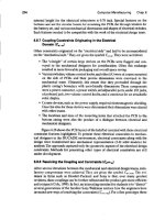
The complete layout for the procedure needed to find the hemoglobin genes is
shown in Figure 9.23, Once found and isolated, the genes are grown in bulk from bac-
terial hosts.
9.8.3 Step 1: Obtaining DNA CTop Right of Figure 9.231
Cells can be opened up to release the DNA and proteins. Subsequently, other pro-
cedures can separate the DNA from the proteins. However, many other steps,
described in this section, are then needed to separate specific genes, such as those
encoding hemoglobin, from the millions of copies of thousands of other types of
genes in the DNA. The experiments described in the following sections, used the
livers of rabbits to obtain the hemoglobin cells, but most kinds of body tissue could
have been broken up to obtain the raw material.
Practical Techniques
Mechanical stirring broke down the cell walls, releasing the DNA and the proteins.
Detergents and enzymes were added to the solution to break down the proteins and
also to inactivate any nucleases that could have cut the DNA. Phenol was then
added, and vigorous stirring moved any remaining protein into the phenol. The mix-
ture was allowed to stand so that a DNA-rich layer could separate out in a test tube,
subsequently to be removed by a pipette. Additional centrifuging further purified
the DNA, which was syringed out for Step 2.
9.8.4 Step 2: Packaging into Bacteriophages (Center Right
of Figure 9.231
Rabbit DNA and phage DNA are first individually cut by a restriction enzyme. The
fragments of rabbit DNA are then mixed with the phage DNA, and the enzyme
DNA ligase is added to join the DNA fragments.
392
HemoglobinmRNA
DNA I~ Reverse transcriptase
synthesis
r-
Nucleotide trtphosphates
_ HemoglobincDNA
Cut and
!-
(QJ
Plasmid DNA
joinDNAs
0
(confers.
antibiotic
resistance)
o
eDNA plasmid
Select and
!-
Bacteria
amplify. (antib.iotic-
eDNAplasmid sensitive)
OOO
~"Y"Pi~
eDNA
plasmid
0"'"
t
radioactively
label eDNA
plasmid
Denature!
Phage containing genonllc
clones of hemoglobin genes
Btotechnoloav Chap. 9
_RabbitDNA
1
(whole genome)
_ Fragmented
r-
"bbnDNA
Phag:DNA
ligase
,
===-==
Recombinant
~ phage DNA
Packaging
Into
phage
particles
r-""'on,
Plaques
Hybridization to
identity clones
FlJllft 9.%3 Obtaining clones of hemoglobin in bulk for later studies on sickle-cell anemia. On tile
right side, fragmented rabbit DNA was packaged into the phage vector. On the left side, radioactive
probes of eDNA were prepared and made in bulk by splicing into ptescnos ana growing them in E.
colt
At the bottom, the phage particles were grown in bulk, and then those containing tbe desired hemoglobin
genes were isolated with the probes using complementary base pairing (from Understanding DNA and
Gene Cloning
by
Drllca, Copyright © 1992. Reprinted by permission of John Wiley
&
SOns,Inc.).
At this point, some of the phages contain rabbit DNA after mixing; the phages are
then coated with phage proteins, packaging the DNA inside phage particles, as shown
on the right of Figure 9.23. The phages are then used to infect
E. coli
hosts on agar.
Unfortunately, only a very small number of the plaques fonned during bacte-
rial growth contain the hemoglobin genes. The challenge is to eventually locate this
9.8 Genetic Engineering 11:A Case Study on Gene Cloning of Hemoglobin
393
extremely small number of the desired plaques. Radioactively labeled nucleic acid
probes ean be used for this purpose. The next two steps are both concerned with the
preparation of these probes, and doing so in such a way as to have enough material
to test all the plaques. Probe preparation is shown on the left of Figure 9.23.
9.8.5
Step 3: Preparing mRNA as a Template for Probes
(Upper Left Side of Figure
9.231
One potential source for radioactive probes is messenger RNA from the gene being
sought. Hemoglobin represents a special case, because the mRNA in red blood cells
is mainly hemoglobin mRNA.
Practical Techniques
Red blood cells from a rabbit were centrifuged to obtain a pellet from which the
RNA could be isolated from the other cell constituents, with phenol and alcohol
treatments and further centrifuging to further purify the RNA. To separate the
mRNA from the ribosomal and transfer RNA, the mixture was passed through a
glass column filled with cellulose. A feature unique to mRNA molecules is that a
sequence of only adenines (A) is added on to the end of their chains. This character-
istic can thus be used to separate out the mRNA molecules from the other types of
RNA present-tRNA and rRNA. Single-stranded DNA composed of thymidines
was attached to the cellulose, and, through base pairing, these attached to adenines
on the mRNA only, allowing the other types of RNA to pass through the column,
thus separating them from the mRNA. The hemoglobin mRNA was then removed
from the column by warming, which broke the A-T base pairs.
9.8.6
Step 4: Increasing the Supply of Probes by Cloning
with
Plasm Ids
(Center
left
Side of Figure
9.231
The mRNA is then converted into cDNA using nucleotides and reverse transcriptase-c-
an enzyme that can use RNA as a template for DNA synthesis. However, to obtain
a larger supply of cDNA,it is first desirable to clone the eDNA into circular plasmids
to be replicated in E. coli, as shown on the center left side of Figure 9.23.
Practical Techniques
The splicing of eDNA with a plasmid, followed by cloning in E. coli hosts, took place
as in Step 2. However, it was still necessary to isolate the particular E. coli cells that
had taken up plasmid DNA into which the rabbit cDNA had been inserted. The pro-
cedure that allows this is described in Figure 9.24. The procedure deliberately chose
a plasmid with two genes for antibiotic resistance: one for tetracycline resistance (ret R)
and the other for ampicillin resistance (amp R). The plasmid was cut within the amp
R gene such that those plasmids that contain the eDNA insert no longer have a tunc-
tional amp R gene. Then, for example, if tetracycline was present, it killed those cells
that had not taken up any of the plasmid. Next, the colonies that had been formed
on the tetracycline-containing agar plate (Figure 9.24d) were tested to find ones that
failed to grow on an ampicillin-containing eger.This distinguished E. coli cells con-
taining plasmids joined to rabbit eDNA from those that did not have rabbit DNA
attached to them. Figures 9.24e and 9.24f show the two types.