Phẫu thuật chỉnh hình chân và mắt cá doc
Bạn đang xem bản rút gọn của tài liệu. Xem và tải ngay bản đầy đủ của tài liệu tại đây (519.26 KB, 11 trang )
Process and Tubercle
Fractures of the Hindfoot
Abstract
Process and tubercle fractures of the talus and calcaneus can be a
source of significant pain and dysfunction. Successful management
requires extensive knowledge of the complex osseoligamentous
anatomy of the hindfoot. The large posterior process of the talus is
composed of a medial and a lateral tubercle; an os trigonum may
exist posterior to the lateral tubercle. The talus has a lateral
process that articulates with the fibula and subtalar joint; the
calcaneus possesses a frequently injured anterior process that
articulates with the cuboid. Injury to these hindfoot structures is
caused by inversion and eversion of the ankle, which can occur
during athletic activity. These injuries often are misdiagnosed as
ankle sprains. A high degree of clinical suspicion is warranted, and
specialized radiographs or other imaging modalities may be
required for accurate diagnosis. Nonsurgical management with cast
immobilization is frequently successful when the fracture is
correctly diagnosed acutely. Large fragments may be amenable to
open reduction and internal fixation. Untreated, chronic injuries
can cause significant pain and functional impairment that may be
improved substantially with late surgical intervention.
T
he calcaneus and talus are the
most frequently fractured tarsal
bones.
1
Most attention in the ortho-
paedic literature has been devoted to
fractures of the neck of the talus and
the posterior facet of the calcaneus.
Other hindfoot fractures have not
been as well studied; fractures in-
volving the peripheral processes and
tubercles of the talus and calcaneus
have been relatively neglected. Con-
sequently, questions persist regard-
ing these fractures. The mechanisms
of injury remain incompletely un-
derstood, and misdiagnosis is not
uncommon. Uncertainty persists re-
garding optimal treatment and prog-
nosis. In treating patients with hind-
foot symptomatology, it is helpful to
have an organized approach to eval-
uating and managing the most com-
mon process and tubercle fractures
of the hindfoot, including the later-
al and posterior processes of the ta-
lus, the medial and lateral tubercles
of the posterior talus, the os tri-
gonum, and the anterior process of
the calcaneus.
Anatomy
The osseoligamentous anatomy of
the hindfoot is complex and often
confusing (Figure 1). Several process-
es and tubercles project from the
main body of the talus and calca-
neus. These structures serve as sites
of ligamentous attachment and con-
tribute to the subtalar and calca-
neocuboid articulations. Because
Mark J. Berkowitz, MD, MAJ,
MC, USA, and
DavidH.Kim,MD
Dr. Berkowitz is Chief, Foot and Ankle
Section, Orthopaedic Surgery Service,
Tripler Army Medical Center, Honolulu,
HI. Dr. Kim is Assistant Clinical
Professor, Orthopaedic Surgery,
University of Colorado School of
Medicine, Denver, CO, and Orthopaedic
Foot and Ankle Surgeon, Colorado
Permanente Medical Group, Denver.
None of the following authors or the
departments with which they are
affiliated has received anything of value
from or owns stock in a commercial
company or institution related directly or
indirectly to the subject of this article:
Dr. Berkowitz and Dr. Kim.
Reprint requests: Dr. Berkowitz, Tripler
Army Medical Center, 1 Jarrett-White
Road, Honolulu, HI 96859-5000.
J Am Acad Orthop Surg 2005;13:492-
502
Copyright 2005 by the American
Academy of Orthopaedic Surgeons.
492 Journal of the American Academy of Orthopaedic Surgeons
these osseous projections are located
along the periphery of the talus and
calcaneus, they are referred to as the
peripheral structures of the hind-
foot.
The talus has five peripheral
structures that may be fractured.
The two talar processes, the lateral
and the posterior, project from the
body of the talus. The lateral process
is a wide, wedge-shaped prominence
extending from the lateral aspect of
the body of the talus.
2
It possesses
two distinct articular facets: the dor-
solateral and the inferomedial. The
dorsolateral facet articulates with
the distal fibula; the inferomedial
facet forms the anterolateral portion
of the subtalar joint. The lateral pro-
cess is the site of insertion of the lat-
eral talocalcaneal ligament.
The posterior process is relative-
ly large (Figure 1, B), and its inferior
surface composes the posterior 25%
of the subtalar articulation.
3
It is the
most variable aspect of hindfoot
anatomy.
4
The posterior process is
composed of two tubercles: the me-
dial and the lateral. These tubercles
are separated by a groove within
which lies the flexor hallucis longus
tendon. Forming a roof over this
groove is the Y-shaped, bifurcate
talocalcaneal ligament, which in-
serts onto each tubercle.
5
The later-
al tubercle (ie, Stieda’s process) is
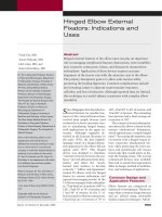
Figure 1
Osseous anatomy of the hindfoot. A, Lateral view of the talus and calcaneus. B, Posterior view of the ankle and hindfoot.
C, Superior view of the talus.
Mark J. Berkowitz, MD, MAJ, MC, USA, and David H. Kim, MD
Volume 13, Number 8, December 2005 493
larger than the medial tubercle and
projects more posteriorly. The poste-
rior talofibular ligament inserts onto
the lateral tubercle of the talus. The
posterior talotibial portion of the
deltoid ligament attaches to the me-
dial tubercle.
The os trigonum is located direct-
ly posterior to the lateral tubercle
(Figure 1, C). It is an accessory bone
of variable size and shape that arises
from a secondary ossification center
between the ages of 8 and 11 years.
In most persons, it fuses to the later-
al tubercle within 1 year of its ap-
pearance. However, it may persist as
a separate ossicle, attached to the ta-
lus by a cartilaginous synchondro-
sis.
4
Burman and Lapidus
6
observed
a distinct os trigonum in 64 of 1,000
radiographs of feet as well as a
“fused os trigonum” in the form of
an elongated lateral tubercle in 429
of the 1,000 radiographs.
The anterior process is the most
commonly fractured peripheral
structure of the calcaneus.
1
This pro-
cess is a saddle-shaped projection of
bone at the superior aspect of the
calcaneal body that extends toward
the navicular
7
(Figure 1, A). Its infe-
rior surface articulates with the
cuboid. The bifurcate ligament in-
serts on the anterior process and
connects the cuboid and navicular
bones. Additionally, the extensor
digitorum brevis muscle takes at
least a portion of its origin from the
anterior process.
Other peripheral structures of the
calcaneus include the sustentacu-
lum tali, the peroneal tubercle, and
the medial and lateral calcaneal tu-
bercles.
8
Injuries to these structures
are rare.
Mechanism of Injury
Process and tubercle fractures of the
hindfoot occur in two distinct pat-
terns. These fractures may be caused
by high-energy trauma, such as a fall
from a height or a motor vehicle ac-
cident.
9
In this setting, they are o ften
found concomitantly with fracture-
dislocations of the subtalar and an-
kle joints.
10
Process and tubercle
fractures also may be caused by low-
energy sprains, such as those occur-
ring during athletic participation.
11
In both types, the position o f the foot
and the vector of the forces applied
to it are critical in producing the
fracture.
Fractures of the lateral talar pro-
cess initially were believed to occur
after forced ankle dorsiflexion and
inversion, usually because of a fall o r
motor vehicle accident.
9,12,13
Snow-
boarding now accounts for most
lateral talar process fractures, with
approximately 2,000 occur ring an-
nually.
14
The mechanism of injury
appears to be related to the forces
transferred to the ankle while the
foot is strapped to the board. Boon et
al
2
produced cadaveric “snowboard-
er’s fractures” only when external
rotation force was applied to the dor-
siflexed, inverted foot. Similarly,
Funk et al
14
demonstrated that ever-
sion of an axially loaded, dorsiflexed
ankle may produce a lateral process
fracture.
The medial tubercle of the poste-
rior process may be fractured when
the foot is suddenly forced into a po-
sition of combined dorsiflexion and
pronation.
11,15
This places the poste-
rior talotibial component of the del-
toid ligament under tension, causing
avulsion of the tubercle. In 1974,
Cedell
15
originally described this in-
jury and its mechanism in four pa-
tients injured during sports activity.
This injury also has been reported af-
ter motor vehicle accidents and falls
in association with subtalar disloca-
tion, talar neck fracture, and total t a-
lar dislocation.
16,17
Fractures of the posterior talar
process most likely are caused by
forceful plantar flexion of the ankle.
Maximum plantar flexion produces
a nutcracker-like compression of the
posterior process between the poste-
rior malleolus and the calcaneus
18,19
(Figure 2). The entire posterior pro-
cess, the lateral talar tubercle, or the
os trigonum may be injured in this
way.
The os trigonum may be injured
by repetitive plantar flexion, similar
to a stress fracture.
20
This injury is
often refer red to as either os trigo-
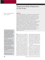
Figure 2
Axial (A) and sagittal (B) computed tomography scans demonstrating fracture of
the entire posterior process of the talus. Forceful plantar flexion compresses the
posterior process of the talus between the calcaneus and the tibia, resulting in
fracture (arrows).
Process and Tubercle Fractures of the Hindfoot
494 Journal of the American Academy of Orthopaedic Surgeons
num syndrome or posterior ankle
impingement. It is seen most often
in professional ballet dancers, soccer
players, and runners.
19,21,22
More
commonly, the synchondrosis is
acutely disrupted by a plantar flex-
ion–inversion mechanism, similar
to an ankle sprain.
21,23
This same
mechanism also may cause a frac-
ture of the lateral tubercle of the pos-
terior talar process. Ankle inversion
tensions the posterior talofibular lig-
ament, producing avulsion of the lat-
eral tubercle (Figure 3). Shepherd
first described this fracture and
mechanism in 1883, and some au-
thors still refer to it as “Shepherd’s
fracture.”
24
Fractures of the anterior calcaneal
process also occur after inversion of
the plantarflexed ankle.
7
This mech-
anism of injury stretches the bifur-
cate ligament and avulses the anteri-
or process. Alternatively, forced
dorsiflexion and eversion may com-
press the anterior process between
the cuboid and the talus, resulting in
a shear fracture
7
(Figure 4).
Diagnosis
A high level of suspicion is required
when diagnosing process and tuber-
cle fractures of the hindfoot. These
fractures can be challenging to dis-
cern on standard radiographs and
physical examination. Unfortunate-
ly, misdiagnosis and delayed diagno-
sis are frequent complications.
25
Lateral talar process fractures
mimic lateral ankle sprains.
13,19,26
Missed lateral talar process fractures
were found retrospectively in 0.86%
of patients initially diagnosed as
having a lateral ankle sprain (13/
1,500 patients).
13
In a series of 25 an-
terior calcaneal process fractures,
the diagnosis was initially incorrect
in 9 patients, with 7 initially mis-
diagnosed with a sprain of the an-
terior talofibular ligament.
7
In
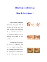
Figure 3
A, Lateral view of the ankle (taken for unrelated reasons before injury) demonstrat-
ing an intact lateral tubercle of the posterior process of the talus. B, Repeat lateral
radiograph of the same patient after a plantar flexion–inversion injury demonstrating
fracture of the lateral tubercle with angulation of the fragment (arrow).
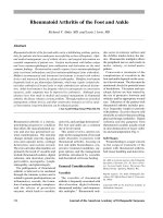
Figure 4
Mechanism of injury of a fracture of the anterior process of the calcaneus. A, Inversion of the plantarflexed ankle (arrow) results
in anterior process avulsion by the bifurcate ligament (inset). B, Dorsiflexion and eversion (arrow) may create a shear fracture
of the anterior process of the calcaneus (inset). (Courtesy of the Mayo Foundation. Copyright 1980.)
Mark J. Berkowitz, MD, MAJ, MC, USA, and David H. Kim, MD
Volume 13, Number 8, December 2005 495
Cedell’s
15
original article on frac-
tures of the medial tubercle of the
posterior talar process, each of the
four patients was initially misdiag-
nosed with a sprain and treated with
a compression bandage and rest.
Paulos et al
5
reported on 20 patients
with avulsion fractures of the poste-
rior talus, all of which were initially
diagnosed as ankle sprain. The aver-
age number of physician visits per
patient before correct diagnosis was
made was 5.8; one patient was seen
17 times.
Failure to diagnose also occurs
with multiple trauma. Process and
tubercle fractures occur in associa-
tion with significant lower extremi-
ty injuries, such as subtalar and an-
kle fracture-dislocations, total talar
dislocations, and lower extremity
long bone fractures.
17,27,28
In treating
these injuries, process or tubercle
fractures may be easily missed. Un-
fortunately, delayed detection of
these fractures increases the likeli-
hood of painful nonunion and ar-
throsis.
7,11,17
Physical Examination
The initial step in diagnosing
these fractures is eliciting the mech-
anism of injury. The position of the
foot and the force applied should
raise suspicion that a particular frac-
ture may have occurred. Once the
mechanism of injury has been deter-
mined, a focused physical examina-
tion is performed to elicit the maxi-
mal point of tenderness. The
maximal point of tenderness repre-
sents the most important diagnostic
feature in distinguishing a peripher-
al fracture from an uncomplicated
ankle sprain.
11
It can be difficult to
discern in a patient with an acute,
swollen ankle, and the patient may
need to be reexamined 10 to 14 days
later.
Lateral talar process fractures can
be particularly difficult to differenti-
ate from sprains on physical exami-
nation. However, careful palpation
just anterior and inferior to the later-
al malleolus should raise suspicion
of this injury. The patient with pos-
terior process fracture demonstrates
deep tenderness anterior to the
Achilles tendon but posterior to the
talus. Fracture of the lateral tubercle
of the posterior talar process and of
the os trigonum provokes point ten-
derness over the posterolateral an-
kle, just medial to the peroneal ten-
dons. Fracture of the medial tubercle
of the posterior talar process demon-
strates localized tenderness medial-
ly, just posterior to the medial mal-
leolus.
11
Forced plantar flexion is another
important test. The patient with
posterior talar process or os trigo-
num fracture frequently reports pain
when the posterior talus is com-
pressed against the tibia during this
maneuver. Likewise, resisted mo-
tion of the great toe can elicit pain as
the flexor hallucis longus tendon
slides past a medial or lateral tuber-
cle fracture of the posterior talar pro-
cess.
5
Anterior calcaneal process frac-
ture usually produces tenderness in
an area approximately 2 cm anterior
and 1 cm inferior to the anterior
talofibular ligament.
7
Swelling and
ecchymosis localized to this area are
signs of anterior calcaneal process
fracture.
Radiographic Evaluation
Radiographs must be carefully
scrutinized to ensure prompt and ac-
curate diagnosis of process and tu-
bercle fractures. As mentioned,
these fractures are difficult to detect
on standard plain radiographs; there-
fore, the use of specialized oblique
radiographs, computed tomography
(CT), magnetic resonance imaging
(MRI), or bone scans may be re-
quired.
Plain Radiographs
Lateral talar process fractures are
best seen on mortise or anteroposte-
rior ankle radiographs, in which a
fragment just inferior to the lateral
malleolus can be visualized
2,13
(Fig-
ure 5). Occasionally, an avulsed frag-
ment is visible on a lateral radio-
graph. Dorsiflexing and inverting the
ankle while taking the lateral radio-
graph may further improve visual-
ization of the fragment.
29
Posterior talar fractures are par-
ticularly difficult to detect and dif-
ferentiate on standard radiographs.
Large posterior process fractures
may demonstrate a prominent frac-
ture line on a standard lateral radio-
graph, but distinguishing between
medial and lateral tubercle frac-
tures and differentiating them from
a normal os trigonum can be chal-
lenging.
17
Paulos et al
5
described us-
ing a special 30° subtalar oblique
view to better visualize lateral tu-
bercle and os trigonum fractures.
Kim et al
30
likewise used a medial
oblique view to evaluate suspected
posteromedial talus fractures (Fig-
ure 6).
Anterior calcaneal process frac-
tures are not well visualized on stan-
dard anteroposterior views of the
foot or ankle. A lateral radiograph
may reveal the fracture, but direct-
ing the beam 20° superior and poste-
rior to the midportion of the foot can
project the anterior process away
from the neck of the talus, enabling
Figure 5
Anteroposterior radiograph of the ankle
demonstrating a fracture of the lateral
process of the talus, which is visible
just inferior to the lateral malleolus.
Process and Tubercle Fractures of the Hindfoot
496 Journal of the American Academy of Orthopaedic Surgeons
visualization of the fracture
7
(Fig-
ure 7).
Computed Tomography
When clinical suspicion is high
but radiographs are negative, CT
scans are very useful for detecting
hindfoot process and tubercle frac-
tures. Multiplanar CT imaging with
fine 1-mm cuts allows accurate as-
sessment of fragment location, size,
displacement, and comminution
10
(Figure 8). Additionally, CT provides
adequate cortical detail to distin-
guish the smooth, sclerotic margins
of an os trigonum from the jagged, ir-
regular contour of an acute lateral
tubercle fracture. This important
distinction is frequently not possible
with standard lateral radiographs.
CT is also sensitive for early degen-
erative changes that may not be de-
tectable on plain radiographs.
21,31
CT
especially should be considered
when subtalar dislocation is suspect-
ed.
28
Subtalar dislocation rarely oc-
curs in isolation, and CT often re-
veals associated process or tubercle
fractures not visualized on plain ra-
diographs.
17
Ebraheim et al
10
used CT to eval-
uate 10 patients with fracture of the
talus. Eight process or tubercle frac-
tures were initially identified on
plain radiographs, but CT was re-
quired to determine size, displace-
ment, subtalar joint involvement,
and treatment. Two fractures initial-
ly missed on plain radiographs were
diagnosed using CT 6 months and 1
year, respectively, after injury. In
several cases, the surgical approach
was determined based on the CT
findings.
Magnetic Resonance Imaging
The ability of fluid-sensitive mag-
netic resonance sequences to dem-
onstrate edema adjacent to injured
structures makes it a useful modal-
ity, particularly in the chronic set-
ting
32,33
(Figure 9). Wakeley et al
32
performed sagittal and coronal spin-
echo sequences on three patients
with chronic posterior ankle pain.
Based on the MRI results, os trigo-
num syndrome was accurately diag-
nosed in each case. Sanders et al
33
re-
ported on a 59-year-old man who
underwent MRI for evaluation of
Figure 8
Axial computed tomography scan
demonstrating a fragment medial to the
flexor hallucis longus groove (arrow),
consistent with a fracture of the medial
tubercle of the posterior process of
the talus. Compare with the lateral
radiograph in Figure 6, A, in which it is
difficult to determine whether there
is an os trigonum, a lateral tubercle
fracture, a medial tubercle fracture, or a
posterior process fracture.
Figure 6
A, Lateral radiograph demonstrating nonspecific fracture of the posterior process
of the talus. B, Medial oblique view demonstrating avulsion fracture of the medial
tubercle fracture of the posterior process of the talus (arrow). (Reproduced with
permission from Kim DH, Hrutkay JM, Samson MM: Fracture of the medial tubercle
of the posterior process of the talus: A case report and literature review. Foot Ankle
Int 1996;17:186-188.)
Figure 7
Oblique lateral view allowing
visualization of fracture of the anterior
process of the calcaneus (arrow).
Mark J. Berkowitz, MD, MAJ, MC, USA, and David H. Kim, MD
Volume 13, Number 8, December 2005 497
chronic lateral ankle pain. MRI re-
vealed a previously undetected later-
al talar process fracture for which
the patient eventually underwent
surgical excision. MRI also may pro-
vide useful information regarding
adjacent soft-tissue structures, such
as tenosynovitis of the flexor hallu-
cis longus tendon or peroneal tendi-
nopathy.
Nuclear Medicine Imaging
Technetium Tc-99m bone scan-
ning is another important technique
for evaluation of hindfoot process
and tubercle fractures.
21,22
In the
presence of an acute or chronically
symptomatic fracture, a bone scan
demonstrates an area of focal radio-
isotope uptake. This may be useful
in detecting occult fractures and in
distinguishing fractures from nor-
mal ossicles
34
(Figure 10).
Paulos et al
5
consider technetium
Tc-99m bone scanning to be the de-
finitive test for diagnosing occult
fractures of the posterior talus. They
found it particularly useful for differ-
entiating an acute lateral tubercle
fracture from a normal os trigonum.
Abramowitz et al
21
likewise report-
ed that 32 of 35 patients with os trig-
onum injury demonstrated increased
focal uptake in the posterolateral as-
pect of the talus on bone scan.
However, bone scanning may in-
dicate false positives and false nega-
tives. Sopov et al
35
evaluated the
scintigraphic findings of 100 consec-
utive soldiers. Of 200 feet, 27
(13.5%) demonstrated uptake in the
region of the os trigonum; however,
only 10 of the 27 feet (37%) were
symptomatic. They concluded that a
positive Tc-99m bone scan is a fre-
quent finding in active individuals
and may even be considered a nor-
mal variant in this population. Sim-
ilarly, i n three patients with negative
bone scans, Abramowitz et al
21
iden-
tified and removed symptomatic os
trigonum with excellent results,
leading the authors to reject the no-
tion that a normal bone scan elimi-
nates the possibility of os trigonum
injury.
Fluoroscopic Injection
Injection of lidocaine under fluo-
roscopic guidance is another useful
diagnostic tool.
34
When physical ex-
amination reveals a point of maxi-
mum tenderness suspicious of a
process or tubercle fracture, a fluoro-
scope can be used to precisely guide
the placement of local anesthetic.
Significant relief of symptoms after
injection strongly points to that
structure as the source of pain. A flu-
oroscopically guided injection also
may have predictive value with re-
spect to surgical treatment. Jones et
al
36
used fluoroscopic injection of
lidocaine into the synchondrosis of
Figure 9
Sagittal T2-weighted magnetic
resonance image of a patient with
chronic posterior ankle pain
demonstrating intraosseous edema in
the os trigonum and adjacent talus and
calcaneus (asterisks), which is
consistent with posterior ankle
impingement.
Figure 10
Lateral projection in a patient with an os trigonum demonstrating focal intense
radioisotope Tc-99m uptake in the posterior aspect of the talus and adjacent tibia
as well as in the calcaneus, consistent with os trigonum syndrome.
Process and Tubercle Fractures of the Hindfoot
498 Journal of the American Academy of Orthopaedic Surgeons
an os trigonum in four patients with
chronic posterior ankle pain. Each
patient experienced transient pain
relief and subsequently underwent
excision of the os trigonum with
complete resolution of symptoms.
Management
The optimal management of process
and tubercle fractures remains con-
troversial. Relatively simple classifi-
cation schemes have been proposed
to help guide treatment (Tables 1
and 2). The most critical factors in-
clude the size of the fragment, dis-
placement, comminution, and de-
gree of articular involvement.
7,14
Nonsurgical Management
Nonsurgical management should
be considered for acute process and
tubercle fractures with small (<1
cm), minimally displaced (<2 mm)
fragments.
1,8
The small size of the
fragment leaves the adjacent articu-
lar surface almost completely intact.
Therefore, talofibular, subtalar, or
calcaneocuboid incongruity usually
is not a problem. Nonsurgical man-
agement also is appropriate for larg-
er fragments that are either non-
displaced or minimally displaced.
These fractures are likely to heal or
result in a stable, asymptomatic fi-
brous union.
11
When these requirements are
met, immobilization in a below-
knee, non–weight-bearing cast can
result in a favorable outcome.
5,7,9,30
Generally, 6 weeks of non–weight-
bearing and cast immobilization is
recommended. When the patient is
asymptomatic after 6 weeks, transi-
tion into a removable walking boot
and progressive weight bearing with
crutches is allowed. When the pa-
tient remains symptomatic after 6
weeks of protected weight bearing
and immobilization, continued re-
striction of activity may be warrant-
ed for several months.
Early diagnosis and management
of hindfoot process and tubercle frac-
tures appear to be critical factors af-
fecting the success of nonsurgical
management.
1,5,7,9,11,26,29,30
Degan et
al
7
successfully used immobilization
for a mean of 5.4 weeks to treat 18 of
25 patients with early diagnosed
acute anterior calcaneal process frac-
tures. A satisfactory result consist-
ing of no or minimal pain and full re-
turn to activity was achieved in
those 18 patients.
Kim and colleagues
11,30
described
successful management of acutely
diagnosed posterior medial talar tu-
bercle fractures. The patients under-
went immediate immobilization in a
non–weight-bearing cast for an aver-
age of 6 weeks. At 2-year follow-up,
the average AOFAS ankle-hindfoot
score was 95 of a total of 100 points.
One patient who healed with a radio-
graphic fibrous union nevertheless
achieved a score of 97.
11
Certain peripheral hindfoot frac-
tures do less well with nonsurgical
management.
5,7,9-11,13,16,17,23,37,38
Later-
al talar process fractures have been
reported to result in generally poor
outcomes when managed with
casting alone.
9,13,26,39
For this rea-
son, Kirkpatrick et al
38
recommend
against nonsurgical management of
all but the truly nondisplaced later-
al talar process fracture. Neverthe-
less, fractures that are acute, extra-
articular, smaller than 1 cm, and
displaced <2 mm may be considered
for conservative management with
6 to 8 weeks of immobilization in a
non–weight-bearing cast
14
(Figure
11).
Large fragments, particularly
those resulting from high-energy
trauma, do not reliably respond to
nonsurgical management. Although
Kim and colleagues
11,30
reported suc-
cess with nonsurgical treatment of
patients with acute posterior medial
talar tubercle fractures, Giuffrida et
al
17
reported failure in each of their
patients despite prompt cast immo-
bilization. A comparison of the two
series, however, highlights impor-
tant differences. Each of the patients
in the report by Kim et al
11
sustained
low-energy athletic injuries that re-
sulted in small avulsion fragments
with minimal articular disruption.
In the report by Giuffrida et al,
17
six
fractures occur red in association
with medial subtalar dislocation.
This high-energy mechanism pro-
duced much larger fracture frag-
ments and a high rate of subtalar
subluxation and incongruity. This
comparison emphasizes that, even
for a single type of fracture, no uni-
versal treatment prescription can be
given.
Chronic injuries seem to have the
worst outcome when managed with
casting. Failure to promptly diag-
nose and initiate proper immobiliza-
tion frequently results in a chroni-
cally painful nonunion. Paulos et
al
5
found that only 6 of 17 chronic
lateral talar tubercle and os trigo-
num injuries responded to a regi-
men of rest, nonsteroidal anti-
inflammatory drugs, stretching, and
activity restriction. In the series of
anterior calcaneal process fractures
reported on by Degan et al,
7
the
Table 1
Classification of Fractures of the
Anterior Process of the Calcaneus
7
Type I Nondisplaced tip avulsion
Type II Displaced avulsion fracture
not involving the
calcaneocuboid
articulation
Type III Displaced, larger
fragments involving the
calcaneocuboid joint
Table 2
Classification of Fractures of the
Lateral Process of the Talus
14
Type A Small, minimally displaced,
extra-articular avulsion
Type B Medium-sized fracture
involving only the
talocalcaneal articular
surface
Type C Larger fracture involving
both talocalcaneal and
talofibular articulations
Mark J. Berkowitz, MD, MAJ, MC, USA, and David H. Kim, MD
Volume 13, Number 8, December 2005 499
worst outcomes were found in pa-
tients with the longest delay in diag-
nosis and treatment.
Surgical Management
Surgical management of hindfoot
process and tubercle fractures
should be strongly considered for
large (>1 cm), displaced (>2 mm) frag-
ments with significant articular in-
volvement.
1,8
Surgery generally con-
sists of open reduction and internal
fixation (ORIF) for large fragments,
primary excision for highly commi-
nuted fractures, and delayed exci-
sion for chronic nonunions. The sur-
gical approach is tailored to the
particular fracture being treated.
Open Reduction and Internal
Fixation
Noncomminuted, displaced frac-
tures that compromise articular con-
gruity should be considered for pri-
mary ORIF. Unreduced large,
displaced, articular fragments have a
high propensity for nonunion, and
the subsequent articular malunion
may progress to arthrosis.
12,16,17,29,40
Accurate assessment of fragment
size and comminution is necessary
to determine whether ORIF is appro-
priate and feasible. CT is frequently
required to make this determina-
tion. CT also precisely localizes the
fracture and helps determine the
most appropriate surgical ap-
proach.
10
Fractures most commonly ame-
nable to ORIF include large lateral
talar process fractures, medial talar
tubercle fractures, and fractures of
the entire posterior talar process.
Stable fixation usually may be
achieved with small or mini-
fragment screws or with Kirschner
wires. Although anterior calcaneal
process fractures may be considered
for ORIF, they are rarely of sufficient
size to warrant this approach.
7
ORIF has been recommended in
several small case series for large,
noncomminuted fractures of the lat-
eral talar process that disrupt either
the talocalcaneal or talofibular artic-
ulations.
9,13,26,29,38
Although results
of ORIF are considered to be superi-
or to those of nonsurgical manage-
ment, persistence of symptoms is
not uncommon even in fractures
managed surgically.
1
The fracture is
exposed via an incision over the tar-
sal sinus, with distal reflection of the
extensor digitorum brevis muscle.
ORIF is also the best treatment
for medial tubercle fractures of the
posterior talus that affect a signifi-
cant amount of the subtalar joint.
Kanbe et al
41
performed ORIF on two
patients with posterior talar medial
tubercle fractures. Neither patient
reported pain at 2-year follow-up,
and radiographs demonstrated no
subtalar arthrosis. Conversely, fail-
ure to anatomically restore this frac-
ture may result in subtalar sublux-
ation and arthrosis.
17
This fracture is
approached through a posteromedial
dissection between the flexor digi-
torum longus tendon anteriorly and
the neurovascular bundle posterior-
ly.
11
The fractured tubercle is visual-
ized medial to the tendon of the flex-
or hallucis longus.
Fracture of the entire posterior
talar process is rare, but it frequently
requires ORIF because of the
relatively large size and signifi-
cant involvement of the subtalar
joint.
18,42-44
Several case reports doc-
ument good results after anatomic
fixation of these fractures.
3,18,42-45
Ei-
ther a posteromedial or posterolat-
eral approach may be used. When the
major displacement is posterome-
dial, the fracture is approached
through a posteromedial dissection
between the flexor digitorum longus
tendon anteriorly and the neurovas-
cular bundle posteriorly.
3
When the
major displacement is posterolateral,
an approach between the peroneal
tendons and the Achilles tendon
should be performed.
43
This ap-
proach requires identification and
protection of the sural nerve. Like-
wise, dissection medial to the flexor
hallucis longus tendon should be
performed cautiously to avoid injury
to the medial neurovascular bundle.
Figure 11
A, Initial coronal computed tomography scan of a fracture of the lateral process of
the talus shown in Figure 5 demonstrating moderate comminution, minimal
displacement, and no significant subtalar incongruity. B, Coronal computed
tomography scan 6 months later demonstrating healing of the fracture fragments
with preservation of subtalar congruity. The patient was asymptomatic.
Process and Tubercle Fractures of the Hindfoot
500 Journal of the American Academy of Orthopaedic Surgeons
Primary Excision
Displaced, intra-articular process
and tubercle fractures that are too
comminuted to fix internally can be
considered for primary excision. Pri-
mary excision allows early mobiliza-
tion without the risk of developing
painful nonunion.
1,8
This approach
has been recommended primarily for
comminuted fractures of the lateral
talar process.
9,13,29
The surgical ap-
proach is identical to that for ORIF,
except that all loose articular frag-
ments are removed. Immobilization
usually consists of 2 to 3 weeks in a
weight-bearing cast or a removable
boot.
Late Excision
Patients who develop symptom-
atic nonunion of a peripheral hind-
foot fracture may improve signifi-
cantly with late fragment excision.
Abramowitz et al
21
excised the os
trigonum via a posterolateral ap-
proach in 41 patients who had failed
nonsurgical management. Improve-
ment in the 100-point AOFAS ankle-
hindfoot score averaged 36 points,
with the best results in patients who
had been symptomatic for fewer
than 2 years. Marumoto and Fer-
kel
31
documented an average 41-
point improvement in the AOFAS
ankle-hindfoot score after arthro-
scopic excision in 11 patients with
os trigonum syndrome. Similar im-
provement has been repor ted after
late excision of the medial tubercle
of the posterior talar process and
anterior calcaneal process frac-
tures.
7,11,23
Results of late excision
seem to deteriorate the longer symp-
toms have been present.
7,21
Complications
The primary complications associat-
ed with process and tubercle frac-
tures of the hindfoot are chronic
pain and late arthrosis. Chronic
symptomatic nonunion is particu-
larly likely when these fractures are
not diagnosed and treated acute-
ly.
5,7,11,30
Even when treated appropri-
ately, patients may remain symp-
tomatic for a prolonged period (up to
2 years in one report
7
). Persistence of
mild pain and stiffness after union
also may occur.
1
Although small
fractures frequently respond favor-
ably to excision, large fracture frag-
ments tend to produce articular in-
congruity, and arthrosis of the
subtalar joint can develop (Figure
12). In these cases, subtalar arthro-
desis may be required.
9,16,17
Summary
Process and tubercle fractures of the
hindfoot are challenging to diagnose
and manage. An understanding of
the complex anatomy of the hind-
foot is required. The clinician must
be diligent and knowledgeable in the
interpretation of plain radiographs
and in the use of additional studies,
such as specialized oblique views,
CT, MRI, and bone scanning. The
most critical prognostic factor is cor-
rect initial diagnosis. Prompt man-
agement, whether cast immobiliza-
tion, ORIF, or primary excision,
provides the best opportunity for
complete recovery. Delay in diagno-
sis increases the likelihood of chron-
ic pain and disability. In these pa-
tients, late excision can provide
significant improvement in symp-
toms, but arthrodesis of the involved
joints also may be considered. Im-
proved understanding of peripheral
hindfoot anatomy and injury pat-
terns should increase physician
awareness of and vigilance for these
fractures.
References
1. Heckman J: Fractures of the talus, in
Bucholz R, Heckman J (eds): Rock-
wood and Green’s Fractures in
Adults, ed 5. Philadelphia,PA: Lippin-
cott Williams & Wilkins, 2001, vol 2,
pp 2091-2132.
2. Boon AJ, Smith J, Zobitz ME, Amrami
KM: Snowboarder’s talus fracture:
Mechanism of injury. Am J Sports
Med 2001;29:333-338.
3. Nadim Y, Tosic A, Ebraheim N: Open
reduction and internal fixation of
fracture of the posterior process of the
talus: A case report and review of the
literature. Foot Ankle Int 1999;20:
50-52.
4. Grogan DP, Walling AK, Ogden JA:
Anatomy of the os trigonum.
J Pediatr Orthop 1990;10:618-622.
5. Paulos LE, Johnson CL, Noyes FR:
Posterior compartment fractures of
the ankle: A commonly missed ath-
letic injury. Am J Sports Med 1983;
11:439-443.
6. Burman MS, Lapidus PW: The func-
tional disturbances caused by the in-
constant bones and sesamoids of the
foot. Arch Surg 1931;22:936-975.
7. Degan TJ, Morrey BF, Braun DP: Sur-
gical excision for anterior-process
fractures of the calcaneus. J Bone
Joint Surg Am 1982;64:519-524.
8. Fitzgibbons T, McMullen S, Mormino
M: Fractures and dislocations of the
calcaneus, in Bucholz R, Heckman J
(eds): Rockwood and Green’s Frac-
tures in Adults, ed 5. Philadelphia,
PA: Lippincott Williams & Wilkins,
2001, vol 2, pp 2131-2179.
9. Hawkins LG: Fracture of the lateral
process of the talus. J Bone Joint Surg
Figure 12
Coronal computed tomography image
demonstrating sclerosis, subchondral
cysts, and irregularity of the subtalar
and talofibular articulations of the
lateral process of the talus. These
findings are consistent with
degenerative arthrosis (arrows).
Mark J. Berkowitz, MD, MAJ, MC, USA, and David H. Kim, MD
Volume 13, Number 8, December 2005 501
Am 1965;47:1170-1175.
10. Ebraheim NA, Skie MC, Podeszwa
DA, Jackson WT: Evaluation of pro-
cess fractures of the talus using com-
puted tomography. J Orthop Trauma
1994;8:332-337.
11. Kim DH, Berkowitz MJ, Pressman
DN: Avulsion fractures of the medial
tubercle of the posterior process of the
talus. Foot Ankle Int 2003;24:172-
175.
12. Fjeldborg O: Fracture of the lateral
process of the talus:Supination-dorsal
flexion fracture. Acta Orthop Scand
1968;39:407-412.
13. Mukherjee SK, Pringle RM, Baxter
AD: Fracture of the lateral process of
the talus: A report of thirteen cases.
J Bone Joint Surg Br 1974;56:263-273.
14. Funk JR, Srinivasan SC, Crandall JR:
Snowboarder’s talus fractures experi-
mentally produced by eversion and
dorsiflexion. Am J Sports Med 2003;
31:921-928.
15. Cedell CA: Rupture of the posterior
talotibial ligament with the avulsion
of a bone fragment from the talus.
Acta Orthop Scand 1974;45:454-461.
16. Ebraheim NA, Padanilam TG, Wong
FY: Posteromedialprocess fractures of
the talus. Foot Ankle Int 1995;16:
734-739.
17. Giuffrida AY, Lin SS, Abidi N, Berbe-
rian W, Berkman A, Behrens FF: Pseu-
do os trigonum sign: Missed postero-
medial talar facet fracture. Foot
Ankle Int 2003;24:642-649.
18. Nasser S, Manoli A II: Fracture of the
entire posterior process of the talus: A
case report. Foot Ankle 1990;10:235-
238.
19. Hedrick MR, McBryde AM: Posterior
ankle impingement. Foot Ankle Int
1994;15:2-8.
20. McDougall A: The os trigonum.
J Bone Joint Surg Br 1955;37:257-265.
21. Abramowitz Y, Wollstein R, Barzilay
Y, et al: Outcome of resection of a
symptomatic os trigonum. J Bone
Joint Surg Am 2003;85:1051-1057.
22. Johnson RP, Collier BD, Carrera GF:
The os trigonum syndrome: Use of
bone scan in the diagnosis. J Trauma
1984;24:761-764.
23. Veazey BL, Heckman JD, Galindo MJ,
McGanity PL: Excision of ununited
fractures of the posterior process of
the talus: A treatment for chronic pos-
terior ankle pain. Foot Ankle 1992;
13:453-457.
24. Shepherd F: A hitherto undescribed
fracture of the astralgus. Journal of
Anatomy and Physiology 1883;17:
79-81.
25. Judd DB, Kim DH: Foot fractures fre-
quently misdiagnosed as ankle
sprains. Am Fam Physician 2002;66:
785-794.
26. Heckman JD, McLean MR: Fractures
of the lateral process of the talus.
Clin Orthop 1985;199:108-113.
27. Elgafy H, Ebraheim NA, Tile M,
Stephen D, Kase J: Fractures of the ta-
lus: Experience of two level 1 trauma
centers. Foot Ankle Int 2000;21:
1023-1029.
28. Bibbo C, Lin SS, Abidi N, et al: Missed
and associated injuries after subtalar
dislocation: The role of CT. Foot
Ankle Int 2001;22:324-328.
29. Bladin C, McCrory P: Snowboarding
injuries: An overview. Sports Med
1995;19:358-364.
30. Kim DH, Hrutkay JM, Samson MM:
Fracture of the medial tubercle of the
posterior process of the talus: A case
report and literature review. Foot
Ankle Int 1996;17:186-188.
31. Marumoto JM, Ferkel RD: Arthro-
scopic excision of the os trigonum: A
new technique with preliminary clin-
ical results. Foot Ankle Int 1997;18:
777-784.
32. Wakeley CJ, Johnson DP, Watt I: The
value of MR imaging in the diagnosis
of the os trigonum syndrome.
Skeletal Radiol 1996;25:133-136.
33. Sanders TG, Ptaszek AJ, Morrison
WB: Fracture of the lateral process of
the talus: Appearance at MR imaging
and clinical significance. Skeletal
Radiol 1999;28:236-239.
34. Karasick D, Schweitzer ME: The os
trigonum syndrome: Imaging fea-
tures. AJR Am J Roentgenol 1996;
166:125-129.
35. Sopov V, Liberson A, Groshar D: Bone
scintigraphic findings of os trigonum:
A prospective study of 100 soldiers on
active duty. Foot Ankle Int 2000;21:
822-824.
36. Jones DM, Saltzman CL, El-Khoury
G: The diagnosis of the os trigonum
syndrome with a fluoroscopically
controlled injection of local anesthet-
ic. Iowa Orthop J 1999;19:122-126.
37. Stefko RM, Lauerman WC, Heckman
JD: Tarsal tunnel syndrome caused by
an unrecognized fracture of the poste-
rior process of the talus (Cedell frac-
ture): A case report. J Bone Joint Surg
Am 1994;76:116-118.
38. Kirkpatrick DP, Hunter RE, Janes PC,
Mastrangelo J, Nicholas RA: The
snowboarder’s foot and ankle. Am J
Sports Med 1998;26:271-277.
39. McCrory P, Bladin C: Fractures of the
lateral process of the talus: A clinical
review. “Snowboarder’s ankle″. Clin
J Sport Med 1996;6:124-128.
40. Myerson MS, Berger BI: Nonunion of
a fracture of the sustentaculum tali
causing a tarsal tunnel syndrome: A
case report. Foot Ankle Int 1995;16:
740-742.
41. Kanbe K, Kubota H, Hasegawa A,
Udagawa E: Fracture of the posterior
medial tubercle of the talus treated by
internal fixation: A report of two cas-
es. Foot Ankle Int 1995;16:164-166.
42. Ebraheim NA, Skie MC, Podeszwa
DA: Medial subtalar dislocation asso-
ciated with fracture of the posterior
process of the talus: A case report.
Clin Orthop 1994;303:226-230.
43. Iyakutty PP, Singaravadivelu V: Frac-
ture of the entire posterior process of
the talus: A case report. J Foot Ankle
Surg 2000;39:198-201.
44. Naranja RJ Jr, Monaghan BA, Okereke
E, Williams GR Jr:Open medial subta-
lar dislocation associated with frac-
ture of the posterior process of the
talus. J Orthop Trauma 1996;10:142-
144.
45. Chen YJ, Hsu RW, Shih HN, Huang
TJ: Fracture of the entire posterior
process of talus associated with subta-
lar dislocation: A case report. Foot
Ankle Int 1996;17:226-229.
Process and Tubercle Fractures of the Hindfoot
502 Journal of the American Academy of Orthopaedic Surgeons