bioanalytical chemistry icp
Bạn đang xem bản rút gọn của tài liệu. Xem và tải ngay bản đầy đủ của tài liệu tại đây (5.64 MB, 217 trang )
bioa
na
lytica
I
c
h
e
m
i
st
ry
This page intentionally left blank
13
n
o
a
n
a
I
y
t
i
c
a
I
chemistry
Andreas Manz
Nicole Pamme
Dimitri lossif idis
Imperial College
London
Imperial College Press
British Library Cataloguing-in-Publication Data
A catalogue record for this book is available from the British Library.
Published by
Imperial College Press
57 Shelton Street
Covent Garden
London WC2H 9HE
Distributed by
World Scientific Publishing Co. Pte. Ltd.
5 Toh Tuck Link, Singapore 596224
USA office: 27 Warren Street, Suite 401-402, Hackensack, NJ 07601
UK office: 57 Shelton Street, Covent Garden, London WC2H 9HE
Printed in Singapore.
For photocopying of material in this volume, please pay a copying fee through the Copyright
Clearance Center, Inc., 222 Rosewood Drive, Danvers, MA 01923, USA. In this case permission to
photocopy is not required from the publisher.
ISBN 1-86094-370-5
ISBN 1-86094-371-3 (pbk)
Typeset by Stallion Press
Email:
All rights reserved. This book, or parts thereof, may not be reproduced in any form or by any means,
electronic or mechanical, including photocopying, recording or any information storage and retrieval
system now known or to be invented, without written permission from the Publisher.
Copyright © 2004 by Imperial College Press
BIOANALYTICAL CHEMISTRY
“fm” — 2004/2/13 — pagev—#1
CONTENTS
Preface ix
List of Abbreviations xi
Chapter 1 Biomolecules 1
1.1 Amino Acids, Peptides and Proteins 1
1.1.1 Amino Acids 2
1.1.2 Peptides and Proteins 7
1.2 Nucleic Acids 14
1.2.1 The Structure of Nucleic Acids 15
1.2.2 Synthesis of Proteins 20
1.3 Biomolecules in Analytical Chemistry 22
1.3.1 Classical Analytical Chemistry 22
1.3.2 Limitations of Classical Analytical Chemistry 22
1.3.3 Bioanalytical Chemistry 23
Chapter 2 Chromatography 29
2.1 The Principle of Chromatography 29
2.2 Basic Chromatographic Theory 31
2.3 Application of Liquid Chromatography for Bioanalysis 34
2.3.1 Reversed Phase Liquid Chromatography (RP-LC) 34
2.3.2 Ion Exchange Chromatography (IEC) 37
2.3.3 Affinity Chromatography 40
2.3.4 Size Exclusion Chromatography (SEC) 42
Chapter 3 Electrophoresis 47
3.1 Principle and Theory of Electrophoresis 48
3.1.1 Electrophoretic Mobility 49
3.1.2 Joule Heating 50
3.1.3 Electroosmotic Flow (EOF) 50
3.1.4 Separation Efficiency and Resolution 54
3.2 Gel Electrophoresis (GE) 56
3.2.1 Instrumentation for Gel Electrophoresis 57
3.2.2 Modes of Gel Electrophoresis 63
3.2.3 Sodium Dodecyl Sulphate–Polyacrylamide Gel
Electrophoresis (SDS–PAGE) 63
v
“fm” — 2004/2/13 — page vi — #2
vi Contents
3.2.4 Isoelectric Focussing (IEF) 64
3.2.5 Two-Dimensional Gel Electrophoresis (2D-GE) 67
3.3 Capillary Electrophoresis (CE) 69
3.3.1 Capillary Electrophoresis Instrumentation 70
3.3.2 Capillary Zone Electrophoresis (CZE) 75
3.3.3 Capillary Isoelectric Focussing (CIEF) 76
3.3.4 Micellar Electrokinetic Chromatography (MEKC) 77
3.3.5 Capillary Gel Electrophoresis (CGE) 82
Chapter 4 Mass Spectrometry 85
4.1 The Principle of Mass Spectrometry 85
4.1.1 Ionisation 86
4.1.2 Mass Analyser 86
4.1.3 Detector 87
4.2 Matrix Assisted Laser Desorption Ionisation – Time of Flight
Mass Spectrometry (MALDI-TOF/MS) 87
4.2.1 Ionisation Principle 87
4.2.2 Mass Analysis in Time-of-Flight Analyser 90
4.2.3 Detection of Ions 92
4.2.4 Resolution 92
4.2.5 Sample Pretreatment 93
4.2.6 Applications of MALDI 94
4.3 Electrospray Ionisation Mass Spectrometry (ESI-MS) 97
4.3.1 Ionisation Principle 98
4.3.2 ESI – Source and Interface 99
4.3.3 Quadrupole Analyser 100
4.3.4 Applications of ESI-MS 101
Chapter 5 Molecular Recognition:
Bioassays, Biosensors, DNA-Arrays and
Pyrosequencing
109
5.1 Bioassays 110
5.1.1 Antibodies 111
5.1.2 Antigens 113
5.1.3 Antibody-Antigen Complex Formation 114
5.1.4 Assay Formats 115
5.1.5 Home Pregnancy Test 120
5.1.6 Enzyme Immunoassays (EI and ELISA) 121
5.2 Biosensors 125
5.2.1 Bioreceptors 126
“fm” — 2004/4/15 — page vii — #3
Contents vii
5.2.2 Transducers 127
5.2.3 The Blood Glucose Sensor 128
5.3 DNA Binding Arrays 131
5.3.1 The Principle of DNA Arrays 131
5.3.2 Fabrication of DNA Arrays 132
5.3.3 Development and Analysis of a DNA Array 134
5.3.4 DNA Sequencing with Arrays 134
5.3.5 Other Applications of DNA Arrays 136
5.4 DNA Identification by Pyrosequencing 136
5.4.1 The Principle of Pyrosequencing 137
5.4.2 Sample Preparation and Instrumentation 140
5.4.3 Applications of Pyrosequencing 140
Chapter 6 Nucleic Acids:
Amplification and Sequencing 143
6.1 Extraction and Isolation of Nucleic Acids 143
6.1.1 CsCl Density Gradient Centrifugation 144
6.1.2 Total Cellular DNA Isolation 145
6.1.3 RNA Isolation – The Proteinase K method 145
6.2 Nucleic Acid Amplification – The Polymerase Chain
Reaction (PCR) 146
6.2.1 The Principle of PCR 146
6.2.2 The Rate of Amplification During a PCR 149
6.2.3 Reagents for PCR 151
6.2.4 Real-Time PCR 153
6.2.5 Reverse Transcription – PCR (RT-PCR) 155
6.3 Nucleic Acid Sequencing 156
6.3.1 The Use of Restriction Enzymes in Sequencing 156
6.3.2 The Chemical Cleavage method
(The Maxam-Gilbert method) 158
6.3.3 The Chain Terminator method (The Sanger or
Dideoxy method) 162
6.4 RNA Sequencing 166
Chapter 7 Protein Sequencing 169
7.1 Protein Sequencing Strategy 170
7.2 End-group Analysis 170
7.2.1 N-terminal Analysis (Edman Degradation) 171
7.2.2 C-terminal Analysis 172
7.3 Disulfide Bond Cleavage 175
“fm” — 2004/2/13 — page viii — #4
viii Contents
7.4 Separation and Molecular Weight Determination of the Protein
Subunits 177
7.5 Amino Acid Composition 178
7.6 Cleavage of Specific Peptide Bonds 179
7.6.1 Enzymatic Fragmentation 180
7.6.2 Chemical Fragmentation Methods 183
7.7 Sequence Determination 183
7.8 Ordering of Peptide Fragments 186
7.9 Determination of Disulfide Bond Positions 186
7.10 Protein Sequencing by Mass Spectrometry 187
Index 189
“fm” — 2004/2/13 — page ix — #5
Preface
In a time when sequencing the human genome has just recently been completed,
when Nobel prizes are awarded to inventors of bioanalytical instrumentation and
when the reading of journals such as Science or Nature has become ever more dif-
ficult to the chemist due to the flood of molecular biology terminology appearing
in these groundbreaking publications At exactly this time, it seems imperative
to provide a small introductory textbook covering the most frequently used instru-
mental methods of analytical chemistry in molecular biology. The increasingly
interdisciplinary nature of modern research makes it essential for researchers of
different backgrounds to have at least a minimal understanding of neighbouring
sciences if they are to communicate effectively.
For many years, Professor Manz has presented a “bioanalytical chemistry”
course at Imperial College, whilst being acutely aware of the lack of a suitable
textbook for this subject. Of course, each individual subunit could be found in yet
another biochemistry, mass spectrometry, separations or analytical chemistry text-
book. However, considering the importance of biomolecules in recent academic
and industrial research, it is somewhat surprising that this is not yet reflected in
current analytical chemistry textbooks. In the light of these facts, it seems appro-
priate for us to write a new book concerning the various aspects of biomolecular
analysis.
This book is aimed primarily at chemistry students, but is also intended to be
a useful reference for students, lecturers and industrial researchers in biological
and medicinal sciences who are interested in bioanalysis techniques. It is assumed
that the basic principles and instrumental techniques of analytical chemistry are
already common knowledge. An important objective of this book is to give an
appreciation of how analytical methods are influenced by the properties that are
peculiar to biomolecules. The priorities that govern the choice of instrumental
techniques for the analysis of molecules such as DNA and proteins are radically
different to those applicable to classical analytical chemistry (see Summary of
Chapter 1). Whereas samples containing small molecules can be characterised by
gas or liquid chromatography, when it comes to DNA sequencing or proteomic
analysis, there is a sudden need for sheer separation power. Hence, students must
have as clear an understanding of isoelectric focussing or 2D slab gel separation as
they would of conventional chromatography. Other methods described in this book
may be completely new to the chemist. For example, the polymerase chain reaction
ix
“fm” — 2004/2/13 — pagex—#6
x Preface
used for DNA amplification or the Sanger reaction for DNA sequencing, where
low yield chemical reactions are performed to generate hundreds of products.
In the first chapter of this book, a general introduction to biomolecules is given.
This is followed by several chapters describing various instrumental techniques
and bioanalytical methods. These include: electrophoresis, isoelectric focussing,
MALDI-TOF, ESI-MS, immunoassays, biosensors, DNA arrays, PCR, DNA and
protein sequencing. Instead of being a comprehensive reference or textbook, it is
intended that this book should provide introductory reading, perhaps alongside
a taught course. A list of references is given at the end of each chapter, should
further information be required on any particular subject.
Hopefully, this book will be well received by both teachers and students, par-
ticularly in a time when techniques of bioanalysis should be familiar to every
chemistry graduate.
The authors would like to thank Dr. Alexander Iles for his comments on the
manuscript.
Andreas Manz, Nicole Pamme, Dimitri Iossifidis
London, March 12, 2003
“fm” — 2004/4/15 — page xi — #7
List of Abbreviations
2D-GE two-dimensional gel electrophoresis
A Adenine
α selectivity factor
Ab antibody
ABTS 2,2
-azino-bis (ethyl-benzothiazoline-6-sulfonate)
ac alternating current
α-CHCA α-cyano-4-hydroxy-cinnamic acid
AChE acetylcholine esterase
ADT adenosine diphosphate
Ag antigen
AIDS acquired immunodeficiency syndrome
Ala Alanine
AMP adenosine monophosphate
AN aggregation number
AP alkaline phosphatase
APS adenosine phosphosulphate
Arg Arginine
Asn Asparagine
Asp Aspartic acid
ATP adenosine triphosphate
bp base pair
BSA bovine serum albumin
c concentration
C Cytosine
C% degree of cross-linking
CCD charged coupled device
cDNA complementary DNA
CE capillary electrophoresis
CGE capillary gel electrophoresis
CHAPS 3-[(cholamido propyl) dimethyl
ammonio]-1-propane sulphonate
CI chemical ionisation
CID collision-induced dissociation
CIEF capillary isoelectric focussing
CM carboxy methyl
CMC critical micelle concentration
xi
“fm” — 2004/4/15 — page xii — #8
xii List of Abbreviations
CNBr cyanogen bromide
CTAB cetyltrimethylammonium bromide
CTAC cetyltrimethylammonium chloride
Cys Cysteine
CZE capillary zone electrophoresis
D diffusion coefficient
Da Dalton
DAD diode array detector
dATP deoxyadenine triphosphate
dATP-αS deoxyadenine α-thio-triphosphate
dc direct current
dCTP deoxycytosine triphosphate
ddNTP 2’,3’-dideoxynucleotide triphosphate
DEAE diethyl aminoethyl
dGTP deoxyguanine triphosphate
DHBA 2,5-dihydroxy benzoic acid
DMS dimethyl sulphate
DMSO dimethyl sulphoxide
DNA deoxyribonucleic acid
dNDP deoxynucleotide diphosphate
dNMP deoxynucleotide monophosphate
dNTP deoxynucleotide triphosphate
DoTAB dodecyl trimethyl ammonium bromide
pI resolution (in isoelectric focusing)
dsDNA double stranded DNA
DTT dithiothreitol
dTTP deoxythymine triphosphate
dielectric constant
E electric field strength
e electron charge
EI electron impact ionisation
EI enzyme imunoassay
E
kin
kinetic energy
ELISA enzyme-linked immunosorbent assay
EOF electroosmotic flow
ESI electrospray ionisation
Fab antigen binding fragment of Ig
FAB fast atom bombardment
Fc crystallisable fragment of Ig
F
ef
electric force
F
fr
frictional force
FRET fluorescence resonance energy transfer
FWHM full width at half maximum
“fm” — 2004/4/15 — page xiii — #9
List of Abbreviations xiii
G Guanine
GC gas chromatography
GE gel electrophoresis
Gln Glutamine
Glu Glutamic acid
Gly Glycine
GOx glucose oxidase
GPC gel permeation chromatography
H height equivalent of a theoretical plate
η viscosity
hCG human chorionic gonadotropin
His Histidine
HIV human immunodeficiency virus
HPCE high performance capillary electrophoresis
HPG human genome project
HPLC high performance liquid chromatography
HRP horseradish peroxidase
i.d. inner diameter
IEC ion exchange chromatography
IEF isoelectric focussing
Ig Immunoglobulin
Ile Isoleucine
IPG immobilised pH gradient
IR infrared
k
capacity factor
k
3
turnover of an enzyme
K
eq
equilibrium constant of antibody-antigen complex formation
K
m
Michaelis-Menten constant
L length (of capillary, colum or gel)
λ wavelength
LC liquid chromatography
Leu Leucine
LIF laser induced fluorescence
Lys Lysine
m mass
M molar, mol L
−1
m/z mass-to-charge ratio
MALDI matrix assisted laser desorption ionisation
µ
app
apparent mobility
MECC micellar electrokinetic capillary chromatography
MEKC micellar electrokinetic chromatography
µ
EOF
electroosmotic mobility
µ
ep
electrophoretic mobility
“fm” — 2004/4/15 — page xiv — #10
xiv List of Abbreviations
µ
ep,AVE
average electrophoretic mobility of two analytes
Met Methionine
mM millimolar
mRNA messenger RNA
MS mass spetrometry
MS/MS tandem mass spectrometry
µ
tot
total mobility
MW molecular weight
N plate number
N
0
initial number of DNA molecules in PCR
N
m
number of DNA molecules in PCR
NMR nuclear magnetic resonance
ODS octadecyl silane
OPA ortho-phthalaldehyde
ox. oxidised
PA polyacrylamide
PAGE polyacrylamide gel electrophoresis
PCR polymerase chain reaction
PEG polyethylene glycol
pH potentium hydrogenis
Phe Phenylalanine
pI isoelectric point
PICT phenylisothiocyanate
pK dissociation constant
ppb parts per billion
PPi pyrophosphate
ppm parts per million
RP reversed phase
Pro Proline
PSD post source decay
PTH phenylthiohydantoin
q charge of ion
QT-PCR quantitative PCR
r ionic/molecular radius
red. reduced
RNA ribonucleic acid
R
S
resolution
RT reverse transcription
RT-PCR reverse transcription polymerase chain reaction
s signal intensity
s
2
peak dispersion
SA sinapinic acid
“fm” — 2004/4/15 — page xv — #11
List of Abbreviations xv
SC sodium cholate
SDS sodium dodecyl sulphate
SEC size exclusion chromatography
Ser Serine
SLD soft laser desorption
SNP single nucleotide polymorphism
ssDNA single stranded DNA
STC sodium taurocholate
STS sodium tetradecyl sulphate
t migration time
T Thymine
T% total gel concentration
t
0
zero retention time
Taq Thermus aquaticus
TFA trifluoroacetic acid
Thr Threonine
t
mc
retention time of micelles
TOF timeofflight
t
R
retention time
TRIS tris (hydroxylmethyl)-aminomethane
tRNA transfer RNA
Trp Tryptophan
Tyr Tyrosine
u flow rate
U Uracil
UV ultraviolet
V applied voltage
v migration velocity
V
0
inter particle volume
Val Valine
v
EOF
velocity of electroosmotic flow
v
ep
electrophoretic velocity
V
g
volume of gel particles
V
i
intrinsic volume
vis visible
v
MC
velocity of micelles
V
R
retention volume
V
t
total volume
w peak width
z ion charge
ζ zeta potential
“chap01” — 2004/2/13 — page1—#1
Chapter 1
BIOMOLECULES
In this chapter, you will lear n about
♦ the biomolecules that are most commonly analysed in bioanalytical
chemistry: amino acids, proteins and nucleic acids.
♦ the structure of these biomolecules and their physical and chemical
characteristics.
♦ some of the functions of these biomolecules and how they interact with
each other in the cell.
Chemists are likely to be familiar with certain biomolecules such as carbo-
hydrates and lipids from their organic chemistry lectures. However, many
do not have a clear understanding of the composition and function of other
biomolecules such as proteins and DNA. This chapter introduces the biomolecules,
which are the target of the analytical methods described in the following
chapters.
1.1 Amino Acids, Peptides and Proteins
Amino acids are the building blocks for peptides and proteins and play an important
part in metabolism. 20 different amino acids are found in living organisms. They
can connect to each other via peptide bonds to form long chains. Proteins may
consist of thousands of amino acids and can have molecular weights of up to
several million Dalton (Da). Shorter chains of up to a few hundred amino acids
are referred to as peptides. The sequence of the amino acids within the molecule
is essential for the structure and function of proteins and peptides in biological
processes.
1
“chap01” — 2004/2/13 — page2—#2
2 Bioanalytical Chemistry
1.1.1 Amino Acids
The general structure of an amino acid is shown in Fig. 1.1. It consists of a tetrahe-
dral carbon atom (C-alpha) connected to four groups: a basic amino group (–NH
2
),
an acidic carboxyl group (–COOH), a hydrogen atom (–H) and a substituent group
(–R), which varies from one amino acid to another. The amino group is in the alpha
position relative to the carboxyl group, hence the name α-amino acids. Amino acids
are chiral with the exception of glycine, where the R substituent is a hydrogen atom.
All natural amino acids have the same absolute configuration: the L-form in the
Fischer convention or the S-form according to the Cahn-Ingold-Prelog rules, with
the exception of cysteine, which has the R-configuration.
Amino acids can be classified according to their substituent R groups (Fig. 1.2 to
Fig. 1.8): in basic amino acids, R contains a further amino group, whereas in acidic
amino acids, R contains a further carboxyl group. In addition, there are aliphatic,
aromatic, hydroxyl containing and sulfur containing amino acids according to the
nature of the substituent, as well as a secondary amino acid.
For convenience, the names for amino acids are often abbreviated to either a
three symbol or a one symbol short form. For example, Arginine can be referred
H
C
HOOC R
NH
2
α
Fig. 1.1. General structure of an α-L-amino acid.
N
H
NH
2
+
H
NNH
2
NH
2
+
NH
3
+
COO
-
H
+
H
3
N
COO
-
H
+
H
3
N
Histidine
COO
-
H
+
H
3
N
Arginine
Lysine
His
Lys
Arg
Fig. 1.2. Basic amino acids.
“chap01” — 2004/2/13 — page 3 — #3
Biomolecules 3
COO
-
COO
-
H
+
H
3
N
COO
-
H
+
H
3
N
Glutamic acid
COO
-
H
+
H
3
N
Asparagine
Aspartic acid
Glu
Asp
Asn
COO
-
O
NH
2
COO
-
H
+
H
3
N
O
NH
2
Glutamine
Gln
Fig. 1.3. Acidic amino acids.
H
COO
-
H
+
H
3
N
COO
-
H
+
H
3
N
CH
3
Alanine
COO
-
H
+
H
3
N
Valine
Glycine
Ala
Gly
Val
COO
-
H
+
H
3
N
Leucine
Leu
COO
-
H
+
H
3
N
Isoleucine
Ile
Fig. 1.4. Aliphatic amino acids.
COO
-
H
+
H
3
N
COO
-
H
+
H
3
N
Tyrosine Tyr
COO
-
H
+
H
3
N
Tryptophan Trp
Phenylalanine Phe
OH
NH
Fig. 1.5. Aromatic amino acids.
“chap01” — 2004/2/13 — page 4 — #4
4 Bioanalytical Chemistry
COO
-
H
+
H
3
N
COO
-
H
+
H
3
N
Methionine
Cysteine
Met
Cys
SH
S
Fig. 1.6. Sulfur containing amino acids.
COO
-
H
+
H
3
N
COO
-
H
+
H
3
N
Threonine
Serine
Thr
Ser
OH
OH
Fig. 1.7. Amino acids with an alcoholic hydroxyl group.
COO
-
Proline Pro
+
H
2
N
Fig. 1.8. Secondary amino acid.
to as Arg or R and Glycine can be shortened to Gly or G. The abbreviations for
the 20 natural amino acids are listed in Table 1.1. These naturally occurring amino
acids are the building blocks of peptides and proteins. Any particular amino acid
is not likely to exceed 10 % of the total composition of a protein (see Table 1.1).
Amino acids can also be classified according to their polarity and charge at
pH 6 to 7, which corresponds to the pH range found in most biological systems.
This is often referred to as the physiological pH. Non-polar amino acids with no
“chap01” — 2004/2/13 — page 5 — #5
Biomolecules 5
Table 1.1. Natural amino acids.
Name Three and
one letter
symbols
M
r
(Da)
found
(1)
(%)
pK
(2)
1
α-COOH
pK
(2)
2
α-NH
+
3
pK
(2)
R
side-chain
basic amino acids
Lysine
Lys K 146.2 5.9 2.16 9.06 10.54
ε-NH
+
3
Histidine His H 155.2 2.3 1.8 9.33 6.04
imidazole
Arginine
Arg R 174.2 5.1 1.82 8.99 12.48
guanidino
acidic amino acids
Aspartic acid
Asp D 133.1 5.3 1.99 9.90 3.90
β-COOH
Glutamic acid
Glu E 147.1 6.3 2.10 9.47 4.07
γ -COOH
Asparagine
Asn N 132.1 4.3 2.14 8.72
Glutamine Gln Q 146.2 4.3 2.17 9.13
aliphatic amino acids
Glycine
Gly G 75.1 7.2 2.35 9.78
Alanine Ala A 89.1
7.8 2.35 9.87
Valine Val V 117.2 6.6 2.29 9.74
Leucine Leu L 131.2 9.1 2.33 9.74
Isoleucine Ile I 131.2 5.3 2.32 9.76
aromatic amino acids
Phenylalanine
Phe F 165.2 3.9 2.20 9.31
Tyrosine Tyr Y 181.2 3.2 2.20 9.21 10.46
phenol
Trytophan
Trp W 204.2 1.4 2.46 9.41
sulfur containing amino acids
Cysteine
Cys C 121.2 1.9 1.92 10.70 8.37
sulfhydryl
Methionine
Mel M 149.2 2.2 2.31 9.28
amino acids with alcoholic hydroxyl groups
Serine
Ser S 105.1 6.8 2.19 9.21
Threonine Thr T 119.1 5.9 2.09 9.10
amino acid with secondary amino group
Proline
Pro P 115.1 5.2 1.95 10.64
Sources:
(1) R. F. Doolittle, Database of nonredundant proteins, in G. D. Fasman (Ed.), Predictions of
Protein Structure and the Principles of Protein Conformation, Plenum Press, 1989.
(2) R. M. C. Dawson, D. C. Elliott, W. H. Elliott, K. M. Jones, Data for Biochemical Research,
3rd edition, Oxford Science Publications, 1986.
“chap01” — 2004/2/13 — page 6 — #6
6 Bioanalytical Chemistry
net charge are Alanine, Valine, Leucine, Isoleucine, Phenylalanine, Tryptophan,
Methionine and Proline. Polar amino acids have no net charge but carry a polar
group in the substituent R. Glycine, Asparagine, Glutamine, Tyrosine, Cysteine,
Serine and Threonine fall into this category. Positively charged amino acids at
physiological pH are Lysine, Histidine and Arginine; whereas negatively charged
amino acids are Aspartic acid and Glutamic acid.
In addition to the 20 natural amino acids, there are other amino acids, which
occur in biologically active peptides and as constituents of proteins. These will
not be covered in this textbook.
1.1.1.1 Zwitterionic character, pK and pI
As amino acids contain a basic and an acidic functional group, they are amphoteric.
The carboxyl group of an amino acid has a pK between 1.8 and 2.5, the amino
group has a pK between 8.7 and 10.7 (see Table 1.1). At the pH found under
physiological conditions, pH 6 to 7, the amino group is ionised to –NH
+
3
and the
carboxyl group is ionised to –COO
−
. Hence, at physiological pH amino acids are
zwitterionic. At low pH values, the carboxyl group is protonated to –COOH and
the amino acid becomes positively charged. At high pH values, the amino group is
deprotonated to –NH
2
and the amino acid becomes negatively charged (Fig. 1.9).
Functional groups in the substituents may have different pK values as well (see
Table 1.1).
For every amino acid, there is a specific pH value at which it exhibits no net
charge. This is called the isoelectric point, pI. At its isoelectric point, an amino
acid remains stationary in an applied electric field, i.e. it does not move to the
positive or negative pole. The isoelectric point can be estimated via the Henderson-
Hasselbalch equation:
pI =
1
2
(pK
i
+ pK
j
) (equation 1.1)
where pK
i
and pK
j
are the dissociation constants of the ionisation steps involved.
This calculation is straightforward for mono-amino and mono-carboxylic acids,
where pK
i
and pK
j
are the pK values of the amino group and the carboxylic
group, respectively. For amino acids with ionisable side chains, the calculation of
the pI value is more complex. The pI values for the natural amino acids are listed in
Table 1.2, and in Table 1.3 pI values are given for some proteins. Differences in pI
can be utilised to separate amino acids or proteins in an electric field. This technique
is called isoelectric focussing and will be discussed in detail in sections 3.2.4
and 3.3.3.
“chap01” —2004/4/15 —pag e 7 —#7
Biomolecules 7
C
NH
3
+
COOH
R
H
C
NH
3
+
COO
-
R
H
C
NH
2
COO
-
R
H
pH 1
pH 7 pH 11
OH
-
H
3
O
+
OH
-
H
3
O
+
Fig. 1.9. Charge of an amino acid at different pH values: zwitterionic character at pH 7,
positive charge at low pH and negative charge at high pH.
Table 1.2. pI values of natural amino acids.
Amino acids
Non-polar chain
pI Amino acids
Polar chain
pI Amino acids
Charged chain
pI
Alanine 6.02 Glycine 5.97 Lysine 9.74
Valine
5.97 Asparagine 5.41 Histidine 7.58
Leucine
5.98 Glutamine 5.65 Arginine 10.76
Isoleucine
6.02 Tyrosine 5.65 Aspartic acid 2.87
Phenylalanine
5.98 Cysteine 5.02 Glutamic acid 3.22
Tryptophan
5.88 Serine 5.68
Methionine 5.75 Threonine 6.53
Proline 6.10
Table 1.3. pI values of some proteins.
Protein pI Protein pI
Pepsin <1.0 Myoglobin (horse) 7.0
Ovalbumin (hen)
4.6 Haemoglobin (human) 7.1
Serum albumin (human)
4.0 Ribonuclease A (bovine) 7.8
Tropomyosin
5.1 Cytochrome c (horse) 10.6
Insulin (bovine)
5.4 Histone (bovine) 10.8
Fibrinogen (human)
5.8 Lysozyme (hen) 11.0
γ -Globuline (human)
6.6 Salmine (salmon) 12.1
Collagen
6.6
1.1.2 Peptides and Proteins
Peptides and proteins are macromolecules made up from long chains of amino
acids joined head-to-tail via peptide bonds. The three-dimensional structure of a
protein is very well defined and is essential for it to function. Proteins are found
“chap01” — 2004/2/13 — page 8 — #8
8 Bioanalytical Chemistry
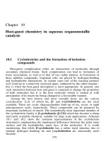
Fig. 1.10. Globular proteins like enzymes and antibodies have a specific surface that
recognises only specific substrates.
in all forms of living organisms and perform a wide variety of tasks. The function
and structure of proteins are outlined in the following sections.
1.1.2.1 The biological function of proteins
In general, there are two types of protein structures: (1) fibrous, elongated proteins
which are not soluble in water and provide structural support and (2) globular
spherical proteins which are water soluble and have specific functions in the
immune system and metabolism.
Globular proteins have a compact, spherical structure with very characteristic
grooves and peaks on their surface. Analogous to a key fitting into a lock, other
molecules fit into these grooves and peaks. This makes globular proteins specific
when it comes to interacting with or recognising other molecules (Fig. 1.10).
Enzymes are an example of such specific proteins. They are biochemical catalysts,
which lower the activation energy and, thus, accelerate immensely the reaction rate
of biological reactions. An enzyme can only react with a substrate if the location of
its functional groups and hydrogen bonds as well as its shape matches the active site
of the enzyme. Ribonuclease for example is an enzyme secreted by the pancreas
to specifically digest ribonucleic acid (RNA). Antibodies are another example of
highly specific globular proteins. They can recognise intruders, antigens, and bind
to them in a key-lock mechanism. Enzymes and antibodies are used as molecular
recognition elements in bioassays (section 5.1) and biosensors (section 5.2).
In the body, proteins also function as transport and storage media. For example,
haemoglobin is responsible for the transport of oxygen in the blood stream, trans-
ferrin for the transport of iron. Ferritin is an example of a protein with a storage
function, which can be found in the liver. It forms a complex with iron, and thus
binds and stores the metal. In the form of hormones, polypeptides can also act
as chemical messengers. By interacting with a matching receptor, usually found
in the cell membrane, they regulate a wide variety of tasks in metabolism. For
“chap01” — 2004/2/13 — page 9 — #9
Biomolecules 9
example, three hormones found in the pancreas, glucagon, insulin and somato-
statin, regulate the storage and release of glucose and fatty acids. Other hormones
control digestion, growth and cell differentiation. Hormones form a large class of
chemical substances. Most hormones are polypeptides, however, some are amino
acid derivates or steroids.
Fibrous proteins have a high tensile strength and mechanical stability. Their
function is to provide structural support to tissues. Collagen, for example, gives
connective strength to skin, bones, teeth and tendons. Ceratin is the major
component of hair and nails.
1.1.2.2 The structure of proteins
Proteins are not just randomly coiled chains of amino acids. A variety of
intramolecular interactions enables the amino acid chain to fold in a specific way
to give the protein a three-dimensional structure and shape. This structure is crit-
ical for its activity and function. Several amino acid strings can be entangled and
connected to each other via disulfide bridges. Parts of the amino acid chain can be
organised into helices or sheets. Globular proteins like enzymes and antibodies are
more folded and coiled whereas fibrous proteins are more filamentous and elon-
gated. To describe the complex structure of proteins, four levels of organisation
are distinguished: primary, secondary, tertiary and quaternary structures.
Primary structure
The sequence of amino acids determines the primary structure of a protein. Chang-
ing just a single amino acid in a critical position of the protein can significantly
alter its activity and function and be the cause of disease and disorders. The amino
acids are connected to each other in a head-to-tail fashion by formation of a peptide
bond (Fig. 1.11), the condensation of a carboxylic and an amino group with the
elimination of water.
Two amino acids connected via a peptide bond are called a dipeptide, three acids
a tripeptide and so on. With an increasing number of acids in the sequence, the
molecules are referred to as oligopeptides and polypeptides. The C
−−
N bond cannot
C
N
O
C
NH
3
+
O
O
-
H
R
1
+
H
3
N
COO
-
R
2
H
+
HNH
3
+
COO
-
H
R
1
R
2
H
+ H
2
O
Fig. 1.11. Peptide bond formation from two amino acids.