bioluminescence methods and protocols
Bạn đang xem bản rút gọn của tài liệu. Xem và tải ngay bản đầy đủ của tài liệu tại đây (17.93 MB, 286 trang )
1
Improvements in the Application
of Firefly Luciferase Assays
Sharon R. Ford and Franklin R. Leach
1. Introduction
1.1. Firefly Luciferase Assay Differs from Usual Enzyme Assays
The firefly luciferase-based assay differs from most familiar enzyme-based
determinations. Most enzyme assays are based either on the production of a
product or the disappearance of a substrate. Usually the compound measured is
stable so that its concentration can be determined after a specific time. At low
adenosine Striphosphate (ATP) concentrations, firefly luciferase is a stoichio-
metric reactant rather than a catalyst. In the case of the firefly luciferase reac-
tion, AMP, PPi, CO*, and oxyluciferin are typical products that accumulate,
but the product that is most often and most easily determined is light. The
photons of light are not accumulated in the measuring technique unless film or
some electronic summation procedure is used in photon counting.
The two-step firefly luciferase reaction sequence is shown below. Step one
forms an enzyme-bound luciferyl adenylate. Either MgATP or LH, (luciferin)
can add first to the enzyme LUC.
LH2 + MgATP + LUC c ) LUC-LH,-AMP + MgPP,
(1)
Step two is the oxidative decarboxylation of luciferin with the production of
light on decay of the excited form of oxyluciferin.
LUGLH2-AMP + O2 + OH-+ LUC-OL + CO2 + AMP
+ light + Hz0 (2)
The oxyluciferin product, OL, is released slowly from the enzyme-product
complex. This gives the flash kinetic pattern observed with high ATP concen-
trations, under which conditions firefly luciferase acts catalytically. The initial
flash of light emission observed with high ATP concentration is owing to a
From h48thods m Molecular Biology, Vol 102 B/olummescenc8 Methods and Protocols
Edlted by R A LaRossa 0 Humana Press Inc , Totowa, NJ
3
ford and Leach
.
8
3
8
P
e
&I
800
600
400
200
0
0 20 40
60
Time, set
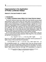
Fig 1, Time-courses with nanomolar ATP.
0 20
40 60
Time, set
Fig. 2. Time-courses
with micromolar ATP.
“first round” of enzyme activity. This flash rapidly decays to a relatively constant
light emission, similar to that seen at low ATP concentrations, which is thought to
be the result of the enzyme slowly turning over by releasmg the oxylucifenn.
1.2.
Kinetic
Pattern Varies with ATP Concentration
The two kinetic patterns of light production are shown in Figs. 1 and 2. This
property can be a source of experimental difficulties. When measurmg light
Application of Firefly Luciferase Assays 5
emission usmg high ATP concentrations, the delay between starting the
reaction and starting the measurement of light emitted, as well as the length of
time that the light emission is measured become critical. In this case, tt is
essential that the reaction be initiated while the sample is within the counting
chamber of the lummometer, that the initiating reagent be rapidly and com-
pletely mixed with the components already in the reaction cuvet, and that the
light emission always be measured over the same period of time.
1.3. Origin of the Use of Firefly Luciferase to Determine ATP
Firefly luctferase was first applied to the determination of ATP in 1947 by
McElroy (I). Given the status of instrumentation available for the measure-
ment of light in the 1940s and 195Os, some procedural compromises evolved.
One was the use of arsenate buffer m the reaction mixture, which reduced light
emitted and changed the time-course of the reaction. In 1952 Strehler and Trot-
ter (2) recommended the use of arsenate buffer to prevent precipitation that
occurred when phosphate buffer and Mg were used. The application of firefly
luciferase to the assay of ATP was described by Strehler and McElroy (3) and
further amplified by Strehler (4).
1.4. Modern Development
New instrumentation with fast response times IS now readily available, and
many ATP determinattons requrre great sensitivity. Those two factors obviate
the need to use arsenate-based assay systems and, in fact, make them undesn-
able. The use of arsenate-inhibited systems persists because of precedence and
the fact that some commercial suppliers still provide firefly luciferase m an
arsenate buffer. McElroy (5) cautions against usmg the commercially prepared
luciferase with arsenate, because it lowers sensitivity, is an inhibitor, and 1s not
required with current instrumentation.
1.5. The Response Is Determined by the Ratio of Reactants
Since the reaction occurs m a defined volume, increasing the concentration of
either luciferase or luciferin increases the light production achieved with a given
concentration of ATP. This concentration increase makes collisions of molecules
more likely. Thus, a change in the ratio of the components changes light productton,
shifting the light ermssion vs ATP concentration standard curve either to the right
(reduced sensitivity) or left (enhanced sensitivity). This 1s illustrated m Table 1.
When using a reaction mixture that contains both luciferase and luciferm added
together in a single volume (such as in a commercially available mix), the
counts observed decrease as the square of any dilution of the reaction mix (7).
The reaction requires three substrates: lucrferm, MgATP, and oxygen. In addt-
tion, several stabilizing compounds are added to a typical assay system. Table 2
Ford and Leach
Table 1
Effect of Changing of Reactant
Proportions on Light ProductioV
Firefly luciferase, nM Luciferin, fl KRLU
54 110 5.0
54 280 66
108 110 86
108 280 12
216 110 16
216 280 23
%gma lucrferase (L 5256) and o-lucrferm (L 6882) were used m a
300~pL vol m the Model 2010A Biocounter [ATP] = 67 pA4 KRLU =
1 ,OOO,OOO counts Modrfied from ref. (6).
Table 2
Reaction Requirements for Firefly Luciferasee
Component omttted Light productton, light untts/lOs
None 5204 1.1
-MgSO,, 5 n&I 2.1 It 0.2
-DTT, 0 5 mA4 52.5 + 0.7
-EDTA, 0 5 mA4 54.0 + 1.2
-Luciferm, 0.358 mA4 0002
-ATP, 321 nM 0.002
Qystallme natrve lucrferase from Sigma was used m a 300~pL vol
The effect of omtsston of the mdtcated component was determmed m trtp-
locate assays on a Model 2010A Blocounter. A light unit 1s 1000 counts
produced. [ATP] = 32 1 nM Modified from
ref. (8)
shows what occurs with the omission of each component. The buffer maintains
the enzyme at its optimum pH of 7.8 (9). -SH compounds are added to ensure
that the cysteine residues of firefly luciferase are not oxidized (there are no
disulfide linkages present in the protein). EDTA is added to prevent any metal
ions from interfering with the reaction. The presence of metals can change the
wavelength of light produced. Firefly luciferase preparations (particularly those
sold in kit form) are often stabilized by the addition of bovine serum albumin,
trehalose, glycerol, or other compound(s).
As shown in Table 2, light production by firefly luciferase is completely
dependent on the presence of Mg2+, ATP, and luciferin in the reaction mixture.
Dithiothreitol (DTT) and ethylenediaminetetraacetic actd (EDTA) are added
to the reaction mixture to prevent inhibitton of the reaction.
Application of Firefly Luciferase Assays
7
0.001 0.01 0.1 1 10
100
ATP, PM
Fig. 3. Light production as a function of ATP concentratton. Note that the plot has
log vs log scales.
The light productron response from firefly luciferase is linear over a range of four to
five logs of ATP concentration (Fig. 3). As little as 50 fg of ATP was measured (IO).
1.6. Optimum Assay Conditions
1.6.1.
pH
The
optimum pH
for the
reaction is
pH 7.8 (9). We have shown that Tricine
buffer, which has a pK, of 8.15 and offers the greatest buffering capacity of
any common buffer, works well for firefly luciferase (II). Table 3 shows the
functionality of several buffers with firefly lucrferase.
The necessity for pH maintenance was clearly demonstrated by the follow-
ing experiment. When ATP solutions were not neutralized, we observed that
10 mM ATP inactivated luciferase during incubation before addition of
luciferin and assay. This occurred when 6 r&I Tris-succinate buffer was used.
When ATP was prepared in a buffer, incubation of firefly luciferase with 10 mM-
concentrations of ATP did not inactivate the enzyme.
l-6.2. Temperature
The optimum temperature for the firefly luciferase is 25OC. At temperatures
>3O”C, native
Photinuspyralis
luciferase is rapidly inactivated. Mutants of luci-
ferase have been isolated with increased temperature stability, but most cornmer-
cially available firefly luciferases are based on the native
P. pyralis
enzyme.
8
Ford and Leach
Table 3
Effect of Buffer on Light Productiona
Buffer, 25 mM pK. 20°C Act relattve to HEPES
MOPS 7 20 0.65
Phosphate 7.21 0.09
TES
7 50
0 54
HEPES 7 55
1 .oo
HEPPS 8.00 0 68
Trtcine 8 15 1.25
Glycine amide
8 20
0 80
Tris 8.30 1 .oo
Glycylglycme 8.40 0 72
“The assays were done a Model 20 10A Blocounter Values obtamed with three
different ATP concentrations were averaged and expressed relative to the value
obtamed with HEPES. All were assayed at pH 7 8 From
ref. (6)
1.6.3. Effect of Products on the Reaction
PP, has little effect at low concentratrons (-0.13, @I), activates when used
at moderate concentrations (-1.3-l 3 @4), and mhrbits at high concentrations
(>1.3 mM) (12). AMP at 1 mM mhtblts firefly luciferase. At low ATP
concentration (0.24 @Y), light production is inhibited by about 70%. At high
ATP concentratron (0.24 n&I), the peak of light production IS inhibited by about
30%, but there 1s little effect on light production at times greater than 1 mm.
1.6.4. Effect of Additives on the Reaction
Several substances have been found that change the flash of light production
mto a linear production of light that lasts for at least a minute as shown in
Figs.
4 and 5.
1. Coenzyme A (CoA). Atrth and colleagues (13) found that CoA addition to a reac-
tion mixture after the flash stimulated light productton; this was presumably
through removal of oxyluciferin from luciferase. The observed enhancement of
light production was proportional to CoA concentratton
(14)
The effect of
CoA
was recently reinvestigated by Wood
(15-l
7), who observed that addition of CoA
prevented the rapid inhibition of light productron and ehcrted a nearly constant
production of light. He found that dethroCoA was a compettttve mhibttor,
suggesting that the sulfhydryl group of CoA was required. Pazzagli et al. (18)
observed no effect of CoA on peak light intensity, but found that 0 66 mA4 CoA
stgmficantly modified the kmettcs of light emtsston They concluded that “despite
the present inability to explain the role of CoA in the btolummescent reaction of the
firefly luciferase, the addition of CoA to the reaction mixture for the firefly luct-
Application of Firefly Luciferase Assays
9
0 20 40 60 80 100 120
Time, set
Fig. 4. Effect of CoA on light production by firefly luciferase. Light productton
was mtttated by injection of ATP at 60 s. The trme-course of light production was
determined m an LKB 1251 luminometer. -o- Control, -o- 0.05 mA4CoA.
0 20 40 60 80 100 120
Time, set
Fig. 5. Effect of PP, and periodate-oxidized and sodium borohydrtde-reduced ADP
on light production by firefly luciferase. Light production was initiated by mjectton of
ATP at 60 s. The time-course of light production was determmed m an LKB 1251
luminometer. + 0.013 mA4 PP,, -A- 1 mA4 orADP, -o-Control
ferase assays has allowed assay conditions of enhanced sensitivity, excellent repro-
ducibility, and a maintained linearity of the calibration curve to be established.”
2. Nucleotide analogs: Ford et al. (12,29) found that cytidine triphosphate and other
nucleotides enhanced firefly luciferase activity in a manner srmilar to that of
CoA. DethioCoA inhibited the activation by both cytidine nucleotides and CoA.
The enhancement of light productton with CoA or nucleotides occurred only with
high ATP concentrations
10
Ford and Leach
3. Triton X-100: Gandelman et al. (20) found that 25 mM Triton X-100 increased
both luciferase light production and the rate of destruction of the enzyme. It pre-
sumably allows formation of a more active, though more labile, enzyme con-
formation. An additive effect of CoA and Triton X-100 has been observed by
Wang and Andrade (21).
4. Other detergents: Simpson and Hammond (22) found that anionic detergents
mhibrted firefly luciferase, catiomc detergents stimulated activity with a sharply
defined concentration optimum, but they also inactivated the enzyme, and non-
ionic and zwittenomc detergents increased reaction rate without affecting stabil-
ity until high concentrations were used. Stability of the enzyme was measured
during a 20-s incubation. Kricka and DeLuca (23) found that a number of sol-
vents stimulated the firefly luciferase reaction by promoting the dissociation of
inhibitory products. These experiments were done in a phosphate-buffered reac-
tion mixture (phosphate inhibits activity), and the time-course of light produc-
tion was not significantly altered. There is no clear evidence that detergents can
improve the routine assay of ATP.
5. PP, and L-luciferin combination: Lundin (24) has shown that addition of 1 I.&’ PP,
and 16 pA4 t-luciferin (Note: this is not the normal substrate) to a firefly luci-
ferase reaction mixture containmg 1 l.uV ATP stabilized light production for -2 mm
This reagent was available from LKB (Stockholm, Sweden), and is now avail-
able from BioOrbit Oy (Turku, Finland), and BioThema (Dalorii, Sweden).
6. Polyphosphates Lundm (25) reported that 20 l&V PP, gives an optimum sus-
tained light emission over an extended period of time (up to 12 mm) at 0.2 mM
ATP. We (Ford et al. [12]) found similar results using 13 @4 PP,. Lower and
higher PP, concentrations were less effective. We also found that tripolyphos-
phate, tetrapolyphosphate, and trlmetaphosphate (all at 1 mM) gave a sustamed
enhanced light emission.
1.7. Use of Additives in Quantitation of Firefly Luciferase
When using the firefly luciferase assay to measure the amount of enzyme m
a sample, maximum sensitivity is needed. Thus, the assay must be done using
high ATP concentrations (-0.2 mA4) and preferably with additives to increase
the light production. Several methods to do this have been developed. Lundm
(25) established an optimized assay for firefly luciferase using 20 mA4 PP, as
an additive to enhance light productron. Boehringer Mannheim (Mannheim,
Germany) sells a kit (cat. no. 1669 893) containing CoA, that yields a con-
stant rate of light production for at least 60 s, and allows the detection of 5 fg
of firefly
luciferase. Promega’s (Madison, WI) luciferase assay system (cat.
no. E1500) contains 270 @4 CoA. Ford et al. (19) report that 0.18 mM
periodate oxidized CTP increased the sensitivity of luciferase determinatron
fourfold and were able to measure 1.5 pg of luciferase. Prolonged incubation
of luciferase with periodate oxidized CTP (>5 mm) inactivated the enzyme.
However, Ford et al. (12) found that the activating activity of perrodate-oxi-
Application of Firefly Luciferase Assays
11
dized and then sodium borohydride-reduced ADP was retained for at least a
150-min incubation of additive with firefly luciferase.
1.8. Mechanisms of Action
Ford et al.
(12)
interpreted that the increased turnover of firefly luciferase
through release of oxyluciferin is the mechanism by which the nucleotide ana-
logs and CoA enhance firefly luciferase activity. There was an increase from
0.97 to -5.23 photons of light produced/mm/molecule of luciferase with 0.24
mMATP. McElroy et al. (26) had previously ascribed the mechamsm of action
of pyrophosphate to the same phenomenon.
2. Materials
2.7. Water and Glassware
Water quality is of paramount importance. Minute contamination of reagents
(especially bacterial contamination) will cause high background luminescence
because of the sensitivity of the technique. We routinely prepare the water
used in all reagents as follows: The building’s reverse osmosis and UV-treated
water is passed through two mixed-bed ion-exchange resins (Barnstead/
Thermolyne D 8902 Ultrapure Cartridges, Dubuque, IA, glass-distilled, pres-
sure-filtered through a sterile 0.45pm Millipore@ (Bedford, MA) filter into
sterile bottles, and then autoclaved. After opening, a bottle of water can be
used for several days if handled using good sterile technique.
We recommend as a minimum standard that “Milli-Q-quality” water be
additionally filtered through a sterile 0.45pm filter and autoclaved before use.
Backgrounds in the standard ATP assay containing 100 pL of Firelight@ and
no ATP in a 500~pL total volume should be cl00 counts/IO s m a Lumac
Model 201 OA Biocounter. If backgrounds are high, the “Milli-Q” water should
be distilled before filtering and autoclaving.
We recommend that all glassware used for reagents for these assays be
washed in phosphate-free detergent, soaked in Pierce (Rockford, IL) brand RBS-
pfs’, rinsed in reverse-omosis-treated (RO) or deionized water, and sterilized.
2.2. Chemicals
Prepare all stocks in sterile glass- or plasticware using sterile water as
described in Subheading 2.1., and store frozen to reduce the chance of bacte-
rial contamination.
1, Tricine: We find that Tricine buffer yields a system giving the greatest light pro-
duction under our laboratory conditions. The optimum pH is 7.8. We use Sigma
(St. Louis, MO) T 9784. Prepare stock solution of 1 .O M, and dilute as needed to
make Tricine-containing reagents.
12
Ford and Leach
2. Bovine serum albumin (BSA): Fraction V Powder (296%) is adequate. We use
Sigma A 2153. BSA is present in many commercial preparations to stabilize fire-
fly luciferase by reducing proteolytic degradation and adsorption to surfaces. The
stock solution is 100 mg/mL in water
3. MgS04: Use ACS-grade salts. A 50-d stock is prepared m water.
4. m-Dithiothreitol (Cleland’s reagent, DTT). Use the highest purity available We
use Sigma D 5545 to prepare a 50-mA4 stock.
5. EDTA: Use the highest grade available. We use Sigma E 1644, disodium salt When
preparing the 50-&stock solution, check pH, and titrate to neutrahty with NaOH
6. Luciferin: n-Luciferin is the natural, functional configuration We recommend Sigma L
6882 sodium salt, because it is readily soluble m water. Alternatively, the free acid form
(Sigma L 9504) is more econormcal, but it must be titrated with NaOH Dissolve the
free acid form at 5.0 mg/mL m 20 mMTricine, pH 7 8, titrate with NaOH to return the
pH to 7.8, and ensure that all the lucifenn is m solution. Protect luciferm from hght
while the solutions are bemg prepared. Purge the atmosphere above the solution with
N2, and store frozen and protected from light (we store m brown bottles, capped with
Parafilm@ and wrapped in foil) For use, dilute the luciferin to 1 .O mg/mL m
20 mMTricme, pH 7.8. Unused diluted lucifenn can be purged with N2 and stored frozen
L-Luciferin supports light production only under special conditions This iso-
mer competes with the natural form. It has been used to lmeanze the time-course
of light production. This is one of the components used in the LKB ATP Mom-
toring reagent, produced now by BioOrbit Oy (25).
7. ATP: Use crystalline, 99-100% pure, dtsodium salt (Cl ppm vanadmm). We use Sigma
A 5394. ATP solutions can be prepared either in 20 mMTncme buffer, pH 7.8, or m
water. Check the pH of ATP solutions and neutralize, if necessary, with NaOH.
8. Pyrophosphate. Use the highest purity available, such as Sigma P 9146 or Sigma
S 9515 tetrasodium salts (decahydrate), 1 mA4 stock pyrophosphate solutions
must be titrated to neutrality
9 CoA. Use either the lithium or the sodium salt (Sigma C 30 19 or C 3 144, respec-
tively). We have always prepared only enough of the 5-mM stock to satisfy a
single day’s need by dtssolvmg in water We have not determined the stability of
CoA solutions on storage.
10. Nucleotide analogs* Periodate-oxidized CTP (Sigma C 5 150, oCTP) and
periodate-oxidized, sodium borohydride-reduced ADP (Sigma A 69 10, orADP),
among others, can be used to linearize the assay. Prepare only enough of the analogs
for a single day of use by dissolving in water. These are prepared as lo-mMstocks
11. Enzyme stabilizer: AuthentiZyme TM Enzyme Stabtltzer from Innovative Chem-
istry (Marshfield, MA) is a proprietary product that protects enzymes from mac-
tivation by oxidation and heavy metals Make solutions accordmg to the
manufacturer’s instructions.
2.3. Firefly Luciferase
We recommend Firelight@, catalog no. 2005 from Analytical Luminescence
Laboratory (Ann Arbor, MI) for routine assays. Dissolve enzyme in 50 mM
Application of Firefly Luciferase Assays
13
Tricine, pH 7.8, containing 10 mA4 MgS04, 1 rnA4 DTT, 1 mM EDTA, and
1 mg/mL BSA. Let enzyme “age” for 21 h at 0-4”C before use. Unused enzyme
can be stored at 4°C overnight, with some loss of activity (see
Note 1).
When purified firefly luctferase is needed, we use Sigma L 5256, crystal-
lized and lyophilized powder. This preparation is no longer available, but IS
replaced by L 2533, which is prepared without arsenate. Dissolve it at 0.1 to
1 mg/mL in 50 mM Tricme, pH 7.8, containmg 10 mA4 MgS04, 1 mM DTT,
1 mM EDTA, and 1 mg/mL BSA or in a 1: 1 mixture of 250 mM Tricme,
pH 7.8, containing 50 mM MgS04, 5 mM DTT, 5 mM EDTA, and Authenti-
Zyme@ Enzyme Stabilizer (see
Note
2). This preparation is not easily soluble:
To dissolve the protein, add the desired solvent and let sit on ice, with occa-
sional gentle mixing, for at least 1 h. Visually check that the protein has all
gone into solution before use. Alternatively, Sigma L 9009 and L 1759 are
soluble preparations containing buffer and salts.
2.4. Luminometer
A high-quality luminometer that allows mjection of reactant mto the sample
while the sample is m the measurmg chamber is needed. We recommend the
Lumac Model 2010A Biocounter (Luma, Landgraf, The Netherlands; recently
purchased by Celsls, Cambridge, UK) or equivalent (see
Note 3).
3. Methods
3.1. Caution
The great sensitivity (50 fg) and wide dynamic range (four decades) of the
firefly luciferase determmation of ATP make a robotic application of the
procedure relatively easy. Numbers can be obtamed, but their meaning could
be misleading. It IS our contentlon that the operator needs to know the nuances
of the assay components and instrumentation to obtain maximally reliable data.
The mind needs to be engaged while doing the measurements. A monograph
on
Biolumznescence Analyszs
has been written by Brolin and Wettermark that
outlines and discusses the particularities of the technique (27).
3.2. Basic Reaction Components
Depending on the parameters of the instrument to be used, we recommend a
reaction volume of from 200-500 pL contammg the following:
25 mMTricine buffer, pH 7 8;
5 mMMgSO&
0.5 mM EDTA;
05mMDTT;
1 mg/mL BSA;
14 Ford and Leach
0.05 mg/mL o-luciferin (if using purified firefly luciferase);
ATP as reqmred,
Firefly luciferase/luciferin (Firelight@) or purified firefly luciferase as required,
Water to desired total volume
A 10X reaction mixture containing 250 mMTricme buffer, pH 7.8; 50 mM
MgS04; 5 rmJ4 EDTA; and 5 mM DTT
IS convenrent to use.
This
mixture
can be prepared ahead, aliquoted in amounts to be used in a single day, and
stored frozen. We recommend using Firelight instead of purified luctferase
plus luciferin for routine assays because of the ease of use and consistency
of results.
3.3. General Protocol
The reaction is carried out at room temperature (25”C), preferably in
semidarkness.
1. Set up reaction cuvets containing for a SOO-pL reaction: 50 pL of 10X reaction
mixture, BSA, and water as needed to brmg the final volume (after subsequent
addition of ATP, luciferin, and enzyme) to 500 pL. These components can be
added to all cuvets before starting the assays
2. Just before placing the cuvet into the countmg chamber, add ATP (at room tem-
perature) and luctferm (kept on ice) tf needed
3 Mix by vortexing, place cuvet into the instrument and start the reaction by mject-
ing the enzyme preparation (at room temperature). Alternatively, enzyme can be
added to the cuvet before placmg rt m the sample chamber and the reaction imtt-
ated by the injection of ATP This is more economrcal if usmg a luminometer
with an automatrc dispenser because of losses of reagent m the lines of the auto-
matic dispenser
4. Determine light emitted for desired time. For routme assays, a 10-s counting time
is usually sufficient The Lumac instrument gives the rate of counting averaged
over the time period selected
Thus, a 30-s countmg time will give the same value
as a 1 O-s counting, but with improved precision (see Note 4).
To measure ATP in biological samples, replace ATP in the general protocol
with the biological sample for which the ATP content is to be determmed. If tt
is necessary to keep the samples cold until just before they are assayed (when
they are warmed to room temperature), the volume of sample assayed should
be kept to a mmimum (no more than 10% of the total reaction volume). For
each biological sample assayed, run a second determination wtth 0.1-0.5 ng of
ATP added to the biological sample to determine the extent of inhibition, if
any, of the assay itself. Inhibition is calculated by comparmg the difference m
light emitted in the biological sample with and without added ATP to the light
emitted from the same concentratton of ATP m the absence of biologtcal
Application of Firefly Luciferase Assays
15
sample. For ATP determinations, it is usually most practical to start the reac-
tion by injecting enzyme. An ATP standard curve must be run each day to
determine the absolute amount of ATP in samples.
3.5. Firefly Luciferase Determination
To measure firefly luciferase in biological samples, replace the Firelight or
purified firefly luciferase in the general protocol with the biological sample to
be assayed, If the biological sample must be kept cold, keep the volume of the
sample to no more than 10% of the total reaction volume. Include o-luciferin
(0.05 mg/mL) in the assay mixture. Assay with a high concentration of ATP
(0.5 mM). Add the biological sample to the assay tube before placing in the
luminometer, and begin the reaction by injecting the ATP.
3.6. Supplementation to Linearize Light Production
When high concentrations of ATP are measured, a flash of light followed by
a decay of light emitted is the normal pattern. This pattern can be converted to
a linear production of light at the high rate of the flash by addition of any
number of compounds as discussed in Subheading 1. To linearize light pro-
duction, add one of the following supplements to the basic reaction mixture:
13-20 p~I4 PP, (used by Lundm and this laboratory);
0.18 m&I oCTP (used in this laboratory);
1 m44 orADP (used m this laboratory);
270-500 p&I CoA (used by Analytical Lummescence and Promega),
1 pA4 PP, and 16 pJ4 L-luciferin (used by BioOrbit Oy).
4. Notes
1. Firefly luciferase: Three grades of firefly luciferase with drfferent degrees of
purity are commercially available. Crude lantern extracts contain sufficient pyro-
phosphatase, so that PP, does not accumulate (28). These preparatrons also
contain adenylate kinase, and nucleoside diphosphate kinase, which enable
nucleotides other than ATP to be enzymatically converted to ATP and thus pro-
duce light in the assay system. These preparations are not recommended for sen-
sitive determination of ATP. Purification procedures have been developed that
remove the adenylate kmase, pyrophosphatase, and nucleoside diphosphate
kinase. These preparations can be used for the sensrtive determination of ATP
Many are supplemented with sufficient luciferin, so that no addittonal lucrferm IS
required. Crystalline luciferase is purer, but is somewhat more difficult to handle
There IS little difference between crystalline native and recombinant firefly lucr-
ferases. The slight differences in conformation and lability to proteolytic enzymes
that exist for these two luciferases are not significant (8).
Although firefly luciferase can be fairly stable when stored properly after
making a solution (29), we recommend the use of a commercial preparation (such
16 Ford and Leach
as Analytical Lummescence Laboratory’s Firelight) made fresh and pooled each
day. The use of a commercial preparation wtth its stabthzers and qualtty control
means that the individual laboratory does not need its own reagent quality-con-
trol program. This laboratory has operated both systems and finds the use of
commercial kits better for routine studies. The use of commerctal kits is now
much more accepted with the advent of molecular biology’s cloning kit-it IS
more time-efficient to let the suppher provtde the quality control. This means
carefully selecting a supplier of reagents. This laboratory evaluated the commer-
cially available reagents in 1986 (6). Much progress has been made in commercial
firefly luciferase reagent kits during the subsequent decade. Many of the suppli-
ers listed in Table 1 of our compartson no longer supply the reagents, and there
are also many new suppliers. The techniques and experiments used m the com-
parative evaluations are still appropriate to evaluate those products The com-
mercial firms whose products have survtved probably have done so because of
good quality. Beginning m 1993, Stanley has pubhshed lists of commercial firms
providmg luminescence kits based on mformatton provided by the supplier (30-34)
There is no experimental comparison of the kits and reagents in Stanley’s listing.
Wang and Andrade (35” have added 100 mg/mL of trehalose to stabilize solu-
tions of firefly luciferase particularly when preparing films.
2. Enzyme stabilizer: Firefly luciferase dtssolved m a mixture of salts and Authen-
tiZymeTM Enzyme Stabilizer is stable frozen for several months, even with
repeated thawing and freezing (29).
3. Instrumentatton-luminometer: Although relatively expensive and specialized,
we recommend the use of an instrument designed for btoluminescent/chemtlumi-
nescent measurements These instruments have a wide range of specific proper-
ties (such as geometry of the detector) and design criteria (temperature control
and sample size). Some permit vartatton of the high voltage supplied to the
photomultrplier, whereas others have fixed voltage, some allow temperature regu-
lation, but others operate at room temperature Ten commercially available
instruments have been experimentally compared by Jago and associates (36) The
most sensitive instruments were the Lumac Model 20 1 OA and the Turner 20 TD
photometers, which had actual hmits of 0.09 and 0.12 pg ATP/sample, respec-
tively. George Turner (37) presents a provocative assessment of instrument
development from the viewpoint of a person trained m physics and electronics
trying to get the most out of the mstrument/reagent system Van Dyke (38)
reviews the manufacturers’ provided information for photometers that were avail-
able in 1985. Further review of the commercial instrumentation has been made
by Phil Stanley in a continuing series of articles (3k343p-41).
If the investigator desires to construct a photometer, Anderson et al. (42) give
complete mstructions. These instructions were updated in 1985 (43) with “the
strong recommendation that in most cases a researcher would be better served to
purchase a commercial mstrument.”
For calibration of light productton, please refer to the methods described by
O’Kane and coworkers (44) and by Lee and Sehger (45).
Application of Firefly Luciferase Assays
17
4. Protocol: We recommend that preliminary experimentation be done to establish
that the reagents, instruments, and protocols are working in your laboratory, and
meet the desired quality-control characteristics. What is the instrument back-
ground, and what are the reagent backgrounds? Is the response to known (stan-
dard) amounts of ATP and/or luciferase in line with published values? Is the
response linear over several orders of magnitude? Is the slope of the standard
curve one? Are the reagents stable over the desired assay period? What is the
response when a know standard amount of either ATP or luciferase is added to an
experimental reaction mixture (m other words, what IS the extent of inhibition m
the assay mix itself)?
Several of the commercial manufacturers have published detailed protocols or
quality-control information for the use of their reagents These include:
Luciferase Assay Guide Book, Protocols and Information for Measuring Fve-
fly Luciferase Expressed in Cells, Analytical Luminescence Laboratory,
1180 Ellsworth Road, Ann Arbor, MI 48108 (l-800-854-7050).
Luminescence Analysis, Application Note 100; and The Bioluminescent
Assay of ATP, Application Note 201 Bio-Orbit Oy, Box 36 SF-20521
Turku, Finland, Vorce +358 2 1 5 10666; Fax +358 2 15 10150.
Luciferase, ATP Biolummescence Assay Kit HS II, and Luciferase Reporter
Gene Assay protocol are available from Boehrmger Mannheim Bio-
chemicals, P 0 Box 50816, Indianapolis, IN 46250 (I-800-428-5437)
(Internet* )
Luciferase Assay System (Part# TB 101) Promega, 2800 Woods Hollow
Road, Madtson, WI, 53711-5399 (I-800-356-9526) (Internet http://
www.promega.com) Protocols and application notes are available on-lme.
Sigma Quality Control Test Procedure for Products Ll759, L5256, and L9009,
available at Internet: http.//www.sigma.sial.com/slgma/enzymes/lucifera.htm.
Luciferase protocol, Tropix, Inc (l-800-542-2369) Internet http llwww.
tropix com/luciptl.htm
Turner Instrument Literature (
Acknowledgments
This research was supported in part by the Oklahoma Agricultural Experi-
ment Station (Project 1806) and IS published with the approval of the Direc-
tor. Robert Matts and E. C. Nelson read the manuscript and made useful
suggestions.
References
1. McElroy, W. D. (1947) The energy source for bioluminescence in an isolated
system. Proc. Nat. Acad Scz. USA. 33,342-345.
2. Strehler, B. L. and Trotter, J. R (1952) Firefly luminescence in the study of energy
transfer mechanism. I. Substrate and enzyme determination. Arch Biochem.
Bzophys 40,284 1
78 Ford and leach
3. Strehler, B. L. and McElroy, W D. (1957) Assay of adenosine triphosphate. Met/r-
ods Enzymol. 3,871-873.
4. Strehler, B. L. (1968) Bioluminescence assay. principles and practice. Methods
Biochem. Anal. 16,99-l 8 1.
5. McElroy, W. D. (1977) Comments on the history of the firefly system, in 2nd
Bt-Annual ATP Methodology Sympostum (G. A. Borun, ed ), SAI Technology,
San Diego, CA, pp. 405-4 13
6. Leach, F. R. and Webster, J. J. (1986) Commercially available firefly luciferase
reagents. Methods Enzymol. 133,5 l-70.
7. Webster, J J. and Leach, F. R. (1980) Optimization of the firefly luicferase assay
for ATP. J Appl Biochem 2,469479.
8. Ford, S. R., Hall, M. L., and Leach, F. R. (1992) Comparison of properties of
commercially available crystallme native and recombinant firefly luciferase J
Btolumtn Chemdumtn. 7, 185-l 93.
9 DeLuca, M. (1976) Firefly luciferase. Adv. Enzymol. 44, 37-63.
10 Webster, J. J , Chang, J C., and Leach, F. R (1980) Sensitivity of ATP determi-
nation. J Appl. Btochem 2,5 16, 5 17
11. Webster, J. J., Chang, J. C., Manley, E. R., Splvey, H O., and Leach, F R. (1980)
Buffer effects on ATP analysis by firefly luciferase Anal Btochem 106,7-l 1
12. Ford, S R., Chenault, K. H., Bunton, L. S., Hampton, G. J., McCarthy, J., Hall, M. S ,
Pangburn, S J., and Leach, F. R. (1996) Use of firefly luciferase for ATP measure-
ment other nucleotides enhance turnover. J Btolumm. Chemtlumrn 11, 149-167
13 Arrth, R. L., Rhodes, W. C., and McElroy, W. D. (1958) The function of coen-
zyme A m luminescence. Btochrm. Btophys Acta 27,5 19-532.
14 McElroy, W D. (1957) Chemistry and physiology of blolummescence, m The
Harvey Lectures, 1955-56 Academic, NY, pp 240-266.
15 Wood, K. V. (1990) Novel assay of firefly luciferase providing greater sensitivity
and ease of use. J Cell Btol 111,380a
16. Wood, K. V. (1991) The origin of beetle luciferases, m Biolumznescence and
Chemzluminescence Current Status (Stanley, P. E. and Kricka, L. J., eds.) John
Wiley, Chtchester, UK, pp. 11-14.
17 Wood, K V. (199 1) Recent advances and prospects for use of beetle luciferase as
genetic reporter, in Btolumtnescence and Chemtlummescence. Current Status
(Stanley, P. E. and Kricka, L J , eds.), John Wiley, Chichester, UK, pp 543-546.
18. Pazzagh, M., Devine, J H., Peterson, D. 0 , and Baldwin, T. 0. (1992) Use of
bacterial and firefly luciferases as reporter genes in DEAE-dextran-mediated
transfection of mammalian cells Anal. Btochem 204,3 15-323
19 Ford, S. R., Hall, M. S., and Leach, F. R. (1992) Enhancement of firefly luciferase
activity by cytidine nucleotides Anal Biochem 204, 283-29 1
20. Gandelman, 0. A., Brovko, L. Y., Bowers, K. C., Cobbold, P. H., Polenova, T.
Y., and Ugarova, N. N. (1993) Kinetics of enzymic oxidation of firefly luciferm
in vitro and m cytoplasm, in Btolumutescence and Chemtlumtnescence Status
Report (Szalay, A. A., Kricka, L J , and Stanley, P E , eds.) John Wiley,
Chichester, UK, pp. 84-88
Apphcation of F/refly Luciferase Assays
79
21, Wang, C. Y. and Andrade, J. D. (1996) Surfactants and coenzyme A as cooperative
enhancers of the activity of firefly luciferase. J. Biolumin Chemtlumtn
11,25.
22. Simpson, W. J. and Hammond, J. R. M. (1991) The effect of detergents on firefly
luciferase reactions. J. Biolumtn. Chemilumtn. 6,97-108.
23. Kricka, L J., and DeLuca, M. (1982) Effect of solvent on the catalytic activity of
firefly luciferase. Arch Biochem Biophys 217,674-681
24. Lundin, A. (1982) Application of firefly luciferease, in Lumtnescent Assays: Per-
specttves tn Endocrtnology and Cltnical Chemtstry (Servo, M. and Pazzagh, M.,
eds.), Raven, New York, NY, pp. 29-45.
25. Lundm, A (1993) Optimised assay of firefly luciferase wrth stable light emtssion,
in Biolumtnescence and Chemilumtnescence: Status Report (Szalay, A. A ,
Krxka, L. J., and Stanley, P., eds), John Wiley, Chichester, UK, pp. 291-295.
26. McElroy, W. D , Hastings, J. W., Couloombm, J., and Sonnenfield, V. (1953) The mechanism
of action of pyrophosphate m ftrefly luminescence. Arch. Btochem. Btophys. 46,399416.
27 Brolin, S. and Wettermark, G. (199 1) Btoluminescence Analysts. VCH Wemheim,
Germany, 151 pp.
28. DeLuca, M and McElroy, W D. (1978) Purification and properties of firefly luci-
ferase. Methods Enzymol 57,3-l 5.
29. Hall, M. S. and Leach, F R (1988) Stability of firefly luciferase in Tricme buffer
and m a commercial enzyme stabilizer. J. Biolumtn Chemtlumtn 2,41-44.
30 Stanley, P E (1993) A survey of some commercially available kits and reagents
which include bioluminescence or chemiluminescence for their operation J
Btolumtn Chemtlumtn 8,5 1-63
3 1 Stanley, P. E. (1993) Commercially avatlable luminometers and imaging devices
for low-light measurements and kits and reagents utthzmg chemiluminescence or
biolummescence: Survey update 1. J. Btolumin. Chemdumm 8,234240.
32. Stanley, P. E. (1993) Commerctally available lummometers and imaging devices
for low-light measurements and kits and reagents utilizing chemiluminescence or
bioluminescence: Survey update 2. J Biolumtn Chemilumtn 9,5 l-53.
33. Stanley, P. E. (1993) Commercially available lummometers and imaging devices
for low-light measurements and kits and reagents utihzmg chemiluminescence or
biolummescence: Survey update 3. J Btolumin Chemilumm 9, 123-125.
34. Stanley, P. E. (1993) Commercially available lummometers and imaging devices
for low-light measurements and kits and reagents utilizmg chemiluminescence or
bioluminescence Survey update 4 J Biolumtn Chemrlumin 11, 175-l 9 1.
35. Wang, C Y., and Andrade, J. D. (1994) Purification and preservation of firefly
luciferase, rn Btolumtnescence and Chemtluminescence Fundamental and
Applied Aspects (Campbell, A. K , Kricka, L. J., and Stanley, P. E., eds.), John
Wiley, Chichester, UK, pp 423-426.
36. Jago, P H., Simpson, W J., Denyer, S. P., Evans, A W., Griffiths, M W.,
Hammond, J. R M., Ingram, T. P , Lacey, R. F., Macey, N W., McCarthy, B. J.,
Salusbury, T. T., Semor, P. S., Sidorowicz, S., Smithers, R., Stanfield, G., and
Stanley, P. E. (1989) An evaluation of the performance of ten commercial
luminometers J Btolumm. Chemtlumrn 3, 131-145
20 Ford and Leach
37. Turner, G. K. (1985) Measurement of light from chemical or biochemical reac-
tions, in Blolummescence and Chemdumlnescence* Instruments and Appllcatlon,
vol. I (Van Dyke, K., ed.), CRC, Boca Raton, FL, pp 43-78.
38. Van Dyke, K. (1985) Commercial mstruments, m Bzoluminescence and Chemzlu-
mwescence: Instruments and Applrcation, vol. I (Van Dyke, K., ed.), CRC, Boca
Raton, FL, pp. 83-128.
39 Stanley, P. E. (1985) Characteristics of commercial radiometers. Methods
Enzymol. 133,587-603.
40. Stanley, P. E. (1992) A survey of more than 90 commerctally available lumi-
nometers and imaging devices for low light measurement of chemilummescence
and bioluminescence, including mstruments for manual, automatic and special-
tzed operation for HPLC, LC, GLC and microplates. Part 1 descriptions. J
Blolumln. Chemdumln. 7,77-108.
41 Stanley, P. E (1992) A survey of more than 90 commercially available lumi-
nometers and imaging devices for low light measurement of chemiluminescence
and btoluminescence, including mstruments for manual, automatic and special-
ized operation for HPLC, LC, GLC and microplates. Part 1 photographs J
Biolumln Chemdumm 7, 157-169
42. Anderson, J. M., Faint, G. J., and Wampler, J. E. (1978) Construction of mstru-
mentation for biolummescence and chemilummescence assays. Methods Enzymol
57,529-540.
43. Wampler, J. E., and Gilbert, J C (1985) The design of custom radiometers, m
Bioluminescence and Chemdumwescence* Instruments and Appllcatlon, vol I
(Van Dyke, K., ed.), CRC, Boca Raton, FL, pp 129-150
44. O’Kane, D J., Ahmad, M , Matheson, I. B. C., and Lee, J (1986) Purification of
bacterial luciferase by high-performance ltquid chromatography Methods
Enzymol. 133, 109-127.
45. Lee, J. and Seliger, H H (1972) Quantum yields ofthe lummol chemdummescence
reaction m aqueous and aprotic solvents. Photochem Photoblol. 15, 109127
2
Visualization of Bioluminescence
Amy Cheng Vollmer
1. Introduction
There are an increasing number of specialized instruments that may be used
for the purpose of measuring biolummescence. Table 1 contains a representa-
tive list of different luminometers and cameras that are available. These mstru-
ments have been used to detect bioluminescence in a number of organisms
using either bacterial luciferase (lux; 1,2) or firefly luciferase (luc; 3,4)
as reporters. Sensitivity of the newer luminometers ranges from six to eight
logs. Options such as temperature control and agitation of samples are usually
available at an extra cost. Most of the systems can be driven by computer with
commercially available or customized software. Storage, display, and analysis
of data mvolve the same or additional software packages. Sample containers
have also become more speciahzed. In the case of the multiplate lummometers,
opaque plates are available m either white or black. Black plates are recom-
mended for bright samples where reflection into neighboring wells results in
“crosstalk.” White plates are recommended for samples that are lower light
emitters, since the reflective surface enhances detection. Opaque plates are also
available with transparent bottoms. Samples in these microplates may be read
m a spectrophotometer (such as an ELISA reader) to measure optical density
of the sample, as an indicator of cell number particularly in the case of bacte-
rial cells. In some applications, opaque microplates containing samples may be
stacked in alternation with transparent microplates, if samples require a light
source. This is essential for many of the studies involving photosynthetic
microorganisms (5,6) as well as for those studying circadian rhythms for which
light entrainment is needed (S-7).
On the other hand, it is possible to measure and document btoluminescence
without purchasing a dedicated instrument. In most laboratories, equipment
From Methods m Molecular Bfology, Vol 102 Blolumrnescence Methods and Protocols
Edlted by R A LaRossa 0 Humana Press Inc , Totowa, NJ
21
Table 1
Commercial Luminometers, Listed by Sample Format
Tube/vial
96-Well Microplate CameraC Manufacturer
Monollght@ 20 10”
1250=, 1251b, 1253”
Monolrght@ 9600
lucy 1
1258 (Galaxy@)
Optocomp@ 1”
Lumstar@
7700 senes
WELLTECH
ML2200, ML2250,
ML3000
Lummoskan@
TD 20e”
TopCount@
LumatTM LB 9507” MlcroLumatm LB96P
Multi-LumaPM LB9507b
McroBetaTM PLUS
MIcroBetam
caml1ght@
Analytical Luminescence Lab
Anthos Labtec, Inc.
BloOrblt Oy
(Man-Tech Assoc.)
BMG Lab Technologies
Cambridge Technology, Inc
Denely Instruments, Inc
Dynatech Laboratories, Inc
TEKCDS 12BIC5
Hamamatsu
ICL90 1
NIghtOWL@ LB 981
Labsystems
MGM Instruments
Prmceton Instruments
Photomcs Co
Troplx, Inc
Turner Designs
Packard Instrument Co
EG&G Berthold/Wallac
LKBiWallac
San Diego, CA
Frederick, MD
Tonawanda, NY
Durham, NC
Watertown, MA
Research Tnangle, NC
Chantllly, VA
Needham Heights, MA
Hamden, CT
Trenton, NJ
JAPAN
Bedford, MA
Mountam View, CA
Menden, CT
Turku, Finland
Galthersburg, MD
“Smgle sample
bMultlple sample
cNot all are CCD cameras
Visualization of Bioluminescence
23
and supplies that can be used successfully in many applications already exist.
There are certainly limitations to their sensitivity, especially since these instru-
ments were usually designed with some other application in mind. This chapter
will focus on the use of such instrumentation for visualtzation of biolumin-
escence in the following ways. A liquid scintillation counter can be used to
measure bioluminescence from Escherichia coli strains carrying stress
promoter::Zux fusions on recombinant plasmids. (We have used a 1219
RackBeta@ from LKB/Wallac, Gaithersburg, MD, driven by UTMac software.)
Screening of bioluminescent bacterial colonies can be performed easily using
X-ray film. Photography of bioluminescent bacterial colonies can be accom-
plished with prolonged exposure times using Polarotd type 57 film or Kodak
T-MAX P3200 35-mm roll film with the appropriate cameras and lenses.
2. Materials
1. Fresh biolummescent bactertal cultures, grown on appropriate media: Liquid cul-
tures should be used for measurements in the scintillatton counter, agar media
should be used for photographic documentatton
2 Sterile 1.5-mL microcentrtfuge tubes without caps: These are available commer-
cially, or the caps can be cut off of standard 1 S-mL mtcrocentrrfuge tubes
3. Glass vials (or otherwise transparent ones with tight-fitting lids) suttable for the
scinttllatton counter used: These vials need to be washed and dried one ttme,
smce they will not come mto direct contact with the bacterial sample The vials
must be large enough to accommodate a 1 S-mL microcentnfbge tube without tts cap.
Alternatively, one can use smaller vials and 0.5-mL mtcrocentrtfuge tubes (see
Note 1).
4. X-ray film, such as Kodak XAR or DuPont Reflecttons@
5. Polaroid type 57 film with appropriate film holder and photostand or Kodak T-MAX
P3200 35-mm high-speed roll film and a 35-mm camera with an assortment of
lenses.
3. Methods
3.1. Use of the Scintillation Counter
1. Scintillation counters have programs that can be set by the operator. LKB/Wallac
calls these “parameter groups ” Set one parameter group to read chemtlummes-
cence, a standard setting for most scinttllation counters. Bioluminescent samples
will be read wtth that settmg The time interval over which the sample is to be
counted can be varied between 10 s and several minutes. Set thts interval to meet
the needs of the reporter system that is being used and the amount of light that 1s
emitted. Intervals that are ~1 mm are typical. Set one other parameter group to
read some other window. Be sure the time Interval for this parameter group is
about 1 O-20 min If The LKB/Wallac system assigns numbers to each parameter
group. Each rack of samples can be identified by a code plug chpped to the lead-
mg edge of the rack
24 Vollmer
2. Place one sterile, capless 1 S-mL mtcrocentrifuge tube inside each glass scmtdla-
tion vial (see Note 1).
3. Carefully place ahquots of bacterial samples into the tube. The volume of the
sample placed into each tube can vary from IO-100 pL. (Volumes >lOO Ccs, may
result in a reduced level of oxygenation of the sample. This may or may not be an
important consideration; see Note 2).
4. Place and tighten lids on the scmtillation vials. After tightening the lids, loosen
by one-quarter turn to allow for the exchange of air (see Note 3).
5. Place bacterial samples into a sample rack that bears the correspondingly
numbered identificatron code plug for that parameter group. If there are
more samples than the number of places in the rack, place additional sample
m another rack that bears no identification code plug. The counter will
consider samples in this next rack as components of the first parameter
group mode
6. Place an empty scintillation vial (with a lid) into another rack. This rack should
have a code plug that identities the second parameter group. By inserting this
rack after the bioluminescent samples, a time delay is Introduced so that the
samples will be read once every l&20 min. This reading cycle ~111 contmue
until the counter is stopped by the insertion of a rack bearing stop code plug or by
interrupting the program through a keyboard command to the UTMac software
on the computer.
7. Data saved on UTMac can be most easily formatted as a Simpletext table, which
can be easily exported and “parsed” mto spreadsheets or graphic programs for
analysis. It IS possible to record the actual times that the sample readings took
place. It is also convement to delete data recorded from counting the “dummy”
sample
8. After readmgs are completed, samples may be removed from the scmtillation
vials for plating or disposal (see Note 4).
3.2. Screening Bioluminescent Bacterial Cultures
Using X-Ray Film
1. Plate bacteria on suitable agar medium. Place plates, agar side up, inside of a
light-tight box that has a removable lid. Use transparent tape to secure the plates
to the bottom of the box.
2. Alternatively, a microttter plate containing hquid bacterial cultures m the wells
may be taped to the bottom of the box. Care should be taken not to tilt the plate or
the box.
3. In the darkroom, place one piece of X-ray film on top of the plates. Secure the film
to the side of the box with transparent tape. Be careful not to place the rest of the
unexposed film near the plates. Very bright emitters produce significant amounts
of light and may expose the film if it is too close Using scissors, cut one corner of
the film to help to orient it later. Mark the corresponding comer of the box
4. Place the lid of the box on top and place the box carefully inside a cabmet or
drawer.
Visualization of Bioluminescence
25
5. Exposure times are highly vanable. Bright emitters need only a few seconds of
exposure. Low light emitters require overnight exposure. Exposure time also
depends on the concentration of bacteria inoculated onto the agar.
6. When removing the film from the box, be sure to remove any pieces of transpar-
ent tape that may have been securing the film to the box. Develop the film, and
then orient it with the plates in the box, aligning the marked corner of the box
with the cut corner of the film. Additional exposures may be done subsequently
(see Note 5).
3.3. Photographing Bacteria on Agar Plates
3.3.1. Using Polaroid Film and Camera
1 Place plate with colonies or other visible bacterial growth under the camera, allgn-
ing the plate so that it is centered m the focal field (see Note 6).
2 With visible light illuminating the plate, take a photograph of the plate Insert a
piece of Polaroid type 57 film Expose the film by pulling the protective barrier
away from the film and opening the shutter. Exposure setting should be set to
allow hmited light (f= 32, l/125 s) Develop the film accordmg to manufacturer’s
instructions.
3 Insert a piece of Polaroid type 57 film mto the film holder. Darken the room
4. Expose the film by pulling the protective barrier away from the film and openmg
the shutter. Settings for exposure should allow for maximum light to enter the
lens (f= 4.5); exposure times will range from minutes to hours (see Note 7)
5. After closing the shutter to terminate exposure, develop film as usual (see Note 8)
3.3.2. Usmg High-Speed 35-mm Film and Camera
1 Load Kodak T-MAX P3200 35-mm film into a 35-mm camera (see Note 9).
2 Place plate with colonies or other visible bacterial growth under the camera, align-
mg the plate so that it 1s centered m the focal field (see Note 6).
3. Darken the room, and expose the film. Several different settings should be used.
Adjust thefstop on the camera to allow maximum light to the lens Exposure
times will vary between 1 and 10 min. Differences in lenses, distance, and bnght-
ness of colonies will affect the quality of the photograph
4. Develop the film as per manufacturer’s instructions using T-MAX Developer
3.4. Results
Data collected by a scintillation counter are comparable to &hat collected by
luminometers. Kinetics are revealed by plotting relative light units as a func-
tion of time.
A
comparison of the linear ranges of a luminometer and scmttlla-
tlon counter has been made followmg the methods of Burlage and Kuo (8), the
only difference being the range of linear response. Figure 1 shows a photo-
graph (panel A) as well as the exposed X-ray film image (panel B) of E. coli
carrying a plasmid bearmg promoter::lux fusions. The results on the X-ray
film demonstrate a greater level of sensmvlty than those on Polaroid film. Light
26
A
Vollmer
123456789
A
B
C
D
E
F
G
B
Fig. 1. All wells contained 50 pL of bacterial cultures, grown to midexponential
phase (36 klett units) in LB. Rows A-F contained strain DPD 2794, E. coli carrying a
plasmid bearing a recA::Zux fusion. Rows A and B contained successive twofold dilu-
tions of mitomycin C, starting with 1 pg/mL in column 1; column 9 contained no
mitomycin C. Rows C and D contained successive twofold dilutions of CdCl, starting
with 2 mM in column 1; column 9 contained no CdCl,. Row E contained successive
two-fold dilutions of ethidium bromide, starting with 1 mg/mL in column 1; column 9
contained no ethidium bromide. Row F contained successive twofold dilutions of
Visualization of Bioluminescence
27
production is correlated with concentration. It is evident that the 30-s exposure
of the X-ray film was too long to distinguish a dose-dependent recA response
(Fig. 1,
rows A, B). This is owing to the high consitutive expression ofrecA (in
the absence of mitomycin
C [Fig. 1,
column
91). Figure 2
compares a Polaroid
photograph of an agar plate with an X-ray image. The ring of light was pro-
duced by
E,
coli strain DPD2794, which carries a recA promoter fused to
ZuxCDABE
induced by mitomycin C (9). A zone of growth inhibitlon is appar-
ent in the photograph. The ring of light in the X-ray image emanates from cells
growing just beyond the zone of inhibition.
Figure 3
compares a Polaroid pho-
tograph of a spread culture of E.
coli
DPD 2794 on an agar plate illuminated by
room light with a Polaroid photograph of that plate taken in the dark. Once
again, a clear zone of growth inhibition is apparent in the photograph. The
circle of light is produced
by cells
just beyond the edges of that zone.
Figure 4
compares a Polaroid photograph of a streak culture on an agar plate of
E. coli
TV 1058 carrying a
lac::lux
plasmid (10) with a 35-mm photograph of that
plate taken m the dark. Since O2 is required for the production of light by
bacterial luciferase, it is not surprising to see maximal light emitted by colo-
nies that have less competitlon for 0,.
4. Notes
1. Colorless and transparent or nearly transparent vials or tubes should be used m
order to allow maximum
light to be detected. Use of color-tmted microcentrlfuge
tubes reduces sensitivity. Neutral colored microcentrifuge tubes may be purchase
without attached caps. Alternatively, attached caps can be easily removed by
cutting at the hinge area.
2. If exogenous aldehyde substrate needs to be introduced for
EuxAB assays, it is
possible to pipet the substrate mto the scintillation vial, outside of the mlcro-
centrifuge tube. If luciferin 1s to be added, it may be added directly into the 1 S-mL
microcentrifuge sample tube.
3. It is Important to bear m mind that the bacteria in the microcentrifuge tubes are
not necessarily kept at constant temperature unless the chamber in which the
samples are housed can be thermally regulated. Adequate mixing and agitation
do occur when the sample racks are processed in the housmg area
4. If the mlcrocentrifuge tubes are removed carefilly and if no reagents have been added
to the scintillation vials themselves, the vials can be immediately recycled for use
(Fig. 1, continued from previous page) H202, starting with 0.0002%; column 9 con-
tamed no H,02. Row G contained 50 pL of TV 1058, E coli carrying a plasmid bear-
ing a lac::lux fusion with no addition of any other chemicals. The Polaroid photograph
(panel A) and DuPont Reflections film, exposed for 30 s m the dark (panel B) show
corresponding levels of light produced. The film was developed using an automated
film processor.