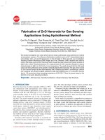
Fabrication of ZnO nanorods decorated Au nanoparticles by hydrothermal and magnetron sputter techniques
Bạn đang xem bản rút gọn của tài liệu. Xem và tải ngay bản đầy đủ của tài liệu tại đây (723.71 KB, 8 trang )
Physics
Fabrication of ZnO nanorods decorated Au nanoparticles
by hydrothermal and magnetron sputter techniques
Mai Thi Ngoc Anh1, Trinh Duc Thien2, Nguyen Thi Minh Hong1, Nguyen Dinh Lam1*
1
Faculty of Engineering Physics and Nanotechnology, VNU University of Engineering and Technology,
Vietnam National University;
2
Faculty of Physics, Hanoi National University of Education.
*
Corresponding author:
Received 02 Aug 2022; Revised 06 Sep 2022; Accepted 07 Nov 2022; Published 18 Nov 2022.
DOI: />
ABSTRACT
In this study, ZnO nanorods decorated with gold (Au) nanoparticles with the desired size
parameters were fabricated using a simple, low-cost, and highly efficient method. The ZnO
nanorod structure was fabricated using a hydrothermal method on a ZnO seed layer with
hydrothermal solution concentrations varying from 20 mM to 90 mM. Au nanoparticles were
coated on the ZnO nanorod structure by magnetron sputtering with a sputtering time from the
40s to 70 s. The characteristics of the fabricated samples were investigated through SEM images
and optical absorption spectroscopy. The results show that the fabricated ZnO nanorods are
relatively uniform, with a cylindrical shape and hexagonal cross-section when the solution
concentration is less than 70 mM. Au nanoparticles were attached to the surface of the ZnO
nanorods with average sizes of 30-50 nm. The optical absorption spectroscopy results showed
that the ZnO nanorods’ absorption edge appeared at a wavelength of approximately 395 nm. In
addition, the exciton absorption peak of Au nanoparticles was between 550 nm and 600 nm and
there was a shift towards shorter wavelengths as the size of the Au nanoparticle decreased. This
result opens up potential applications of this material such as increasing photocatalytic
efficiency and its, use in photonic devices, etc.
Keywords: Au nanoparticles; Hydrothermal; Magnetron sputtering; ZnO nanorods.
1. INTRODUCTION
ZnO is a metal oxide semiconductor that has many applications in life such as sensors
[1-7], photodetectors [8-12], water splitting [13-14], and photocatalytic [15-19]. These
applications of ZnO mainly appear when it exists at the nanometer scale. ZnO on the
nanoscale can be fabricated in many shapes and structures such as nanowires, nanodisks,
nanotubes, and nanoflowers. ZnO nanostructures are fabricated by various methods but
are usually divided into physical and chemical methods. Most physical methods have the
advantages of producing high crystalline and uniform ZnO structures but require
expensive and modern equipment. The chemical methods have the advantages of low
cost, do not require modern equipment, and fast sample manufacturing speed. We choose
hydrothermal as the chemical process in this paper to create ZnO nanostructure.
ZnO materials have significant applications in photocatalytic but the efficiency with
visible light is still low due to the large optical bandgap and fast electron-hole pairs
recombination rate. Using the plasmonic effect of Au nanoparticles attached to ZnO
structures, the photocatalytic effect has been enhanced [20-22]. In this paper, we show
how to make ZnO nanorods embellished with Au nanoparticles, describe them, and then
adjust their size to meet application-specific requirements.
40
M. T. N. Anh, …, N. D. Lam, “Fabrication of ZnO nanorods … magnetron sputter techniques.”
Research
2. FABRICATION PROCESS
2.1. Fabrication of ZnO nanorods
The fabrication process of ZnO nanorods is presented in figure 1. Firstly,
Zn(COOCH3)2 .2H2O and Isopropanol (IPA) were mixed in a magnetic stirring machine
to make a seed solution and then spin-coating on a clean glass substrate to form a ZnO
seed layer. Then, the layer was heated in the air at 500 oC for 1 hour. Besides that,
Zn(NO3)2, C6H12N4, and H2O were mixed in different concentrations to create the ZnO
grow solution. Then, the glass substrate with the ZnO seed layer above was put into the
autoclave with ZnO grow solution. For the purpose of producing ZnO nanorods on a
glass substrate, they were all subjected to the hydrothermal process for two hours at 80
°C. After that, the autoclave was slowly cooled down to room temperature and the
samples were washed with deionized (DI) water and dried.
Figure 1. Schematic of the fabrication process of structured ZnO nanorods
by spin-coating and hydrothermal techniques.
2.2. Fabrication of ZnO nanorods decorate Au nanoparticles
The process of decorating gold nanoparticles on ZnO nanorods was described in
figure 2. A gold nanolayer was deposited onto the ZnO template structures by using the
magnetron sputtering technique. The discharge current was 20 mA and the sputtering
time was changed from 40 s to 70 s. After that, the samples were annealed at 500 oC in
the air for 15 min and naturally cooled down to room temperature. The Au nanolayers
were melted and formed Au nanoparticles well distributed on ZnO nanorods.
Figure 2. Fabrication process of Au nanoparticles decorated ZnO nanorod
by magnetron sputter and thermal annealing.
3. RESULTS AND DISCUSSION
Initially, ZnO nanorods were well-formed on grass substrate. The rods have a
diameter from 0.13 μm to 0.713 μm, a height from 0.63 μm up to 2.093 μm, and are
perpendicular to the base when the seed solution has a concentration of 1M. Varying the
concentration of the solution results in the change in size and density of ZnO nanorods.
Journal of Military Science and Technology, No.83, 11 - 2022
41
Physics
In fact, the diameter and length of the rods decreased to 30 nm and 200 nm, respectively,
when we reduced the concentration of the seed solution to 0.1M. The number of rods
also drastically increased. ZnO nanorod SEM pictures at various seed solution
concentrations are shown in figure 3.
Figure 3. SEM images of ZnO nanorods in different seed solution concentrations.
With a target to create structures at the nanometer scale, we decided to keep the seed
solution concentration of 0.1M and optimize the ZnO structure’s parameters by changing
other reaction conditions. We investigate the influence of hydrothermal solution
concentration on ZnO structures. Figure 4 shows the SEM side-view images of ZnO
nanorods in different hydrothermal solution concentrations. Each sample’s structure is
relatively uniform in bar-shaped, and its size strongly depends on the hydrothermal
solution. When the hydrothermal concentrations increased from 20 mM to 70 mM, the
diameter of the rods increased from 50 nm to 300 nm, and the height of the rods
increased from 200 nm to 900 nm as shown in figure 5.
Figure 4. SEM images of ZnO nanorods side view
in different hydrothermal solution concentrations.
When the hydrothermal solution concentration is 20 mM, the orientation
perpendicular to the substrate surface of the ZnO nanorods is poor, the diameter of the
ZnO nanorods is small and the density of the rods is large. As the hydrothermal solution
concentration increased to 50 mM, the orientation perpendicular to the substrate surface
42
M. T. N. Anh, …, N. D. Lam, “Fabrication of ZnO nanorods … magnetron sputter techniques.”
Research
increased, the diameter and height of the nanorods increased, and the density of the
nanorods decreased. When the hydrothermal solution concentration was 70 mM, the
ZnO nanorods appeared to be voided and filmed due to the large diameter of the rods.
This is more evident when the hydrothermal solution concentration is 90 mM. Therefore,
the ZnO one-dimensional structure begins to break down when the hydrothermal
solution concentration is 70 mM and the best ZnO one-dimensional structure can be
obtained in this study when the hydrothermal solution concentration is 50 mM, the
hydrothermal temperature is 80 oC, hydrothermal time is 2 h, hydrothermal solution
volume is 50 ml.
Figure 5: The change of ZnO nanorods’ height and diameter
with the hydrothermal solution concentration.
We decided to coat a gold nanolayer on the best ZnO nanorods obtained above.
Figure 6 shows the SEM images of ZnO before (a) and after (b-c) coated with a gold
nanolayer by magnetron sputtering technique. The Au nanolayer was covered relatively
uniformly on ZnO nanorods substrate.
Figure 6. SEM images of ZnO before (a) and after (b-c) coated
with a gold nanolayer by magnetron sputtering technique.
After coating, the gold nanolayer was annealed at 500 oC for 15 min to break and
form gold nanoparticles. These particles are evenly distributed on ZnO nanorods as
displayed in figure 7. Au nanoparticle size increases linearly with sputtering time. The
greater the particles, the longer the sputtering time. To concretize, as the magnetron
sputtering time increased from 40 s to 70 s, the average size of the Au nanoparticles
increased from 10 nm to 80 nm.
Journal of Military Science and Technology, No.83, 11 - 2022
43
Physics
Figure 7. SEM images of Au nanoparticles decorated on ZnO nanorods
after annealing, sputtering in the 40 s (a) and 70 s (b).
The influence of gold nanoparticles on the optical properties of the ZnO nanorods
structure was investigated through the optical absorption and photoluminescence
spectrum presented in figures 8, 9, and 10.
Figure 8. Optical absorption spectrum of ZnO nanorods decorated Au nanoparticles
in different sputtering times.
Figure 8 indicated that the absorption edge of ZnO nanorods is 365 nm. The optical
absorbance increased markedly as the Au nanoparticle size increased. The exciton
absorption peak of Au nanoparticles is between 550 nm and 600 nm and there is a shift
towards a short wavelength as the size of Au nanoparticles increases.
In the photoluminescence spectrum of the two samples of ZnO nanorods coated with
Au nanoparticles in figure 9, the sample with Au particles coated in the 40 s higher than
the Au sputtered sample at 70 s different from the optical absorption spectrum in figure
8. The plasmonic effect for smaller particles is higher than for larger particles that agree
with proposed theories about the plasmonic effect from other studies. This also reveals
the ability to enhance the photocatalytic effect of ZnO nanorods decorated Au
nanoparticles compared with ZnO nanorods sample.
44
M. T. N. Anh, …, N. D. Lam, “Fabrication of ZnO nanorods … magnetron sputter techniques.”
Research
Figure 9. Photoluminescence spectrum of Figure 10. Optical absorption spectrum of
ZnO nanorods decorated Au nanoparticles in
ZnO nanorods in different annealing
different sputtering times.
temperatures.
Figure 10 demonstrates the influence of annealing temperature on the optical property
of ZnO samples. There was a significant increase in optical absorbance between the ZnO
nanorods sample and ZnO nanorods coated with a gold nanolayer without annealing due
to the impact of the coating gold film. The ZnO nanorods coated with Au nano-thin film
annealing at different temperature of 400 oC and 500 oC also reveals an absorption peak
of Au nanoparticles in the range from 570 nm to 600 nm, and a blue-shift when the size
of Au nanoparticles decreases. The higher the heating temperature, the more the gold
layer melts, breaks, and shrinks to form gold particles of smaller size, and the more
obvious the plasmonic effect appears. This is consistent with the other conclusions and
results mentioned above.
4. CONCLUSIONS
We have successfully fabricated the ZnO nanorods decorated Au nanoparticles on
glass substrates by hydrothermal and magnetron sputtering methods. The nanorods
reached a hexagonal face, perpendicular to the base, and best size when fabricated with
the hydrothermal solution concentration of 50 mM. Then, the gold nanoparticles were
decorated on top of ZnO nanorods by magnetron sputtering and thermal annealing
techniques. The size of Au nanoparticles depends strongly on sputtering time. Exciton
peaks of Au nanoparticles were shown on the UV-Visible spectrum and there was a
blueshift when the size of Au nanoparticles decreased. The obtained results in this study
are oriented to application in the decomposition of organic compounds of the ZnO onedimensional structure with Au nanoparticles.
Acknowledgment: This work has been supported by the VNU University of Engineering and
Technology under project number CN20.11.
REFERENCES
[1]. Qomaruddin, Olga Casals, Hutomo Suryo Wasisto, Andreas Waag, Joan Daniel Prades,
Cristian Fabrega, "Visible-Light-Driven Room Temperature NO2 Gas Sensor Based on
Localized Surface Plasmon Resonance: The Case of Gold Nanoparticle Decorated Zinc
Oxide Nanorods (ZnO NRs)", Chemosensors; 10 (1): 28, (2022).
Journal of Military Science and Technology, No.83, 11 - 2022
45
Physics
[2]. Sheng-Joue Young, Yu-Long Chen, "Enhancing pH sensors Performance of ZnO nanorods
with Au nanoparticles adsorption", IEEE Sensors Journal; 21 (12): 13068-13073, (2021).
[3]. Hongtao Wang, Yueyue Li, Chenchang Wang, Yuan Li, Jihao Bai, Yueying Liu, Linsheng
Zhou, Fengmin Liu, Kengo Shimanoe, Geyu Lu, "N-pentanol sensor based on ZnO
nanorods functionalized with Au catalysts", Sensors and Actuators B: Chemical; 339:
129888, (2021).
[4]. Sheng-Joue Young, L. T. Lai, "Investigation of a Highly Sensitive Au NanoparticleModified ZnO Nanorod Humidity Sensor", IEEE Transactions on Electron Devices; 68 (2):
775-779, (2021).
[5]. Mingjie Yang, Shendan Zhang, Fengdong Qu, Shu Gong, Chenhao Wang, Lei Qiu, Minghui
Yang, Wenlong Cheng, "High performance acetone sensor based on ZnO nanorods modified
by Au nanoparticles", Journal of Alloys and Compounds; 797: 246-252, (2019).
[6]. Hyeong-Min Kim, Jae-Hyoung Park, Seung-Ki Lee, "Fiber optic sensor based on ZnO
nanowires decorated by Au Nanoparticles for improved plasmonic biosensor", Scientific
Reports; 9: 15605, (2019).
[7]. Wen-Hui Zhou, Hai-Hui Wang, Wei-Tian Li, Xiu-Chun Guo, Dong-Xing Kou, Zheng-Ji
Zhou, Yue-Na Meng, Qing-Wen Tian and Si-Xin Wu, "Gold Nanoparticles Sensitized ZnO
nanorods Arrays for Dopamine Electrochemical Sensing", Journal of The Electrochemical
Society; 165 (12): G3001, (2018).
[8]. Xiaomiao Fei, Dayong Jiang, Nan Wang, Hongping Zhao, Meijiao Xing, and Haoda Li,
"Study on Ultraviolet Photodetector modified by Au nanoparticles on ZnO Nanowires",
Journal of Physics: Conference Series; 1907: 012044, (2021).
[9]. Lalit Goswami, Neha Aggarwal, Manjri Singh, Raiji Verma, Pargam Vashishtha, Shubhendra
K. Jain, Jai Tawale, Rajeshwari Pandey, and Govind Gupta, "GaN Nanotowers Grown on Si
(111) and Functionalized with Au Nanoparticles and ZnO Nanorods for Highly Responsive
UV Photodetectors", ACS Applied Nano Materials; 3 (8): 8104-8116, (2020).
[10]. Rizwan Khan, Periyayya Uthirakumar, Tae Hwan Kim, In-Hwan Lee, "Enhanced
photocurrent performance of partically decorated Au nanoparticles on ZnO nanorods
based UV photodetector", Materials Research Bulletin; 115: 176-181, (2019).
[11]. Cheng-Liang Hsu, Hsin-Yu Wu, Chung-Cheng Fang, Sheng-Po Chang, "Solution-Processed
UV and Visible Photodetectors Based on Y-Doped ZnO Nanowires with TiO2 Nanosheets and
Au Nanoparticles", ACS Applied Energy Materials; 1 (5): 2087-2095, (2018).
[12]. Iwantono Iwantono, Siti Khatijah Md Saad, Fera Anggelina, Awitdrus Awitdrus, Muhamad
Adam Ramli, Akrajas Ali Umar, "Enhanced charge transfer activity in Au nanoparticles
decorated ZnO nanorods photoanode", Physica E: Low-dimensional Systems and
Nanostructures; 111: 44-50, (2019).
[13]. Juyoung Yu, Jongsung Kim, "Preparation of uniform gold nanoparticles of different
quantity deposited on zinc oxide nanorods for photoelectrochemical water splitting",
Chemosphere; 287 (3): 132168, (2022).
[14]. Junhao Cai, Jianrui Cao, Heng Tao, Ruoping Li, Mingju Huang, "Three-dimensional
ZnO@TiO2 core-shell nanostructures decorated with plasmonic Au nanoparticles for
promoting photoelectrochemical water splitting", International Journal of Hydrogen
Energy; 46 (73): 36201-36209, (2021).
[15]. Bharat Baruah, Christopher Kelley, Grace B. Djokoto & Kelly M. Hartnett, "Polymercapped gold nanoparticles and ZnO nanorods form binary photocatalyst on cotton fabrics:
Catalytic breakdown of dye", Frontiers of Materials Science; 15: 431-447, (2021).
46
M. T. N. Anh, …, N. D. Lam, “Fabrication of ZnO nanorods … magnetron sputter techniques.”
Research
[16]. Menekse Sakir, Samaa Salem, Senem Tapan Sanduvac, Ertugrul Sahmetlioglu, Gokhan
Sarp, M. Serdar Onses, Erkan Yilmaz, "Photocatalytic green fabrication of Au
nanoparticles on ZnO nanorods modified membrane as flexible and photocatalytic active
reusable SERS substrates", Colloids and Surfaces A: Physicochemical and Engineering
Aspects; 585: 124088, (2020).
[17]. Quan Truong Hoang, Vasanthan Ravichandran, Thuy Giang Nguyen Cao, Ji Hee Kang,
Young Tag Ko, Tae Il Lee, Min Suk Shim, "Piezoelectric Au-decorated ZnO nanorods:
Ultrasound-triggered generation of ROS for piezocatalytic cancer therapy", Chemical
Engineering Journal; 435 (3): 135039, (2022).
[18]. Y.Zhang, S. Wang, Y. Zhao, Y. Ding, Z. Zhang, T. Jiang, Z. L. Wang, L. Li, "Piezophototronic effect boosted catalysis in plasmonic bimetallic ZnO heterostructure with
guided fermi level alignment", Materials Today Nano; 18: 100177, (2022).
[19]. Shuzhen Dou, Juan Du, Qunyan Zhu, Zongshun Wang, Yalei Wang, Qiye Chen, Nan Lu,
"Au nanoparticles/ ZnO nanorods as SALDI-MS substrate for on-plate enrichment and
detection of glutathione in real samples", Sensors and Actuators B: Chemical; 335: 129709,
(2022).
[20]. Haolin Li, Jianwei Ding, Shuangfei Cai, Wei Zhang, Xining Zhang, Ting Wu, Chen Wang,
Morten Foss, Rong Yang, "Plasmon-enhanced photocatalytic properties of Au/ZnO
nanowires", Applied Surface Science; 583: 152539, (2022).
[21]. Mohit Tiwari, Aditya Singh, Samit Dureja, Suddhasatwa Basu, Sudip K. Pattanayek, “Au
nanoparticles decorated ZnO/ZnFe2O4 composite SERS-active substrate for melanine
detection”, Talanta; 236: 122819, (2022).
[22]. Quoc Khoa Doan, Manh Hong Nguyen, Cong Doanh Sai, Van Thanh Pham, Hong Hanh
Mai, Nguyen Hai Pham, Thanh Cong Bach, Viet Tuyen Nguyen, Trong Tam Nguyen, Khac
Hieu Ho, Thi Ha Tran, “Enhanced optical properties of Zno nanorods decorated with gold
nanoparticles for self cleaning surface enhanced Raman applications”, Applied Surface
Science; 505: 144593, (2020).
TÓM TẮT
Chế tạo cấu trúc một chiều ZnO có đính hạt nano Au
bằng phương pháp thủy nhiệt và phún xạ magnetron
Trong báo cáo này, chúng tơi trình bày phương pháp đơn giản, giá thành thấp
và hiệu quả cao sử dụng để chế tạo vật liệu ZnO nanorods (NRs) có đính hạt nano
vàng (Au) với các thơng số kích thước như mong muốn. Với phương pháp này, cấu
trúc một chiều ZnO được nuôi bằng phương pháp thủy nhiệt trên lớp mầm ZnO,
sau đó được phủ Au bằng phương pháp phún xạ magnetron. Tính chất của mẫu
được khảo sát thông qua các phép đo SEM và UV-Vis. Kết quả cho thấy các thanh
nano ZnO được tạo ra tương đối đồng đều, có tiết diện lục giác và được đính các
hạt nano Au trên bề mặt. Trong điều kiện thủy nhiệt ở 800 oC và nồng độ dung
dịch là 50 mM, các thanh ZnO nanorods được tạo ra với chất lượng tốt nhất có
chiều cao 800 nm và đường kính 100 nm. Các hạt nano Au có kích thước trung
bình trong khoảng từ 30 nm đến 50 nm với thời gian phún xạ là 40 s và 70 s. Kết
quả này mở ra các ứng dụng tiềm năng của vật liệu này như làm tăng hiệu suất
quang xúc tác, sử dụng trong các thiết bị quang – quang tử,…
Từ khoá: ZnO nanorods; Au nanoparticles; Hydrothermal.
Journal of Military Science and Technology, No.83, 11 - 2022
47