loss of vinculin and membrane bound catenin promotes metastasis and predicts poor prognosis in colorectal cancer
Bạn đang xem bản rút gọn của tài liệu. Xem và tải ngay bản đầy đủ của tài liệu tại đây (6.77 MB, 15 trang )
Li et al. Molecular Cancer 2014, 13:263
/>
RESEARCH
Open Access
Loss of vinculin and membrane-bound β-catenin
promotes metastasis and predicts poor prognosis
in colorectal cancer
Ting Li1†, Hanqing Guo2†, Ying Song2†, Xiaodi Zhao1, Yongquan Shi1, Yuanyuan Lu1, Sijun Hu1, Yongzhan Nie1,
Daiming Fan1 and Kaichun Wu1*
Abstract
Background: Loss of cell-cell adhesion is important for the development of cancer invasion and metastasis.
Vinculin, a key adhesion-related protein, can affect metastasis and prognosis in several tumours. Here, we
determined the biological roles of vinculin in the metastasis of colorectal cancer (CRC) and evaluated its clinical
significance as a potential disease biomarker.
Methods: The expression level of vinculin in CRC cell lines and tissues was measured using Real-Time PCR and
western blotting. Moreover, vinculin function was analysed using Transwell assays and in vivo metastasis assays
in gain- and loss-of-function experiments. Furthermore, the impact of vinculin together with membrane-bound
β-catenin on the prognosis of 228 CRC patients was investigated by immunohistochemistry. Additionally, the
expression of epithelial-mesenchymal transition (EMT) indicators was verified by immunohistochemistry in CRC
tissues obtained from these patients.
Result: Vinculin expression was found to be significantly downregulated in highly metastatic CRC cell lines and
metastatic tissues. Both in vitro and in vivo experiments showed that vinculin suppressed invasion, migration
and metastasis in CRC cells and that this suppression could be attenuated by silencing β-catenin. Moreover, the
expression of vinculin and membrane-bound β-catenin were positively correlated in CRC tissues, and lack of
vinculin expression emerged as an independent prognostic factor in patients with CRC. Finally, the loss of
vinculin and membrane-bound β-catenin was associated with node metastasis, organ metastasis and expression
of EMT indicators.
Conclusion: Our results suggest that vinculin may play specific roles in the EMT and metastasis of CRC and that
loss of vinculin could be used as a prognostic factor for CRC.
Keywords: Vinculin, β-catenin, Colorectal cancer, Metastasis, Prognosis, EMT
Background
Colorectal cancer (CRC) is the third most commonly diagnosed cancer in males and the second most commonly diagnosed cancer in females [1]. The CRC death rates have
been decreasing in several Western countries [2], largely
resulting from improved treatment, increased awareness
and early detection [3]. However, an estimated 608,700
* Correspondence:
†
Equal contributors
1
Department of Gastroenterology & State Key Laboratory of Cancer Biology,
Xijing Hospital, The Fourth Military Medical University, Xi’an 710032, China
Full list of author information is available at the end of the article
deaths have still occurred, making CRC the fourth leading
cause of cancer deaths in males and the third leading
cause of cancer deaths in females [1]. The poor prognosis
of CRC is associated with tumour invasion and metastasis,
which often leads to therapeutic failure. Recently, it has
been reported that some loss of cell-cell adhesion may be
important for the development of CRC invasion and
metastasis [4,5].
Vinculin is a ubiquitously expressed, actin-binding protein
that localises to the cytoplasmic face of integrin-mediated
cell-extracellular matrix junctions (focal adhesions) and
cadherin-mediated cell-cell junctions [6]. Normally, vinculin
© 2014 Li et al.; licensee BioMed Central. This is an Open Access article distributed under the terms of the Creative Commons
Attribution License ( which permits unrestricted use, distribution, and
reproduction in any medium, provided the original work is properly credited. The Creative Commons Public Domain
Dedication waiver ( applies to the data made available in this article,
unless otherwise stated.
Li et al. Molecular Cancer 2014, 13:263
/>
plays a key role in focal adhesion formation [7], cell proliferation [8] and regulation of the actin cytoskeleton
[9,10]. Loss of vinculin has been found in the development of many cancers, such as squamous carcinoma
[11,12], rhabdomyosarcoma [13] and breast cancer [14],
implying that vinculin may have anti-tumour effects.
Recent studies confirmed this finding, as vinculin inhibited multiple processes associated with malignant tumours, including invasion, metastasis and apoptosis
[8,15]. Specifically, overexpression of vinculin caused
reduced cell migration, whereas knockdown of vinculin
enhanced cell motility [16-18]. Researchers also found
that vinculin-null cells had upregulated activity of extracellular signal-regulated kinase (ERK), leading to enhanced survival and motility, which are important for
metastasis [8]. In addition, low levels of vinculin may
predict poor survival in squamous cell cancer [12], but
the biological role of vinculin and its prognostic value
in CRC have not been fully investigated.
Vinculin activation is a key event in its coordination
with focal adhesion formation, which is important for
the suppression of cell mobility. This activation requires
various binding partners, such as talin [19], α-actinin
[20], α-catenin [21], β-catenin [22] and paxillin [8], to
unmask the binding sites of vinculin and continue its
localisation to focal adhesions. Because it is an important cell-cell adhesion protein, β-catenin can bind to vinculin to stabilise E-cadherin (E-cad) at the cell surface
[22,23]. This important stabilising function requires the
binding of vinculin to the N-terminus of β-catenin [23],
which can bind a number of proteins that regulate the
transition of cancer cells from an epithelial to a more
mesenchymal phenotype [24,25]. Recently, the epithelialmesenchymal transition (EMT), a critical process in tumour
invasion and metastasis, was found to be associated
with the translocation of β-catenin from the membrane
to the nucleus [26]. Upregulated levels of β-catenin in
the nuclei of CRC cells were found to induce the activity
of the transcription factor ZEB1, leading to EMT and a
more invasive phenotype [27]. The EMT process is
characterised by decreased levels of epithelial cell-cell
adhesion molecules, such as E-cad [28], and increased
levels of mesenchymal cell-cell adhesion molecules, such
as vimentin (VIM) [29]. In addition, reduced membranebound β-catenin expression and increased cytoplasmic
E-cad expression predict poor survival in gastric cancer
[30]. Decreased levels of β-catenin and E-cad on the cell
membrane were also observed in CRC in a recent study
[31]. Based on these findings and our results that reveal
the diminished levels of vinculin in CRC, we hypothesised that the loss of vinculin and β-catenin at the cell
surface could be advantageous for the development of
EMT and metastasis and may predict poor survival in
CRC patients.
Page 2 of 15
In this study, we investigated the biological function of
vinculin and its prognostic value in CRC. We identified
significant downregulation of vinculin in metastatic CRC
cells and tissues. Furthermore, restoration of vinculin suppresses CRC metastasis in vitro and in vivo, whereas loss
of vinculin promotes CRC invasion and migration. In
addition, we found that vinculin may regulate CRC invasion and migration at least partially through β-catenin.
We further verified the positive correlation between the
expression of vinculin and membrane-bound β-catenin
and their correlation with an EMT indicator. More importantly, our data provide novel evidence that vinculin
and membrane-bound β-catenin expression can serve as
predictive biomarkers of poor prognosis in CRC patients.
Results
Vinculin expression is downregulated in CRC cell lines
and inversely correlated with CRC metastasis
To examine the significance of vinculin in CRC carcinogenesis, we measured the expression of vinculin in five
human CRC cell lines (HCT116, Caco2, HT29, SW620
and SW480) and in HIEC, an immortalised colon epithelial cell line. Western blotting showed that vinculin expression was significantly decreased in all five CRC cell
lines compared with HIEC (Figure 1A). Interestingly,
compared with SW480, vinculin expression was significantly decreased in SW620, a cell line established from
the lymph node metastasis of the same patient as
SW480 [32]. qRT-PCR also showed that mRNA expression of vinculin was relatively lost in various CRC cell
lines (Figure 1B). Furthermore, tissues from lymph node
metastases expressed lower levels of vinculin compared
with primary CRC tissues and the adjacent normal tissues, indicating the inverse relationship between the expression of vinculin and the metastatic status of CRC
tissues (Figure 1C, D). Taken together, these results suggest that downregulation of vinculin is correlated with
increased CRC metastasis and that vinculin might inhibit
CRC progression.
Vinculin suppresses CRC cell invasion and metastasis
in vitro and in vivo
To investigate whether vinculin regulates CRC cell invasion
and migration, we performed in vitro gain-of-function analyses by overexpressing vinculin with a lentiviral vector encoding vinculin in SW620 cells. Conversely, SW480 cells
were transfected with lentiviral vectors encoding vinculin
siRNA or control siRNA. After cell transfection and antibiotic screening for 6 weeks, extracts from SW480 and
SW620 cells transfected with the vinculin vector, siRNA or
control vector were submitted to western blotting and
compared (Figure 2A, B). Transwell assays showed that ectopic expression of vinculin significantly suppressed the invasion and migration of SW620 cells (Figure 2C). In
Li et al. Molecular Cancer 2014, 13:263
/>
Page 3 of 15
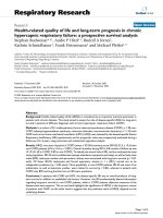
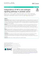
Figure 1 Expression of vinculin in CRC cell lines and tissue samples. (A) The expression level of vinculin in five CRC cell lines and HIEC cells
was measured by western blotting and quantified using Quantity One v4.6.2 software (one of three similar blots is shown). β-actin was used as
a loading control. (B) The mRNA level of vinculin in five CRC cell lines and HIEC cells was measured by qRT–PCR using GAPDH as an internal
control. (C) The expression of vinculin in adjacent non-cancerous colon tissues (N), primary CRC tissues (C) and lymph node metastatic tissues
(M) from 6 patients was examined by western blotting. (D) The mRNA level of vinculin in paired tissues was measured using qRT-PCR. Vinculin
expression was determined in tumour tissue relative to the patient’s adjacent normal tissue, and the relative expression of vinculin in the CRC cell
lines was normalised to that in HIEC cells. Each sample was analysed in triplicate (*P < 0.05, **P < 0.01).
contrast, the migration and invasion of SW480 cells sharply
increased when endogenous vinculin was silenced by
siRNA (Figure 2D). These results suggest that vinculin suppresses CRC cell invasion and migration in vitro.
To further validate whether vinculin could regulate the
metastatic phenotype of CRC in vivo, we injected SW620vinculin cells, which stably express vinculin, into nude mice
through the lateral tail vein. Liver and lung metastasis of
CRC was apparent in mice injected with SW620-vinculincontrol cells, while few metastatic tumours were detected
in mice injected with SW620-vinculin cells (Figure 2E). In
contrast, inhibition of vinculin in SW480 cells increased
the rate of metastasis to liver and lung (Figure 2F). Taken
together, these results indicate that vinculin has a suppressor role in CRC metastasis.
Vinculin regulates CRC metastasis through β-catenin
To understand the underlying molecular mechanism by
which vinculin suppresses CRC invasion and metastasis,
we further investigated whether β-catenin, an important
Li et al. Molecular Cancer 2014, 13:263
/>
Page 4 of 15
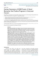
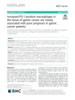
Figure 2 Vinculin suppresses CRC cell invasion and metastasis in vitro and in vivo. (A) Western blot showing vinculin expression in SW620
cells infected with lentiviral vector encoding vinculin and in SW480 cells infected with vinculin siRNA (B). β-actin was used as an internal control.
(C) Transwell migration and invasion assays using SW620 cells stably expressing vinculin or control vector. Representative images are shown on
the left, and the quantification of 10 randomly selected fields is shown on the right. (D) Transwell migration and invasion assays using SW480
cells stably infected with vinculin siRNA or control siRNA. (E) Representative H&E staining of livers and lungs isolated from mice that received
injections of SW620-control-vector or SW620-vinculin-vector cells. Black arrows indicate metastatic intrahepatic or lung tumours. The incidences
of liver and lung metastasis in 10 mice are presented on the right. (F) Representative H&E staining of livers and lungs isolated from mice that
received injections of SW480-control-siRNA or SW480-vinculin-siRNA cells.
Li et al. Molecular Cancer 2014, 13:263
/>
binding partner of vinculin and a key activator of cancer
malignant phenotypes, was involved in this process. Immunofluorescence assays showed that β-catenin was
primarily located in the plasma membrane in SW480
cells; however, following vinculin siRNA infection,
β-catenin showed less localisation in the nucleus and instead localised in the nucleus and cytoplasm (Figure 3A).
On the other hand, the expression of membrane-bound
β-catenin in SW620 cells significantly increased after
vinculin restoration, while nuclear β-catenin was almost
absent (Figure 3B).
To further verify whether β-catenin accounted for the
change in CRC invasion and metastasis induced by vinculin, we transfected β-catenin siRNA into SW480 cells previously transfected with the vinculin siRNA vector. Transwell
assays showed that the β-catenin siRNA significantly reduced vinculin-siRNA-induced CRC cell invasion and migration (Figure 3C). By contrast, restoration of β-catenin
significantly abrogated the inhibition of invasion and migration that was induced by vinculin overexpression in SW620
cells (Figure 3D). Collectively, these results suggest that vinculin may regulate CRC invasion and migration at least
partially through β-catenin.
Vinculin positively correlates with membrane-bound
β-catenin in CRC
To further investigate the expression levels and possible
associations between vinculin and β-catenin in CRC, we
measured the expression of vinculin and β-catenin in primary CRC tissue arrays containing 228 cases. Decreased
levels of vinculin and β-catenin on the cell membrane
were observed in CRC tissues compared to adjacent
normal tissues, and immunohistochemistry showed low
expression of vinculin in 145 of 228 CRC tissues
(63.6%), as opposed to 59 of 228 adjacent non-cancerous
tissues (25.9%) (Figure 4A, B, Additional file 1: Table S1).
Moreover, diminished expression of membrane-bound
β-catenin was also detected in 138 of 228 CRC tissues
(60.5%), whereas lack of membrane-bound β-catenin was
found in only 36 of 228 adjacent non-cancerous tissues
(15.8%) (Figure 4A, C, Additional file 1: Table S1). Furthermore, membrane-bound β-catenin was correlated
with high vinculin expression (Figure 4D, Additional file 2:
Table S2). Taken together, these results indicated that a
low level of vinculin was significantly correlated with the
absence of membrane-bound β-catenin.
Low vinculin expression and lack of membrane-bound
β-catenin are associated with tumour malignancy in CRC
The correlations between vinculin or β-catenin expression and various clinicopathological features of CRC are
summarised in Table 1. There was a statistically significant correlation between differentiation and vinculin
expression (P = 0.0021) or β-catenin expression (P = 0.0163).
Page 5 of 15
More importantly, the loss of vinculin was associated with
lymph node metastasis and organ metastasis (P = 0.0273,
P = 0.01078). Node or organ metastasis was also related
to the absence of membrane-bound β-catenin expression
(P = 0.0027, P = 0.0159), further supporting the relationship
between decreased vinculin and the absence of membranebound β-catenin in CRC tissues. Interestingly, we found that
decreased vinculin expression was significantly associated
with vascular invasion (P = 0.0371). Tumour depth was also
found to be associated with the absence of membranebound β-catenin expression (P = 0.0121). There were no significant differences in these molecules with regard to patient
gender, age, tumour stage or location.
Low vinculin expression is an independent prognostic factor
We evaluated the three-year survival rates using the
Kaplan-Meier method. Our results showed that vinculin
loss was confirmed to be an independent prognosticator for
low survival of CRC patients (Figure 5A, P = 0.001). Because our results indicated that vinculin and β-catenin are
co-expressed in CRC, we set out to detect whether the impact of vinculin on the prognosis of CRC patients was affected by β-catenin expression. Thus, the patients were
divided into four groups according to their expression patterns of vinculin and β-catenin: vinculin(High, H)/β-catenin
(Membrane-bound, M), vinculin(Low, L)/β-catenin(NonMembrane-bound, NM), vinculin (H)/β-catenin(NM) and
vinculin(L)/β-catenin(M). Survival analysis showed that
patients with vinculin(L)/β-catenin(NM) expression endured the lowest overall survival (Figure 5B, P < 0.001). Furthermore, in patients with low and high expression of
vinculin, β-catenin(NM) patients showed a decreased survival time compared to β-catenin(M) patients (Figure 5C,
P < 0.001; Figure 5D, P = 0.042). However, low vinculin was
found to result in low survival rates when membranebound β-catenin was absent (Figure 5E, P = 0.037), but not
when membrane-bound β-catenin was detected (Figure 5F,
P = 0.506).
A univariate analysis according to the Cox proportional
hazard regression model further confirmed these results
(Table 2). Low expression of vinculin in the primary
tumour was associated with an increased risk of death (HR:
1.805; 95% confidence interval [CI]: 1.262-2.582). Similarly,
lack of membrane β-catenin was also associated with higher
risk (HR: 2.420; 95% CI: 1.685-3.475). As expected, larger
tumour size, vascular invasion, node metastasis and organ
metastasis at the time of primary surgery were also associated with poorer survival. Taken together, these results suggest that low vinculin expression is an independent
prognostic factor with poor prognosis in colon cancer.
Vinculin inhibits EMT in CRC
Because extensive evidence suggests that translocation
of β-catenin from the cell membrane to the nucleus can
Li et al. Molecular Cancer 2014, 13:263
/>
Page 6 of 15
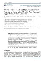
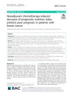
Figure 3 Vinculin regulates CRC metastasis via β-catenin. (A) Immunofluorescence analysis of β-catenin (green) in SW480 cells infected with
vinculin or control siRNA. Merged images represent overlays of β-catenin (green) and nuclear staining by DAPI (red). (B) Immunofluorescence
analysis of β-catenin (green) in SW620 cells infected with the vinculin or control vector. (C) Transwell migration and invasion assays for SW480
cells infected with control or vinculin siRNA along with lentiviral vector expressing β-catenin siRNA. Representative images are shown on the
left, and the quantification of 10 randomly selected fields is shown on the right. (D) Transwell migration and invasion assays using SW620 cells
infected with control or vinculin vectors along with lentiviral vector expressing β-catenin.
Li et al. Molecular Cancer 2014, 13:263
/>
Page 7 of 15
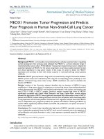
Figure 4 Vinculin positively correlates with membrane-bound β-catenin in CRC. Tissue microarrays of consecutive immunostained sections
were performed using specific antibodies against vinculin and β-catenin. (A) Representative images of vinculin and β-catenin protein expression
levels in normal and cancer specimens. (B) The expression level of vinculin was significantly lower in CRC tissue species than in normal colorectal
tissue species (P < 0.0001, χ2 test). The same result was observed for membrane-bound β-catenin (P < 0.0001, χ2 test) (C). (D) Membrane-bound
β-catenin was positively correlated with vinculin expression in CRC tissues (P < 0.0001, χ2 test).
initiate the EMT process [26], we speculated that loss of
vinculin might impact EMT in CRC. To investigate this
hypothesis, we detected the expression of the epithelial
differentiation marker E-cad and the mesenchymal marker
VIM in CRC cells and tissues. Western blots showed that
membrane-bound β-catenin expression as well as E-cad
dramatically increased in SW620 cells infected with vinculin vectors, whereas the expression of nuclear β-catenin
and VIM were inhibited. In contrast, silencing vinculin
in SW480 cells increased the subcellular expression of
β-catenin, suppressed the expression of E-cad and upregulated the expression of VIM (Figure 6A, B).
To evaluate whether those results could be translated
to the clinical setting, immunohistochemistry on tissue
arrays was further conducted. The results showed that
positive, strong expression of E-cad was detected in vinculin(H)/β-catenin(M) CRC tissues, but not in vinculin
(L)/β-catenin(NM) tissues, whereas VIM expression was
present in the latter group of CRC tissues, but not in
the former (Figure 6C, D). Moreover, the results showed
that the absence of both vinculin and membrane-bound
β-catenin were correlated with decreased E-cad and increased VIM (Figure 6 E-H, Additional file 3: Table S3),
indicating that loss of vinculin and membrane-bound
β-catenin may benefit the process of EMT in CRC.
Discussion
Metastasis is one of the most distinguished phenotypes
of a malignant tumour, and it results in extremely poor
prognosis and relatively high recurrence. The phenotypic
Li et al. Molecular Cancer 2014, 13:263
/>
Page 8 of 15
Table 1 Correlations of Clinico-pathological variables with vinculin and β-catenin expression
Vinculin
P value
Membrane-bound β-catenin
Low
High
Absent
Present
145
83
138
90
Male
76
49
71
54
Female
69
34
67
36
<60
55
37
>60
90
46
Well
59
51
Morderate
62
Poor
24
T1-T2
47
32
T3-T4
98
51
I - II
82
51
III - IV
63
32
Signoid
38
24
Colon /Rectum
107
59
Negative
77
56
Positive
68
27
Negative
72
54
Positive
73
29
Negative
115
77
Positive
30
6
Total cases
P value
Gender
0.4068
0.2048
Age
0.3304
61
31
77
59
56
54
17
55
24
15
27
12
39
40
99
50
76
57
62
33
40
22
98
68
0.1677
Differentiation
0.0021*
0.0163*
Depth of Tumor
0.3866
0.0121*
Tumor stage
0.4885
0.2716
Location
0.7572
0.5428
Vascular invasion
0.0371*
85
48
53
42
65
61
73
29
110
89
28
8
0.2203
Node Metastasis
0.0273*
0.0027*
Organ metastasis
0.0078*
0.0159*
Analysis by chi-square criterion or Fisher’s exact test.
*P<0.05.
changes of reduced cell adhesion and increased cell motility drive tumour metastasis [33]. Recently, accumulating studies have suggested that the loss of vinculin, a
protein contributing to cell-cell adhesion, is observed in
the development of various cancers and may lead to
metastatic changes. In this study, we investigated the
biological role and prognostic value of vinculin in CRC
metastasis.
Vinculin is a 117-kDa actin-binding protein consisting
of 1066 amino acids and is localised on the cytoplasmic
face of integrin-mediated cell-extracellular matrix junctions (focal adhesions) and cadherin-mediated cell-cell
junctions. Early studies have found that fibroblasts isolated from vinculin-deficient mice showed decreased
adhesion strength and faster migration than control cells
[34], and this result was also verified in vinculin-null carcinoma cells [16]. Recent studies found that poor vinculin labelling in tumours of squamous-epithelial origin
appeared to be related to the metastatic potential of the
tumour [12]. Overexpression of vinculin in cancer cells
was found to suppress their tumourigenic ability and
metaplastic capacity [17]. These results, which revealed
the cell-adhesion reduction and motility increase associated with vinculin loss, indicate the potential involvement of vinculin in tumour metastasis. However, few
studies have been performed to determine the biological
role of vinculin in CRC metastasis. Thus, we were interested as to whether CRC metastasis, a multistep process
Li et al. Molecular Cancer 2014, 13:263
/>
Page 9 of 15
Figure 5 Survival curves of CRC patients according to vinculin and β-catenin expression. (A) Patients with low vinculin expression showed
a significantly poor prognosis than those with high vinculin expression. (B) Subgroup analysis of CRC patients with low or high expression of
vinculin accompanied with the absence or presence of membrane-bound β-catenin. (C) The correlation of membrane-bound β-catenin expression
with overall survival among patients with tumours expressing low or high levels of vinculin (D). (E) The correlation of vinculin expression with overall
survival among patients with tumours with or without membrane-bound β-catenin (F).
that normally begins with the loss of cell-cell adhesion
leading to the detachment of cancer cells and invasion
of the basement membrane, is related to the loss of vinculin. In this study, the expression of vinculin in several
CRC cell lines was determined using qRT–PCR and
western blotting. Vinculin expression was significantly
decreased in all five CRC cell lines and was lower in
SW620 cells compared with the immortal colon epithelial cell line HIEC. Furthermore, the results obtained
from clinical CRC tissue also confirmed that vinculin
was downregulated in advanced stages of CRC, indicating its possible involvement in both oncogenic transformation and tumour metastasis. We subsequently
found that vinculin significantly suppressed CRC cell invasion and metastasis both in vitro and in vivo. Some
studies reported that vinculin-knockout cells were 3-fold
less invasive in three-dimensional collagen matrices
simulating extracellular matrix (ECM) because the
connection between the ECM and the actomyosin cytoskeleton through integrin-type cell-matrix adhesion receptors is facilitated by vinculin [35,36]. These studies
focused on tractions exerted by these cells to the ECM
and cell-ECM adhesions. In our study, however, we explored the effects of vinculin on tumor metastasis, which
often develops through several essential steps including
not only invading through surrounding ECM, but also
Li et al. Molecular Cancer 2014, 13:263
/>
Page 10 of 15
Table 2 Univariate and multivariate analysis for overall survival (Cox proportional hazards regression model)
Factors
Gender (male/female)
Multivariate analysis1
Univariate analysis
HR
95%CI
P
HR
95%CI
P
1.148
0.825-1.596
0.412
-
-
-
Differentiation (well/ moderately, poorly)
1.340
0.966-1.860
0.080
-
-
-
Vascular invasion (absent/present)
1.843
1.327-2.561
0.0002*
1.410
0.996-1.997
0.0527
Depth of Tumor (T1-T2/T3-T4)
1.185
0.850-1.453
0.317
-
-
-
Node Metastasis (absent/present)
2.739
1.963-3.820
0.0015*
1.631
1.139-2.315
0.0062*
Organ metastasis (absent/present)
4.509
2.915-6.975
<0.0001*
5.401
3.379-8.634
<0.0001*
Vinculin expression (high/low)
1.805
1.262-2.582
0.0012*
2.048
1.401-2.995
0.0002*
membrane-bound β-catenin expression (absent/present)
2.420
1.685-3.475
<0.0001*
2.024
1.402-2.923
0.0019*
1
Multivariate analysis performed only for variables significant in the univariate analysis.
*P < 0.05.
loosening of cell-cell adhesions and other steps. Thus it
still requires further investigation to elaborate the specific functions of vinculin on cell-cell adhesion and cellECM adhesion through which tumor invasion and metastasis takes place.
Vinculin exists in two conformations. In the inactive
conformation, the extensive interactions between the
head and tail domains prevent detectable binding to
most of its ligands [19]. In the active conformation, displacement of the head-tail interactions leads to a significant accumulation of ternary complexes [37]. Vinculin
activation requires several binding partners, such as talin
[19], α-actinin [20], α-catenin [21], β-catenin [22] or
paxillin [8], to fully unmask its binding sites and continue its localisation to focal adhesions. Several studies
have indicated the close interaction between vinculin
and β-catenin. Vinculin plays a critical role in maintaining the beta-catenin-MAGI-2 interaction in epithelial
cells [38]. Moreover, β-catenin was found to be required
to recruit vinculin to the cell cortex and to strengthen
the junction’s association with the underlying cytoskeleton in response to tension [39]. Furthermore, researchers found that the interaction between vinculin
and β-catenin is crucial for the stabilisation of E-cad
at the cell surface [23], which is considered to be associated with inhibition of tumour metastasis [40]. In
this study, we found that vinculin modulates CRC metastasis through β-catenin. We confirmed that vinculin
and membrane-bound β-catenin are co-expressed in normal tissues and that their expression is partially decreased
or lost in CRC tissues. Moreover, our results indicated that
vinculin restoration accumulates β-catenin on the cell
membrane and that silencing vinculin benefits the translocation of β-catenin. Finally, vinculin-induced cell migration and invasion were reversed by β-catenin. Taken
together, these results establish a functional connection
between vinculin and β-catenin and confirm that vinculin
functions as an anti-metastatic protein in CRC cells by affecting the subcellular location of β-catenin.
Vinculin expression has been associated with squamous
cell tumours, and vinculin loss might predict high metaplastic ability and poor prognosis [12]. A recent study also
revealed that the expression of cytoskeletal proteins, including vinculin, talin and tensin, was downregulated and
correlated with carcinogenesis, invasion and metastasis of
CRC, irrespective of its relative limited-case capacity [15].
As an important binding partner and a specific activator
of vinculin, β-catenin has been found to be associated with
CRC survival in several studies [31,41-43]. The decreased
localisation of β-catenin on the cell membrane combined
with its increased expression in the cytoplasm and nucleus
may be involved in abnormal E-cad expression. This expression pattern also coincides with the poor clinical prognosis of patients with CRC [31]. Our results indicated that
vinculin was associated with membrane-bound β-catenin
in CRC. Through analysing the relationship between
vinculin or β-catenin and various clinicopathological
features, we found that decreased expression of vinculin
and β-catenin in the cell membrane is correlated with
poor differentiation, extensive tumour invasion and a
high incidence of metastasis. Moreover, our analysis of
survival of the four groups of CRC patients further revealed that the loss of vinculin together with decreased
membrane-bound β-catenin predicted the lowest level
of survival. Most importantly, we found that lack of vinculin expression was independently associated with
poor prognosis in colon cancer.
Recently, the concept of the epithelial–mesenchymal
transition (EMT), which was first reported in early embryonic morphogenesis [44], has been extended to cancer
progression and metastasis [45]. During EMT, non-motile,
polarised epithelial cells embedded via cell-cell junctions
in a cell collective dissolve their cell-cell junctions
and convert into individual, non-polarised, motile and invasive mesenchymal cells [46]. Typically, cancer cells
experiencing this process show decreased levels of epithelial cell-cell adhesion molecules such as E-cad [28] and
increased levels of mesenchymal cell-cell adhesion molecules
Li et al. Molecular Cancer 2014, 13:263
/>
Figure 6 (See legend on next page.)
Page 11 of 15
Li et al. Molecular Cancer 2014, 13:263
/>
Page 12 of 15
(See figure on previous page.)
Figure 6 Vinculin regulates CRC metastasis through β-catenin. (A) Western blot analysis of vinculin, membrane-bound β-catenin and
nuclear β-catenin in SW620 cells infected with vinculin or control vector (left) and SW480 cells transfected with vinculin or control siRNA (right).
(B) Western blot analysis of E-cadherin and vimentin in SW620 cells infected with vinculin or control vector (left) and of SW480 cells transfected
with vinculin or control siRNA (right). (C) CRC tissue with high expression of vinculin, membrane-bound β-catenin, positive staining of E-cadherin
and negative staining of vimentin. (D) CRC tissue with low vinculin expression, the absence of membrane-bound β-catenin, negative staining
of E-cadherin and positive staining of vimentin. (E) E-cadherin expression was positively correlated with vinculin expression as well as (F) with
membrane-bound β-catenin in CRC tissues (P < 0.0001, χ2 test; P < 0.0001, χ2 test). (G) Vimentin expression was negatively correlated with vinculin
expression as well as (H) with membrane-bound β-catenin in CRC tissues (P = 0.0274, χ2 test; P = 0.0153, χ2 test).
such as VIM [29]. Several studies indicated that vinculin
strengthens the mechanical links between adhesion
complexes (containing E-cad, β-catenin and α-catenin)
and the actin cytoskeleton. It has been reported that
vinculin plays a role in the establishment or regulation
of the E-cad-based cell adhesion complex in breast cancer
cells by directly interacting with β-catenin [22]. Recent
studies also suggest that vinculin is part of a protein complex on the cytoplasmic face of E-cad, which includes
β-catenin and its binding partners MAGI-2 and α-catenin
[38]. Researchers further confirmed that this complex is
required to stabilise a famous tumour suppressor PTEN
[39]. Taken together, these results suggest that vinculin,
through interactions with β-catenin on the cell membrane,
may act as an anti-metastatic factor in carcinogenesis.
Based on these findings and our results that suggest a
positive correlation between vinculin and β-catenin, we
hypothesised that the loss of vinculin and β-catenin at the
cell surface may be an advantage for EMT and metastasis
in CRC. To test this hypothesis, two markers, i.e., E-cad
and VIM, which represent epithelial and mesenchymal differentiation, respectively, were examined in CRC cells and
tissues, respectively. We found that restoration of vinculin
induced the upregulation of E-cad and the downregulation
of VIM in CRC cells, while silencing vinculin in CRC cells
decreased E-cad expression and increased VIM expression. Using IHC on CRC microarrays, we further identified positive E-cad staining in cancer tissues stained with
high vinculin expression and membrane-bound β-catenin,
whereas VIM staining was negative. Conversely, the tissues with low expression of vinculin and the absence of
membrane-bound β-catenin showed low levels of E-cad
and extremely high levels of VIM. These phenomena further suggest that vinculin has multiple roles in EMT and
CRC metastasis.
Conclusion
We have demonstrated that vinculin is significantly downregulated in highly metastatic cells and tissues. Vinculin
overexpression can inhibit CRC cell migration, invasion
and metastasis both in vitro and in vivo. Furthermore,
vinculin functions as an anti-metastatic protein, and in
CRC cells, it functions at least partially by regulating the
subcellular location of β-catenin. Furthermore, the loss of
vinculin expression is independently associated with poor
prognosis in CRC. Hence, we believe that vinculin could
be a potential new target in the development of therapies
for CRC.
Materials and methods
Cell culture and tissue collection
HIEC, HCT116, Caco2 and HT29 cells were cultured in
RPMI-1640 medium (Thermo Scientific HyClone, Beijing,
China). SW480 and SW620 cells were maintained in
DMEM (HyClone). Paired samples of primary CRC, adjacent normal tissues and lymph node metastatic tissues
were obtained from patients who had undergone CRC
surgery at Xijing Hospital, Xi’an, China. All samples were
clinically and pathologically shown to be correctly labelled.
This study was approved by the Hospital’s Protection of
Human Subjects Committee, and informed consent was
obtained from every patient.
Patients
This study included 228 patients with CRC who underwent surgical therapy at Tianjin Medical University
Cancer Institute and Hospital between 2004 and 2008.
Paired samples of primary CRC and adjacent normal tissues were obtained from these patients with written informed consent for research purposes. The use of human
tissues in this study was approved by the institutional review board of the Fourth Military Medical University and
was performed in accordance with the international guidelines for the use of human tissues. Clinical data (including
gender, age, grade, stage, tumour depth, differentiation,
lymph node and organ metastasis status) were obtained
from each patient’s medical records. The average age of
the group was 61.4 years (range: 18–85 years). A 3-year
follow-up was performed on the patients for survival analysis from the date of surgery until the date of death or last
follow-up, which ranged from 1 month to 36 months. All
patients received postoperative chemotherapy using a
fluorouracil-based regimen without neoadjuvant chemotherapy, radiation therapy or immunotherapy. The cases
lost to follow-up or those who died of a cause other than
CRC were treated as censored data for the analysis of survival rates. Ethical approval to perform this study was obtained from the local Human Research Ethics Committee.
Li et al. Molecular Cancer 2014, 13:263
/>
Western blot analysis
Total proteins were prepared from fresh frozen tissue or
cultured cell samples by complete cell lysis (Roche,
Mannheim, Germany) with protease and phosphatase inhibitors. Cytoplasmic proteins and nuclear proteins were
isolated using the Nuclear and Cytoplasmic Protein
Extraction Kit (Beyotime, Jiangsu, China). Denatured
proteins (20–50 μg) were separated on SDS-PAGE gels
and transferred to nitrocellulose membranes. The
following primary antibodies were used: vinculin (Millipore,
Darmstadt, Germany), β-actin (Sigma-Aldrich Inc., St. Louis,
MO, US), β-catenin (BD Bioscience, San Jose, CA, US),
Histone (Santa Cruz Biotechnology, Santa Cruz, US),
E-cad (Cell Signaling Technology, Boston, MA, US)
and VIM (Santa Cruz). The bands were scanned using the
ChemiDocXRS+ Imaging System (Bio-Rad) and quantified
using Quantity One v4.6.2 software (Bio-Rad).
Real-time quantitative RT–PCR (qRT-PCR)
Total RNA extraction, quality control and one-step
qRT-PCR were performed as previously reported [47].
The data were normalised using glyceraldehyde-3phosphate dehydrogenase (GAPDH) as a reference gene.
The PCR primers for vinculin and GAPDH were as follows: vinculin-forward 5′- CCAAAACATGTCTCCTAT
ATCCTGG-3′, vinculin-reverse 5′- GAAGTGTCCTTC
AGACAGGG -3′; and GAPDH-forward 5′-ATGTC GT
GGAGTCTACTGGC-3′, GAPDH-reverse 5′-TGACCT
TGCCCACAGCCTTG-3′. Reverse Transcription PCR
was performed using the PrimeScript RT reagent Kit
(TaKaRa, Dalian, China) following the manufacturer′s
instruction. Quantitative Real-time PCR was performed
using the SYBR Premix Ex Taq II (TaKaRa) and measured
using a LightCycler 480 system (Roche, Basel, Switzerland).
Expression of GAPDH was used as an internal control.
2-△△CT referred to the fold of the RNA expression of one
sample compared to the calibration sample.
Lentivirus infection and oligonucleotide transfection
The overexpression and siRNA constructs of vinculin and
β-catenin were purchased from GeneChem (Shanghai,
China). The constructs containing the vinculin and βcatenin CDS or siRNA sequence and 100 bases of the
upstream and downstream regions flanking these sequences were inserted into the pGCSIL-GFP vector.
Target cells (1 × 10⁵) were subcultured in 24-well plates
and infected with 2 × 10⁷ lentivirus-transducing units in
the presence of 5 μg/ml polybrene. Empty lentiviral
vector was used as the negative control.
Migration and invasion assays
For the migration assays, infected cells were harvested and
resuspended in serum-free DMEM medium, and 2 × 10⁵
cells were placed into Boyden chambers (Corning, MA,
Page 13 of 15
USA) with an 8.0-μm pore membrane. For invasion assays,
2 × 10⁵ cells were placed into chambers coated with
150 μg of Matrigel (BD Biosciences, Maryland, USA). The
chambers were then inserted into the wells of a 24-well
plate and incubated for 48 h in DMEM medium with 10%
FBS before examination. The cells remaining on the upper
surface of the membranes were removed, while the cells
adhering to the lower surface were fixed, stained in a dye
solution containing 0.05% crystal violet and counted under
a microscope (Olympus Corp., Tokyo, Japan) to calculate
their relative numbers, as described before [48]. The results of three independent experiments were averaged.
In vivo metastasis assays
For the in vivo metastasis assays, 2 × 10⁶ SW620 cells infected with vinculin vector lentivirus and SW480 cells
infected with vinculin siRNA lentivirus were suspended
in 200 μl PBS and injected into the tail vein of nude
mice (10 in each group, female nu/nu). After 4 weeks,
the mice were sacrificed, and tumour tissues derived
from various organs were dissected and examined histologically. The nude mice were provided by the Experimental Animal Center of the Fourth Military Medical
University. All animal studies complied with the Fourth
Military Medical University animal use guidelines and by
the protocols approved by the Fourth Military Medical
University Animal Care Committee.
Immunofluorescence
Indirect immunofluorescence staining for β-catenin in
stable SW480 and SW620 cells was performed as previously described [49].
Tissue microarrays
Colorectal cancer tissues or adjacent non-cancerous tissues were made into tissue microarrays using a Tissue
Microarrayer (Beecher Instruments, Silver Spring, USA ™)
according to the manufacturer’s instructions. Briefly, core
tissue biopsies (2 mm in diameter) were taken from representative areas of individual, paraffin-embedded tissues.
The staining results of the different areas in these tissue
array blocks showed excellent agreement. Two to three
cores from each case were chosen for analysis. We defined
an adequate case as a tumour that occupied 10% of the
core area.
Immunohistochemistry (IHC)
Immunohistochemistry (IHC) was performed on formalinfixed, paraffin-embedded primary CRC and adjacent normal tissues as described previously [50]. Briefly, the slides
were subjected to antigen retrieval in Target Retrieval Solution, pH 9 (DAKO) with PT Link (DAKO). Tissues were incubated in a mouse monoclonal antibody against vinculin
(Millipore, dilution 1:50), β-catenin (BD, dilution 1:100),
Li et al. Molecular Cancer 2014, 13:263
/>
E-cad (Cell Signal Technology, dilution 1:100) or VIM
(Santa Cruz, dilution 1:100). Negative controls were incubated with mouse or rabbit IgGs (DAKO). The percentage
of positive cells was divided into four grades (percentage
cores) [51]: <1% (0), 1–25% (1), 26–50% (2), 51–75% (3)
and >75% (4). The intensity of staining was divided into
four grades (intensity scores): negative (0), weak (1), moderate (2) and strong (3). The histological score (H-score) was
determined using the following formula: overall scores =
percentage score × intensity score. For the vinculin and
EMT markers (E-cad and VIM), less than 10% positive
staining was deemed negative [51]. For membrane-bound
β-catenin, tumours were considered positive if >50% of the
cells exhibited membrane-bound expression of the protein
and negative if the expression was below 50%. However, in
reality, staining for membrane-bound β-catenin was very
homogenous, with a majority of tumours being either
strongly positive for membrane-bound β-catenin, with
nearly 100% of the cells expressing β-catenin at the membrane, or completely negative, with <5% of cells exhibiting
immune-reactivity for β-catenin at the cell membrane [49].
Statistical methods
Continuous data are presented as the means ± s.e.m.,
and two groups were compared using Student’s unpaired
t-test. The correlation coefficient between the expression
of vinculin and β-catenin was estimated using the
Spearman correlation method. The chi-squared value
was used to confirm the correlation between EMT
markers and vinculin or β-catenin. The association between clinicopathological variables and vinculin/β-catenin
expression were examined by χ2 tests. The categorical data
were analysed by a Fisher’s exact test. Overall survival
(OS) curves were analysed using the Kaplan–Meier
method, and differences were examined using log-rank
tests. Cox’s proportional hazard regression test was used
to estimate univariate and multivariate hazard ratios for
prognosis. P values were two sided, and those <0.05 were
considered statistically significant. All analyses were performed with the SPSS software (version 14.0).
Additional files
Additional file 1: Table S1. Expression of vinculin and β-catenin in
CRC and adjacent tissues(n = 228). Analysis by chi-square criterion test or
Fisher’s exact test.
Additional file 2: Table S2. Correlation of vinculin and β-catenin
expression in CRC and adjacent tissues(n = 228). Analysis by chi-square
criterion test or Fisher’s exact test.
Additional file 3: Table S3. Correlation of vinculin and β-catenin with
EMT indicators in CRC tissues (n = 228). Analysis by chi-square criterion or
Fisher’s exact test.
Abbreviations
CRC: Colorectal cancer; EMT: Epithelial-mesenchymal transition; E-cad: Epithelia
-cadherin; VIM: Vimentin; ERK: Extracellular signal-regulated kinase; ZEB1:
Page 14 of 15
Zinc-finger E-box binding homeobox 1; PCR: Polymerase chain reaction;
GAPDH: Glyceraldehyde- 3-phosphate dehydrogenase; siRNA: short
interfering RNA.
Competing interests
The authors declare that they have no competing interests.
Authors’ contributions
TL, HG, YS and KW participated in the study design, performing the
experiments, data analysis and drafting of the manuscript. HG and TL
performed the in vivo experiments and immunohistochemistry. YS, SH and
BX assisted with the in vitro experiments and provided clinical tissues. DF, YS,
YN and QZ gave suggestions on study design and discussed and interpreted
the data. YK carried out the clinical sample analyses. All authors read and
approved the final manuscript.
Acknowledgements
We acknowledge Dr. Yong Guo and Dr. Yong Gu from Xijing Hospital for
their help with pathological analyses. We acknowledge Dr. Yi Zhou from
Tianjin Medical University Cancer Institute and Hospital and Dr. Dake Chu
from Xijing Hospital for providing the tissue microarray and clinical data. This
work was supported by grants from the National 973 Project of China (No.
2010CB529302) and the National 863 Project of China (No. 2012AA02A504).
Grants support
This work was supported by the National Key and Basic Research
Development Program of China (No. 2010CB529302), the National 863
Project of China (No. 2012AA02A504), and National Natural Science
Foundation of China (no. 81270445, 81370484).
Author details
1
Department of Gastroenterology & State Key Laboratory of Cancer Biology,
Xijing Hospital, The Fourth Military Medical University, Xi’an 710032, China.
2
Department of Gastroenterology, Xi’an Central Hospital, College of
Medicine, Xi’an Jiaotong University, Xi’an, Shanxi, China.
Received: 23 July 2014 Accepted: 27 November 2014
Published: 11 December 2014
References
1. Jemal A, Bray F, Center MM, Ferlay J, Ward E, Forman D: Global cancer
statistics. CA Cancer J Clin 2011, 61:69–90.
2. Center MM, Jemal A, Smith RA, Ward E: Worldwide variations in colorectal
cancer. CA Cancer J Clin 2009, 59:366–378.
3. Mitry E, Bouvier AM, Esteve J, Faivre J: Benefit of operative mortality
reduction on colorectal cancer survival. Br J Surg 2002, 89:1557–1562.
4. Hanahan D, Weinberg RA: Hallmarks of cancer: the next generation.
Cell 2011, 144:646–674.
5. Cavallaro U, Christofori G: Cell adhesion and signalling by cadherins and
Ig-CAMs in cancer. Nat Rev Cancer 2004, 4:118–132.
6. Ziegler WH, Liddington RC, Critchley DR: The structure and regulation of
vinculin. Trends Cell Biol 2006, 16:453–460.
7. Humphries JD, Wang P, Streuli C, Geiger B, Humphries MJ, Ballestrem C:
Vinculin controls focal adhesion formation by direct interactions with
talin and actin. J Cell Biol 2007, 179:1043–1057.
8. Subauste MC, Pertz O, Adamson ED, Turner CE, Junger S, Hahn KM:
Vinculin modulation of paxillin-FAK interactions regulates ERK to
control survival and motility. J Cell Biol 2004, 165:371–381.
9. Wen KK, Rubenstein PA, DeMali KA: Vinculin nucleates actin
polymerization and modifies actin filament structure. J Biol Chem 2009,
284:30463–30473.
10. Carisey A, Tsang R, Greiner AM, Nijenhuis N, Heath N, Nazgiewicz A, Kemkemer R,
Derby B, Spatz J, Ballestrem C: Vinculin regulates the recruitment and release
of core focal adhesion proteins in a force-dependent manner. Curr Biol 2013,
23:271–281.
11. Kawahara E, Tokuda R, Nakanishi I: Migratory phenotypes of HSC-3 squamous
carcinoma cell line induced by EGF and PMA: relevance to migration
of loosening of adhesion and vinculin-associated focal contacts with
prominent filopodia. Cell Biol Int 1999, 23:163–174.
12. Lifschitz-Mercer B, Czernobilsky B, Feldberg E, Geiger B: Expression of the
adherens junction protein vinculin in human basal and squamous cell
Li et al. Molecular Cancer 2014, 13:263
/>
13.
14.
15.
16.
17.
18.
19.
20.
21.
22.
23.
24.
25.
26.
27.
28.
29.
30.
31.
32.
33.
34.
tumors: relationship to invasiveness and metastatic potential.
Hum Pathol 1997, 28:1230–1236.
Meyer T, Brinck U: Immunohistochemical detection of vinculin in human
rhabdomyosarcomas. Gen Diagn Pathol 1997, 142:191–198.
Somiari RI, Sullivan A, Russell S, Somiari S, Hu H, Jordan R, George A,
Katenhusen R, Buchowiecka A, Arciero C, Brzeski H, Hooke J, Shriver C:
High-throughput proteomic analysis of human infiltrating ductal
carcinoma of the breast. Proteomics 2003, 3:1863–1873.
Yang HJ, Chen JZ, Zhang WL, Ding YQ: Focal adhesion plaque associated
cytoskeletons are involved in the invasion and metastasis of human
colorectal carcinoma. Cancer Invest 2010, 28:127–134.
Coll JL, Ben-Ze’Ev A, Ezzell RM, Rodriguez FJ, Baribault H, Oshima RG, Adamson ED:
Targeted disruption of vinculin genes in F9 and embryonic stem cells changes
cell morphology, adhesion, and locomotion. Proc Natl Acad Sci U S A 1995,
92:9161–9165.
Rodriguez FJ, Geiger B, Salomon D, Sabanay I, Zoller M, Ben-Ze’Ev A:
Suppression of tumorigenicity in transformed cells after transfection
with vinculin cDNA. J Cell Biol 1992, 119:427–438.
Gu S, Papadopoulou N: Activation of membrane androgen receptors in
colon cancer inhibits the prosurvival signals Akt/Bad InVitroand InVivoand
blocks migration via Vinculin/Actin signaling. Mol Med 2011, 17:1.
Bakolitsa C, Cohen DM, Bankston LA, Bobkov AA, Cadwell GW, Jennings L,
Critchley DR, Craig SW, Liddington RC: Structural basis for vinculin
activation at sites of cell adhesion. Nature 2004, 430:583–586.
Bois PR, O’Hara BP, Nietlispach D, Kirkpatrick J, Izard T: The vinculin binding
sites of talin and alpha-actinin are sufficient to activate vinculin.
J Biol Chem 2006, 281:7228–7236.
Imamura Y, Itoh M, Maeno Y, Tsukita S, Nagafuchi A: Functional domains
of alpha-catenin required for the strong state of cadherin-based cell adhesion.
J Cell Biol 1999, 144:1311–1322.
Hazan RB, Kang L, Roe S, Borgen PI, Rimm DL: Vinculin is associated with
the E-cadherin adhesion complex. J Biol Chem 1997, 272:32448–32453.
Peng X, Cuff LE, Lawton CD, DeMali KA: Vinculin regulates cell-surface
E-cadherin expression by binding to beta-catenin. J Cell Sci 2010,
123:567–577.
Lal M, Song X, Pluznick JL, Di Giovanni V, Merrick DM, Rosenblum ND,
Chauvet V, Gottardi CJ, Pei Y, Caplan MJ: Polycystin-1 C-terminal tail
associates with beta-catenin and inhibits canonical Wnt signaling.
Hum Mol Genet 2008, 17:3105–3117.
Stemmer V, de Craene B, Berx G, Behrens J: Snail promotes Wnt target
gene expression and interacts with beta-catenin. Oncogene 2008,
27:5075–5080.
Clevers H, Nusse R: Wnt/beta-catenin signaling and disease. Cell 2012,
149:1192–1205.
Sanchez-Tillo E, de Barrios O, Siles L, Cuatrecasas M, Castells A, Postigo A:
beta-catenin/TCF4 complex induces the epithelial-to-mesenchymal
transition (EMT)-activator ZEB1 to regulate tumor invasiveness.
Proc Natl Acad Sci U S A 2011, 108:19204–19209.
Dorudi S, Sheffield JP, Poulsom R, Northover JM, Hart IR: E-cadherin
expression in colorectal cancer: an immunocytochemical and in situ
hybridization study. Am J Pathol 1993, 142:981–986.
Ngan CY, Yamamoto H, Seshimo I, Tsujino T, Man-i M, Ikeda JI, Konishi K,
Takemasa I, Ikeda M, Sekimoto M, Matsuura N, Monden M: Quantitative
evaluation of vimentin expression in tumour stroma of colorectal cancer.
Br J Cancer 2007, 96:986–992.
Ramesh S, Nash J, McCulloch PG: Reduction in membranous expression of
beta-catenin and increased cytoplasmic E-cadherin expression predict
poor survival in gastric cancer. Br J Cancer 1999, 81:1392–1397.
Stanczak A, Stec R, Bodnar L, Olszewski W, Cichowicz M, Kozlowski W,
Szczylik C, Pietrucha T, Wieczorek M, Lamparska-Przybysz M: Prognostic
significance of Wnt-1, beta-catenin and E-cadherin expression in
advanced colorectal carcinoma. Pathol Oncol Res 2011, 17:955–963.
Hewitt RE, McMarlin A, Kleiner D, Wersto R, Martin P, Tsokos M, Stamp GW,
Stetler-Stevenson WG: Validation of a model of colon cancer progression.
J Pathol 2000, 192:446–454.
Valastyan S, Weinberg RA: Tumor metastasis: molecular insights and
evolving paradigms. Cell 2011, 147:275–292.
Xu W, Baribault H, Adamson ED: Vinculin knockout results in heart and
brain defects during embryonic development. Development 1998,
125:327–337.
Page 15 of 15
35. Mierke CT, Kollmannsberger P, Paranhos Zitterbart D, Smith J, Fabry B,
Goldmann WH: Mechano-coupling and regulation of contractility by the
vinculin tail domain. Biophys J 2008, 94:661–670.
36. Mierke CT, Kollmannsberger P, Zitterbart DP, Diez G, Koch TM, Marg S,
Ziegler WH, Goldmann WH, Fabry B: Vinculin facilitates cell invasion into
three-dimensional collagen matrices. J Biol Chem 2010, 285:13121–13130.
37. Chen H, Choudhury DM, Craig SW: Coincidence of actin filaments and
talin is required to activate vinculin. J Biol Chem 2006, 281:40389–40398.
38. Subauste MC, Nalbant P, Adamson ED, Hahn KM: Vinculin controls PTEN
protein level by maintaining the interaction of the adherens junction
protein beta-catenin with the scaffolding protein MAGI-2. J Biol Chem
2005, 280:5676–5681.
39. Ray S, Foote HP, Lechler T: beta-Catenin protects the epidermis from
mechanical stresses. J Cell Biol 2013, 202:45–52.
40. Solanas G, Porta-de-la-Riva M, Agusti C, Casagolda D, Sanchez-Aguilera F,
Larriba MJ, Pons F, Peiro S, Escriva M, Munoz A, Dunach M, De Herreros AG,
Baulida J: E-cadherin controls beta-catenin and NF-kappaB transcriptional
activity in mesenchymal gene expression. J Cell Sci 2008, 121:2224–2234.
41. Kinugasa T, Akagi Y, Ochi T, Tanaka N, Kawahara A, Ishibashi Y, Gotanda Y,
Yamaguchi K, Shiratuchi I, Oka Y, Kage M, Shirouzu K: Increased claudin-1
protein expression in hepatic metastatic lesions of colorectal cancer.
Anticancer Res 2012, 32:2309–2314.
42. Morikawa T, Kuchiba A, Yamauchi M, Meyerhardt JA, Shima K, Nosho K,
Chan AT, Giovannucci E, Fuchs CS, Ogino S: Association of CTNNB1
(beta-catenin) alterations, body mass index, and physical activity with
survival in patients with colorectal cancer. JAMA 2011, 305:1685–1694.
43. Choi HN, Kim KR, Lee JH, Park HS, Jang KY, Chung MJ, Hwang SE, Yu HC,
Moon WS: Serum response factor enhances liver metastasis of colorectal
carcinoma via alteration of the E-cadherin/beta-catenin complex.
Oncol Rep 2009, 21:57–63.
44. Shook D, Keller R: Mechanisms, mechanics and function of epithelialmesenchymal transitions in early development. Mech Dev 2003,
120:1351–1383.
45. Polyak K, Weinberg RA: Transitions between epithelial and mesenchymal
states: acquisition of malignant and stem cell traits. Nat Rev Cancer 2009,
9:265–273.
46. Yilmaz M, Christofori G: EMT, the cytoskeleton, and cancer cell invasion.
Cancer Metastasis Rev 2009, 28:15–33.
47. Li T, Lu YY, Zhao XD, Guo HQ, Liu CH, Li H, Zhou L, Han YN, Wu KC, Nie YZ,
Shi YQ, Fan DM: MicroRNA-296-5p increases proliferation in gastric
cancer through repression of Caudal-related homeobox 1. Oncogene
2014, 33:783–793.
48. Zhao X, Dou W, He L, Liang S, Tie J, Liu C, Li T, Lu Y, Mo P, Shi Y, Wu K,
Nie Y, Fan D: MicroRNA-7 functions as an anti-metastatic microRNA in
gastric cancer by targeting insulin-like growth factor-1 receptor.
Oncogene 2013, 32:1363–1372.
49. Salim T, Sjolander A, Sand-Dejmek J: Nuclear expression of glycogen synthase
kinase-3beta and lack of membranous beta-catenin is correlated with poor
survival in colon cancer. Int J Cancer 2013, 133:807–815.
50. Neumann J, Horst D, Kriegl L, Maatz S, Engel J, Jung A, Kirchner T:
A simple immunohistochemical algorithm predicts the risk of distant
metastases in right-sided colon cancer. Histopathology 2012, 60:416–426.
51. Uchikado Y, Natsugoe S, Okumura H, Setoyama T, Matsumoto M, Ishigami S,
Aikou T: Slug Expression in the E-cadherin preserved tumors is related
to prognosis in patients with esophageal squamous cell carcinoma.
Clin Cancer Res 2005, 11:1174–1180.
doi:10.1186/1476-4598-13-263
Cite this article as: Li et al.: Loss of vinculin and membrane-bound β-catenin
promotes metastasis and predicts poor prognosis in colorectal cancer.
Molecular Cancer 2014 13:263.