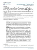
MEOX1 promotes tumor progression and predicts poor prognosis in human non-small-cell lung cancer
Bạn đang xem bản rút gọn của tài liệu. Xem và tải ngay bản đầy đủ của tài liệu tại đây (1.52 MB, 7 trang )
Int. J. Med. Sci. 2019, Vol. 16
Ivyspring
International Publisher
68
International Journal of Medical Sciences
2019; 16(1): 68-74. doi: 10.7150/ijms.27595
Research Paper
MEOX1 Promotes Tumor Progression and Predicts
Poor Prognosis in Human Non-Small-Cell Lung Cancer
Lichao Sun1, Hebao Yuan2, Joseph Burnett2, Mari Gasparyan2, Yuan Zhang1, Feng Zhang1, Zhihua Yang1,
Yuliang Ran1, Duxin Sun2
1.
2.
State Key Laboratory of Molecular Oncology, Cancer Hospital, Chinese Academy of Medical Sciences, Peking Union Medical College, Beijing, 100021, P. R.
China
Department of Pharmaceutical Sciences, University of Michigan, Ann Arbor, MI 48109
Corresponding authors: Lichao Sun, PhD, State Key Laboratory of Molecular Oncology, Cancer Hospital, Chinese Academy of Medical Sciences, Peking
Union Medical College, Beijing, 100021, P. R. China. Email: and Duxin Sun, PhD, Department of Pharmaceutical Sciences, University of
Michigan, Ann Arbor, MI 48109. Email:
© Ivyspring International Publisher. This is an open access article distributed under the terms of the Creative Commons Attribution (CC BY-NC) license
( See for full terms and conditions.
Received: 2018.05.31; Accepted: 2018.11.26; Published: 2019.01.01
Abstract
Background: MEOX1 is a homeobox transcriptional factor, and plays essential roles in regulating
somite development. Our previous study indicated that MEOX1 is a critical molecular target in
mesenchymal-like cancer cells in PTEN-deficient Trastuzumab resistant breast cancer. Despite the
potential implication of MEOX1 for the cancer progression, no previous studies examined its level
and clinical significance in lung cancer tissues. In this study, we aimed to detect the MEOX1
expression and correlate its level with clinical outcome in non-small-cell lung cancer patients
(NSCLC).
Methods: MEOX1 gene expression in lung cancer was examined by using the Oncomine database.
MEOX1 protein levels were evaluated by IHC using the corresponding primary antibody on two
different commercial lung cancer tissue arrays. siRNA knockdown was used to elucidate the
function of MEOX1.
Results: Analysis of the Oncomine datasets identified that an elevation of MEOX1 in gene
amplification in lung cancer tissues in comparison to normal lung tissues. Immunohistochemistical
analysis demonstrated that MEOX1 was localized predominantly in the nucleus, and positive rate
was 67.3% (111/165) in NSCLC samples. Statistical analysis revealed high levels of MEOX1
significantly correlated with Lymph Node Metastasis and Stage. Kaplan-Meier survival analysis
showed that high levels of MEOX1 were significantly associated with unfavorable survival in NSCLC
patients, and MEOX1 nucleus staining had worse survival, than did patients with overall expression
in lung squamous cell carcinoma patients. Multivariate Cox's regression analysis found that MEOX1
was an independent poor prognostic predictor for patients with NSCLC. Silencing of MEOX1 by
specific SiRNA significantly inhibited H460 and H1299 cell proliferation and sphere formation in
serum-free medium.
Conclusions: Our results firstly indentified that high levels of MEOX1 especially nuclear staining
was an independent prognostic factor for NSCLC, and it served a essential roles in the regulation of
cell proliferation and colony formation in vitro. It may represent a potential target for the NSCLC
treatment.
Key words: lung cancer, MEOX1, prognosis
Introduction
Lung cancer is one of the most common cancers
with highest mortality rate in China[1]. Comparing
with other pathological type, Non-small-cell lung
cancer (NSCLC) accounts for approximately 80-85%
Int. J. Med. Sci. 2019, Vol. 16
of lung cancer cases[2]. Despite great improvements
in treatment over recent decades, the outcome of
patients has not significantly improved. Therefore, it
is necessary to identify the novel therapeutic targets
for lung cancer treatment.
MEOX1 is a critical homeobox transcriptional
factor, which could affect the somite formation[3].
Several studies have proved that dysregulation of
MEOX1 is associated with cancer progression. In
ovarian cancer, MEOX1, as a PBX1 cofactor in, mediates the cellular growth signal[4]. In Triple-negative
breast cancer (TNBC), MEOX1 was overexpressed in
mesenchymal stem–like subtype cancer cells, which
were sensitive to NVP-BEZ235 and dasatinib treatment [5]. Our previous study identified that MEOX1
was overexpressed in breast cancer tissues and played
key roles in developmement of trastuzumabresistance and epithelial to mesenchymal transtion. In
addition, down-regulation of MEOX1 could decrease
mammosphere and colony formation in vitro, and
inhibit the tumor growth and breast cancer stem cell
frequency in vivo[6]. Although MEOX1 might play an
important role in carcinogenesis, no previous studies
investigated its expression, clinical significance and
function in lung cancer.
In this study, we not only demonstrated that
MEOX1 was significantly elevated and closely associated with poor prognosis of NSCLC patients, but also
regulated cell proliferation and sphere formation in
vitro for the first time.
Materials and methods
Analysis of Oncomine data
Oncomine was used for the analysis of MEOX1
level in lung cancer ().
Gene expression was evaluated by comparing lung
cancer and normal patient datasets. The analyzing
procedure was performed according to previous
studies[7].
Clinical samples and evaluation of
immunostaining
Two different commercial lung cancer tissue
arrays were constructed by Shanghai Biochip Co. Ltd.
as described[8]. Briefly, lung adenocarcinoma tissue
array contains 90 cases of patients, and lung
squamous cell carcinoma tissue array contains 75
cases respectively. Both of them contained the
correspondent adjacent normal tissues. For all the
specimens, clinicopathological factors and survival
status were available.
The MEOX1 protein levels were evaluated by
standard Avidin-biotin complex peroxidase immunohistochemical staining. Briefly, after deparaffinizatio-
69
nin xylene and graded alcohols, heated antigen
retrieval was done in citrate buffer (10mmol/L pH
6.0) by water-bath kettle heating for 30min. Endogenous peroxidase was blocked in 0.3% hydrogen
peroxide for 10 min. Nonspecific binding was blocked
by incubation in 10% normal animal serum for 10min.
Sections were incubated at 4°C for 24 h with primary
antibodies against MEOX1 (HPA045214, SigmaAldrich). We employed semi-quantitative scoring
system in considering the staining intensity and the
percentage of positive cancer cells, which has been
widely accepted and used in previous studies[6, 9].
Tissues with no or faint staining were rated as 0, with
moderate staining as 1, with strong staining as 2. The
percentage of positive cells was scored as follows: 0 (0
to 25%), 1 (25%-50%), 2 (>50%). The results were
determined using the following formula: overall score
= positive percentage score × intensity score. The
staining were defined as negative (from 0 to 2), and
positive (>2).
Knockdown by siRNA
SiRNA targeting human MEOX1was purchased
from Qiagen (validated FlexiTube siRNA). Lipofectamine® RNAiMAX was used to transfect cells. As a
negative control, a non-targeting sequence siRNA was
employed (Qiagen, 1027281). Knockdown at mRNA
level was confirmed by real-time quantitative RTPCR. MEOX1 Primer: Forward primer, 5-GAAACCC
CCACTCGGAAGG-3, Reverse primer, 5-GGGTGCT
GCTCAGTGAAGAT-3.
MTS Cell Proliferation Assay
Cancer cells were seeded at a density of 2,000
cells per well in 96-well plates. Cell growth was
determined by MTS assay according to manufacturer’s instruction by measuring the absorbance at 490
nm on a Synergy 2 plate reader (Biotek).
Mammosphere Formation Assay
Mammosphere culture was done according to
MammoCult™ Human Medium Kit (05620, STEM
CELL Technologies Inc.) supplemented with Heparin and Hydrocortisone. Single cells were plated in
six-well ultralow attachment plates (Corning) at a
density of 500 cells/ml. After 14 days of culture, the
number of mammospheres was counted on a Nikon
Eclipse TE2000-S microscope and the photos were
acquired with MetaMorph 7.6.0.0.
Statistical Analysis
The SPSS 15 software package (SPSS, Inc.,
Chicago, IL) was used for statistical analysis. The
association between MEOX1 expression and clinicopathologic features was analyzed by using χ2-test or
two-sided t-test as appropriate. Kaplan-Meier analysis
Int. J. Med. Sci. 2019, Vol. 16
and log-rank test were used to evaluate the overall
survival (OS) of patients with lung cancer according
to the expression of MEOX1. Cox's proportional
hazards regression model was used for multivariate
analysis of survival in lung cancer patients. All
comparisons were two-tailed, and p-value of < 0.05
was considered statistically significant.
Results
Gene expression of MEOX1 in lung cancer
By using Oncomine datasets, we investigated the
MEOX1 gene expression in lung cancer. The results
70
showed that MEOX1 gene copy number in lung
cancer tissues was higher than in normal tissues in
TCGA and Weiss Lung datasets (Figure. 1A, 1B).
Moreover, up-regulation of MEOX1 was also found in
Squamous cell lung carcinoma with distant metastasis
(M1 stage) in compared with ones without distant
metastasis (M0 stage) in TCGA dataset (Figure 1C).
Taken together, these data indicated that MEOX1 was
highly expressed in lung cancer tissues especially in
NSCLC, which might play essential roles in lung
cancer progression.
Figure 1. MEOX1 mRNA or DNA expression in human lung cancers using the Oncomine database. A. MEOX1 gene copy number in human lung
cancers vs. normal tissues in TCGA. B. MEOX1 gene copy number in human lung cancers vs. normal tissues in Weiss datasets. C. MEOX1 mRNA expression in
Squamous cell lung carcinoma grouped by M stage.
Int. J. Med. Sci. 2019, Vol. 16
71
Figure 2. MEOX1 expression in lung cancer tissues is determined by immunohistochemistry. A. Representative staining of lung adenocarcinoma
tissues. B. Representative staining of squamous cell lung carcinoma tissues.
Over-expression of MEOX1 protein in NSCLC
tissues by immunohistochemistry analysis
We evaluated the expression of the MEOX1 in
Human NSCLC tissue arrays by immunohistochemistry. The results demonstrated that MEOX1 was
positive in 111/165 primary tumors (67.3%), but there
was no or weak staining of adjacent normal tissues.
Moreover, MEOX1 was predominantly localized to
the nucleus (Figure. 2). Statistical analysis revealed
that whether cytoplasmic or nuclear MEOX1 staining
was significantly associated with Lymph Node
metastasis and Stage. Furthermore, MEOX1 expression was more frequent in adenocarcinoma than
squamous cell carcinoma (P = 0.000). However, no
statistically significant correlations were identified
between the expression of MEOX1 and other
clinicopathologic characteristics (Table 1).
MEOX1 expression level correlated with poor
prognosis of NSCLC
Kaplan-Meier analysis showed that the overall
survival time of lung cancer patients with high levels
of MEOX1 was markedly shorter than those with
MEOX1-negative expression (Figure. 3A, 3B). Further
analysis indicated that MEOX1 nucleus staining had
worse survival, than did patients with overall
expression in squamous cell lung cancer patients
(Figure. 3C). Then, we performed Cox multivariate
regression model to test the independent value of
each variable predicting overall survival. The results
revealed that MEOX1 nuclear staining level
(HR=3.304; 95% CI: 2.115-5.161; P=0.000) and Stage
(HR=1.750; 95% CI: 1.030-2.972; P=0.038) were
statistically independent predictive factors of poorer
prognosis for lung cancer (Table 2).
Table 1. Correlation between MEOX1 expression and
clinicopathological parameters in 165 lung cancer cases
Total MEOX1 staining
negative positive pvalue
43:11
77:34
0.165
Gender (Male:
Female)
Age
62.9±8.0
Type
Squamous cell
35
carcinoma
Adenocarcinoma 19
carcinoma
Tumor size(cm)
<5
37
>5
17
Depth of invasion
T1+T2
45
T3+T4
9
Lymph node involvement
N0
37
N1+N2
17
Distant metastasis
M0
54
M1
0
Grade
Ⅰ+Ⅱ
40
Ⅲ+Ⅳ
14
Stage
Ⅰ+Ⅱ
38
Ⅲ+Ⅳ
16
63.2±10.2
0.836
0.000
MEOX1 nuclear staining
negative positive p-val
ue
59:17
61:28
0.191
40
62.9±9.0 63.3±10.0 0.802
0.000
47
28
71
29
61
54
22
58
31
64
12
76
13
49
27
33
56
75
1
88
1
56
20
69
20
53
23
39
50
0.902
75
36
0.420
0.801
95
16
0.833
0.001
45
66
0.000
0.321
109
2
0.910
0.725
85
26
0.566
0.008
54
57
0.001
Int. J. Med. Sci. 2019, Vol. 16
72
Table 2. Multivariate analysis of Cox Proportional Hazards Model
for lung cancer
Characteristics
B
SE
Wald df
Sig.
Exp
(B)
95.0% CI for
Exp(B)
Lower Upper
1.195 0.228 27.576 1.000 0.000 3.304 2.115 5.161
MEOX1 nuclear
staining
Stage
Gender
Tumor Size
Depth of invasion
Lymph node
metastasis
Distance metastasis
0.560
-0.028
0.179
-0.080
0.268
0.270
0.225
0.217
0.290
0.269
4.285
0.016
0.676
0.076
0.993
0.038
0.900
0.411
0.783
0.319
1.750
0.972
1.196
0.923
1.307
1.000
1.000
1.000
1.000
1.000
1.030
0.625
0.781
0.522
0.772
2.972
1.512
1.830
1.630
2.215
0.396 0.761 0.271 1.000 0.603 1.486 0.334
6.603
mammosphere formation number by 89.1% and 79.6%
in H1299 and H460 cells over 14 days. And,
mammosphere size was significantly decreased as
well (Figure 4A). MTS assay indicated that
knockdown of MEOX1 in H1299 cells grew at a rate
71.2% slower than control cells. Consistently,
inhibition of MEOX1 also attenuated lung cancer cell
H460 proliferation, and the inhibitory rate was
approximately 51.2% (Figure 4B). Together, these
results showed that MEOX1 might play important
roles in lung cancer progression.
Discussion
MEOX1 silencing suppressed the self-renewal
and proliferation of lung cancer cell in vitro
To study the function of MEOX1 in lung cancer
cell line, we employed siRNA knockdown to study
the effect of MEOX1 on cell proliferation and
self-renewal ability in vitro. Silencing of MEOX1
significantly reduced its mRNA as determined by
quantitative real-time PCR. The results showed that
inhibition of MEOX1 dramatically suppressed the
Lung cancer is a leading cause of cancer death
worldwide[10]. Approximately, non-small cell lung
cancer (NSCLC) accounts for 85% lung cases, which is
typically classified into two subtypes including
adenocarcinoma (AD) and squamous cell carcinoma
(SCC) [7, 11]. Although novel treatment strategies
have been developed, the lung cancer death rate still
remains high[12-14]. Therefore, it is necessary to
identify other novel targets for clinical practice.
Figure 3. Survival curves for NSCLC cancer using the Kaplan-Meier method and the log-rank test. A. Overall survival curves for patients with negative
MEOX1 expression (dot line) and patients with positive total or nuclear staining MEOX1 (solid line); B. Overall survival curves for lung adenocarcinoma patients with
negative MEOX1 expression (dot line) and patients with positive total or nuclear staining (solid line); C. Overall survival curves for lung squamous cell carcinoma
patients with negative MEOX1 expression (dot line) and patients with positive total or nuclear staining MEOX1 (solid line).
Int. J. Med. Sci. 2019, Vol. 16
73
Figure 4. MEOX1 regulates lung cancer cell mammosphere formation and proliferation in vitro. A. Representative images of mammosphere formed 14
days after siRNA knockdown of MEOX1 in H1299 and H460 cells in comparison with parental or Nc-control cells. B. The growth of H1299 and H460 cell following
MEOX1 knockdown as determined by MTS in comparison with parental or Nc-control cells.
MEOX1 is a critical homeobox gene, which is
essential for the development of somite formation.
Mutations in MEOX1 are associated with Klippel-Feil
Syndrome[15]. It has been reported that aberrant
expression of MEOX1 might involve in cancer
progression. In ovarian cancer, MEOX1 was identified
as a direct target gene of PBX1 involving in cancer cell
growth[4]. Moreover, it has reported that MEOX1 was
overexpressed in mesenchymal stem – like subtype
cancer cells in TNBC [5]. Our previous study also
demonstrated that MEOX1 might play key roles in
mesenchymal-like breast cancer cell proliferation and
BCSC self-renewal ability. Moreover, suppression of
MEOX1 by sulforaphane (SF) was able to effectively
inhibit bulk tumor volume and reduce frequency of
BCSCs in an orthotopic xenograft mouse model of
breast cancer Immuunohistochemistry assay showed
that the expression of MEOX1 was specifically
elevated in breast cancer tissues, and nuclear protein
localization of MEOX1 was correlated with poorer
overall survival, an advanced tumor stage, and the
presence of lymph node metastasis[6]. Despite the
potential implication of MEOX1 for the cancer
progression, no previous studies have examined its
level and clinical significance in lung cancer.
In this study, we firstly evaluated MEOX1
expression levels by using Oncomine datasets. We
found that MEOX1 gene copy number was higher
than in normal tissues in TCGA and Weiss Lung
datasets. Interestingly, MEOX1 levels were higher in
Squamous cell lung carcinoma with distant metastasis
(M1 stage) than in ones without distant metastasis
(M0 stage) in TCGA dataset. It is plausible that
MEOX1 might contribute to lung cancer progression.
Then, we detected MEOX1 protein level in human
NSCLC tissue arrays by using IHC. The results
showed that MEOX1 was highly expressed in NSCLC
cancer tissues, with a positive rate of 67.3%.
We do find the MEOX1 cytoplasmic staining in
lung cancer tissues, and the positive rate was 16.0%
(12/75) in lung squamous cell carcinoma tissues and
11.1% (10/90) in lung adenocarcinoma tissues,
respectively. As a transcriptional factor, we found that
MEOX1 staining was predominantly localized to the
nucleus. Clinical relevance analysis showed that
positive expression of MEOX1 in NSCLC tissues was
significantly correlated with Lymph Node Metastasis
and Stage. Kaplan–Meier analysis indicated that lung
cancer patients with high levels of MEOX1 had
significantly shorter time than those with MEOX1
Int. J. Med. Sci. 2019, Vol. 16
negative expression. Moreover, Cox multivariate
regression model revealed that MEOX1 nuclear
staining level and Stage were statistically independent
predictive factors relating to poorer prognosis for
lung cancer.
According to the MEOX1 level in lung cancer
tissues and its clinical relevance, it is plausible that
MEOX1 might promote tumor progression. Then we
employed the SiRNA to knockdown its level in lung
cancer cell line to assess its effects on cell growth in
vitro. The results demonstrated that down-regulation
of MEOX1 led to a significant reduction in lung cancer
cell line H460 and H1299 proliferation and
mammosphere formation in serum-free medium. In
the future, we would further identify its downstream
target genes, and determine the related molecular
mechanism in lung cancer progression.
In conclusion, our study firstly demonstrated
that over-expression of MEOX1 was significantly
associated with poor survival in NSCLC. MEOX1
could be used as a potential novel target for human
NSCLC.
74
11. Faruki H, Mayhew GM, Serody JS, Hayes DN, Perou CM, Lai-Goldman M.
Lung Adenocarcinoma and Squamous Cell Carcinoma Gene Expression
Subtypes Demonstrate Significant Differences in Tumor Immune Landscape.
Journal of Thoracic Oncology. 2017;12: 943-953.
12. Singh PK, Singh H, Silakari O. Kinases inhibitors in lung cancer: From
benchside to bedside. Biochimica et biophysica acta. 2016; 1866: 128-40.
13. Provencio M, Sánchez A, Garrido P, Valcárcel F. New Molecular Targeted
Therapies Integrated With Radiation Therapy in Lung Cancer. Clinical Lung
Cancer. 2010; 11: 91-7.
14. Sun L, Chen L, Sun L, Pan J, Yu L, Han L, et al. Functional Screen for Secreted
Proteins by Monoclonal Antibody Library and Identification of Mac-2 Binding
Protein (Mac-2BP) as a Potential Therapeutic Target and Biomarker for Lung
Cancer. Molecular & Cellular Proteomics. 2013; 12: 395-406.
15. Mohamed Jawahir Y, Faqeih E, Alsiddiky A, Alshammari Muneera J, Ibrahim
Niema A, Alkuraya Fowzan S. Mutations in MEOX1, Encoding Mesenchyme
Homeobox 1, Cause Klippel-Feil Anomaly. American Journal of Human
Genetics. 2013; 92: 157-61.
Acknowledgement
Supported by grant from National Key R&D
Program of China (No: 2017YFC1308702), Beijing
Nova Program (No: Z1511000003150121), Beijing
Talents Fund (No:2015000021223ZK23), Beijing Gao
Chuang Ji Hua (No.G02060050).
Competing Interests
The authors have declared that no competing
interest exists.
References
1.
Chen W, Zheng R, Baade PD, Zhang S, Zeng H, Bray F, et al. Cancer statistics
in China, 2015. CA: a cancer journal for clinicians. 2016; 66: 115-32.
2. Siegel RL, Miller KD, Jemal A. Cancer Statistics, 2017. CA: a cancer journal for
clinicians. 2017; 67: 7-30.
3. Mankoo BS, Skuntz S, Harrigan I, Grigorieva E, Candia A, Wright CVE, et al.
The concerted action of Meox homeobox genes is required upstream of genetic
pathways essential for the formation, patterning and differentiation of
somites. Development. 2003; 130: 4655-64.
4. Thiaville MM, Stoeck A, Chen L, Wu RC, Magnani L, Oidtman J, et al.
Identification of PBX1 target genes in cancer cells by global mapping of PBX1
binding sites. PloS one. 2012; 7: e36054.
5. Lehmann BD, Bauer JA, Chen X, Sanders ME, Chakravarthy AB, Shyr Y, et al.
Identification of human triple-negative breast cancer subtypes and preclinical
models for selection of targeted therapies. The Journal of Clinical
Investigation. 2011; 121: 2750-67.
6. Sun L, Burnett J, Gasparyan M, Xu F, Jiang H, Lin CC, et al. Novel cancer stem
cell targets during epithelial to mesenchymal transition in PTEN-deficient
trastuzumab-resistant breast cancer. Oncotarget. 2016; 7: 51408-22.
7. Sun L, Wang Y, Yuan H, Burnett J, Pan J, Yang Z, et al. CPA4 is a Novel
Diagnostic and Prognostic Marker for Human Non-Small-Cell Lung Cancer.
Journal of Cancer. 2016; 7: 1197-204.
8. Chen L, Wei T, Si X, Wang Q, Li Y, Leng Y, et al. Lysine Acetyltransferase
GCN5 Potentiates the Growth of Non-small Cell Lung Cancer via Promotion
of E2F1, Cyclin D1, and Cyclin E1 Expression. The Journal of Biological
Chemistry. 2013; 288: 14510-21.
9. Sun L, Hu H, Peng L, Zhou Z, Zhao X, Pan J, et al. P-Cadherin Promotes Liver
Metastasis and Is Associated with Poor Prognosis in Colon Cancer. The
American journal of pathology. 2011; 179: 380-90.
10. DeSantis CE, Lin CC, Mariotto AB, Siegel RL, Stein KD, Kramer JL, et al.
Cancer treatment and survivorship statistics, 2014. CA Cancer J Clin. 2014; 64:
252-71.