an integrated proteomics analysis of bone tissues in response to mechanical stimulation
Bạn đang xem bản rút gọn của tài liệu. Xem và tải ngay bản đầy đủ của tài liệu tại đây (1.71 MB, 14 trang )
Li et al. BMC Systems Biology 2011, 5(Suppl 3):S7
/>
RESEARCH
Open Access
An integrated proteomics analysis of bone tissues
in response to mechanical stimulation
Jiliang Li1, Fan Zhang2,3, Jake Y Chen2,3,4*
From BIOCOMP 2010 - The 2010 International Conference on Bioinformatics and Computational Biology
Las Vegas, NV, USA. 12-15 July 2010
Abstract
Bone cells can sense physical forces and convert mechanical stimulation conditions into biochemical signals that
lead to expression of mechanically sensitive genes and proteins. However, it is still poorly understood how genes
and proteins in bone cells are orchestrated to respond to mechanical stimulations. In this research, we applied
integrated proteomics, statistical, and network biology techniques to study proteome-level changes to bone tissue
cells in response to two different conditions, normal loading and fatigue loading. We harvested ulna midshafts and
isolated proteins from the control, loaded, and fatigue loaded Rats. Using a label-free liquid chromatography
tandem mass spectrometry (LC-MS/MS) experimental proteomics technique, we derived a comprehensive list of
1,058 proteins that are differentially expressed among normal loading, fatigue loading, and controls. By carefully
developing protein selection filters and statistical models, we were able to identify 42 proteins representing 21 Rat
genes that were significantly associated with bone cells’ response to quantitative changes between normal loading
and fatigue loading conditions. We further applied network biology techniques by building a fatigue loading
activated protein-protein interaction subnetwork involving 9 of the human-homolog counterpart of the 21 rat
genes in a large connected network component. Our study shows that the combination of decreased antiapoptotic factor, Raf1, and increased pro-apoptotic factor, PDCD8, results in significant increase in the number of
apoptotic osteocytes following fatigue loading. We believe controlling osteoblast differentiation/proliferation and
osteocyte apoptosis could be promising directions for developing future therapeutic solutions for related bone
diseases.
Introduction
Bone tissues are sensitive to its mechanical environment
[1]. It is well accepted that the presence of a reasonable
level of mechanical stress on bones (known as normal
loading) could enhance bone formation and maintain a
healthy bone mass [2]. Prolonged absence of normal
loading on bones–usually associated with extended physical inactivity due to injuries–could decrease bone formation and increase bone resorption, eventually leading
to bone loss and disuse osteoporosis. When the level of
mechanical stimulations exceeds the normal amount for
an extended period of time, a stress condition known as
fatigue loading could occur. In fatigue loading, microdamage such as small cracks in bone tissues may
* Correspondence:
2
Indiana University School of Informatics, Indianapolis, IN 46202, USA
Full list of author information is available at the end of the article
appear, triggering a cascade of bone remodeling processes that attempt to repair damaged bone tissues via
sequential bone resorption and formation [3]. When
fatigue loading conditions are not recognized early and
addressed, the risks for bone injuries and bone diseases
will increase. Therefore, understanding the constituents
and functions of molecular repertoires involved in fatigue loading has been a central focus of study in molecular biology of the bone.
It still remains unknown what all the mechanicallysensitive genes and proteins in bone cells under
mechanical stress are and how their differential expressions are regulated [4]. Past research identified osteoblast as being recruited to bone surfaces to form new
bones in response to loading [5]. In fatigue loading conditions, the migration of osteoblast to the bone surface
is known to co-occur with migrations of osteoblast
© 2011 Li et al. This is an open access article distributed under the terms of the Creative Commons Attribution License (http://
creativecommons.org/licenses/by/2.0), which permits unrestricted use, distribution, and reproduction in any medium, provided the
original work is properly cited.
Li et al. BMC Systems Biology 2011, 5(Suppl 3):S7
/>
progenitors and osteoblast to bone damaged areas, thus
activating bone remodeling process and damage repairs
[6-11]. This process requires temporal coordination of
osteoblast and osteoblast to repair damaged bone tissues. Therefore, osteoblast-associated genes were
reported and presumed to be involved with different
levels of mechanical stimulation signals [12]. Several
biochemical studies have also suggested that anabolic
mechanical stimulation may increase the expression of
c-fos, osteopontin, COX-2, guanosine triphosphatases
(GTPases), adenylate cyclase, phospholipase C (PLC),
and mitogen-activated protein kinases (MAPKs), which
can further lead to elevated expression of bone anabolic
factors such as prostaglandins and Nitric oxide (See
reference [13] for a review).
In this work, we performed the first proteomic study
of mechanical loading of bone tissues using Rat as an
animal model. Prior to our study, large-scale functional
genomics analysis of the activation of bone remodeling
process were performed in a few microarray studies
[14,15]. While these earlier studies suggested osteocyte
apoptosis and Wnt signaling pathways were two critical biological processes involved, proper controls
against normal loading conditions were not performed
in those experimental studies. It was not clear what
mRNA level changes observed in fatigue loading were
shared in common with normal loading. Nor is it clear
whether the biological processes observed at the
mRNA expression level could overlook critical protein
changes, since many recent studies revealed that largescale gene expression and proteomics tend to complement (instead of significantly overlap) with each other
[16,17]. Elucidating proteomics level changes, particularly when integrated with prior findings of genes and
new models developed at the molecular signaling network/pathway level, can lead to new insights on bone
mechanical stress and development of novel molecular
biomarkers.
Experimental procedures
Design of bone loading experiments using rat models
In order to study proteomics profile differences in living
bone tissues, an ulnar axial compression loading system
was chosen (see illustration in Figure 1). The system
allows loading experimentation at different stress levels
for animal models [6,10,11].
Female Sprague-Dawley Rat (age: 6 months; weight:
250-300 grams) were purchased from Harlan (Indianapolis, Indiana, USA). Animals were acclimatized for two
weeks and housed in environmentally controlled rooms
in Laboratory Animal Resource Center (LARC) of Indiana University School of Medicine and fed standard Rat
chow and water ad libitum. All the procedures performed in this study were in accordance with the
Page 2 of 14
Figure 1 An illustration of the ulnar axial compression loading
system to study the effects of different levels of mechanical
stress on bones in animal models.
Indiana University Animal Care and Use Committee
Guideline.
Nine animals were divided randomly into 3 groups:
control (CTRL), loading (L) and fatigue loading (FL)
groups. All the animals were anesthetized with an intraperitoneal injection of ketamine (60 mg/kg; Ketaset®–
Fort Dodge Animal Health, Fort Dodge, IA) and xylazine (7.5 mg/kg; Sedazine®–Fort Dodge Animal Health,
Fort Dodge, IA). The animals in the control group were
sacrificed 96 hours post-injection without being subject
to mechanical loading. The right ulnae of the remained
animals were loaded or overloaded based on treatment
groups. The animals in the loading group were loaded
with a peak force of 20 N for 360 cycles and then sacrificed at 96 hours after the loading session. For the animals in fatigue loading group, one bout of loading with
a peak force of 20 N at 2 Hz was not stopped until 1015% stiffness loss. The overloaded animals were also
sacrificed at 96 hours after the loading session.
Load was applied using a load-controlled, electromagnetic loading device. Total loading cycles was adjusted
through the connected load controller. Stiffness loss
during the loading procedure was observed through
continuous monitoring of displacement of the arm on
the loading device using a CCD Laser Displacement
Sensor (LK Series, Keyence Corp. Osaka, Japan).
Li et al. BMC Systems Biology 2011, 5(Suppl 3):S7
/>
Liquid chromatography coupled tandem mass
spectrometry proteomics analysis
The ulnae were dissected out immediately and cleaned
of all muscle and connective tissue after all the Rats
were sacrificed. Both of 5-mm proximal and distal ends
of the ulnae were removed. The remaining ulna midshafts were snap frozen in liquid nitrogen and stored at
-80°C until protein isolation. For total protein isolation,
Rat ulna midshafts were shattered and ground to a fine
powder under liquid nitrogen using mortars and pestles.
There were three groups (The control, loading and fatigue loading groups), three samples per group, and two
HPLC injections per sample (Table 1).
Label-free protein identification and protein quantitative analysis services were performed by professionals at
the Protein Analysis and Research Center/Proteomics
Core of Indiana University School of Medicine, colocated at Monarch Life Sciences, Inc, Indianapolis. For
a thorough review of the principle and method developed at Monarch, refer to the review by Wang et al
[18].
The protein identification tasks were analyzed using
standard commercial-strength protocols and commercial
software packages developed at Monarch, which have
supported many scientific research case studies in areas
including proteomics studies, biomarker discovery, and
bioinformatics analysis, e.g., [19-21]. Briefly, Tryptic
peptides were analyzed using Thermo-Finnigan linear
ios-trap mass spectrometer (LTQ) coupled with a HPLC
system. Peptides were eluted with a gradient from 5 to
45% Acetonitrile developed over 120 minutes and data
were collected in the triple-play mode (MS Scan, zoom
scan, and MS/MS scan). The acquired raw peak list data
were generated by XCalibur (version 2.0) using default
parameters and further analyzed by an algorithm using
default parameters described by Higgs et al [22]. MS
database searches were performed against the combined
protein data set from International Protein Index (IPI;
version 1.2) [23] and the non-redundant NCBI-nr
human protein database (2005 version), which totaled
22,180 protein records. The resulting MS/MS data were
searched using SEQUEST Cluster from Thermo Scientific (bundled with BioWorks software suite version 2.70
based on the original SEQUEST algorithm [24]). During
Table 1 The experimental design for proteomics analysis
of bone loading in rat
Samples
Replicates
Injection runs (Subtotals)
CTRL
3
2
6
L
3
2
6
FL
3
2
6
The LC-MS/MS experiment consists of 3 groups × 3 samples × 2 replicates =
18 LC/MS injections run in random order. The three groups are: Controls
(CTRL), Loaded (L), and Fully-Loaded (FL).
Page 3 of 14
search, we set the number of missed cleavages permitted
to be 2. We search fixed modifications to be Iodoethanol on Cys and variable modifications to be Oxidation
on Met. The mass tolerance for precursor ions were set
at 2 Da and the mass tolerance for fragment ions were
set at 0.7 Da. For novel protein that could not be positively identified by SEQUEST, we used the de novo
sequencing function of the BioWorks software to obtain
peptide sequence information for the collision-induced
dissociation (CID) spectra. Carious data processing filters for protein identification were applied to keep only
peptides with the XCorr score above 1.5 for singly
charged peptides, 2.5 for doubly charged peptides, and
3.5 for triply charged peptides. These XCorr scores were
set according to linear discriminant analysis similar to
that described in DTASelect (version 2.0) to control
false-positive rate at below 5% levels. These empirical
thresholds were validated in large data sets processed by
Monarch in similar conditions and peptide identification
parameters. The false positive rates of these large-scale
studies under the used parameters were estimated from
the number and quality of spectral matches to the decoy
database.
Protein quantification tasks were also conducted using
software developed at Monarch Life Sciences, Inc. First,
all extracted ion chromatograms (XICs) were aligned by
retention time. Each aligned peak were matched by precursor ion, charge state, fragment ions from MS/MS
data, and retention time within a one-minute window.
Then, after alignment, the area-under-the-curve (AUC)
for each individually aligned peak from each sample was
measured, normalized, and compared for relative abundance–all as described in [22]. The normalization methods by Higgs et al [22] were used, and the data were
then transformed back to the original scale. Here, a linear mixed model generalized from individual ANOVA
(Analysis of Variance) was used to quantify protein
intensities and calculate statistical significance. In principle, the linear mixed model considers three types of
effects when deriving protein intensities based on
weighted average of quantile-normalized peptide intensities: 1) group effect, which refers to the fixed non-random effects caused by the experimental conditions or
treatments that are being compared; 2) sample effect,
which refers to the random effects (including those arising from sample preparations) from individual biological
samples within a group; 3) replicate effect, which refers
to the random effects from replicate injections from the
same sample preparation. Standard statistical data preprocessing techniques, including quantile normalization
and randomization of measurement orders, were applied
first to eliminate technical bias due to random variations
from biological samples and their replicates. The model
fitting was performed in the SAS software (version 9)
Li et al. BMC Systems Biology 2011, 5(Suppl 3):S7
/>
using PROC MIXED. The REML method was used as a
fit mechanism and degrees of freedom were computed
using the Satterthwaite method. The RANDOM statement was used to model the covariance with the
NOBOUND parameter option in the PROC statement.
The p-value estimates the proportion of times a change
at least as big as evaluated will be observed if in fact
there is no real change. All the p-values were then
transformed into q-values that estimate the False Discovery Rate (FDR) [25].
Homologous gene mapping of rat and human proteins
Due to the lack of protein-protein interaction data coverage in Rat, we map all Rat protein-encoding genes to
their human gene homolog to take advantage of large
sets of protein interaction data available in human. The
homologous gene mapping involved four steps. First, we
extracted all the Rat protein identifiers (IPI number and
protein GI accessions) from the sequence annotation
field of the proteomics search results. Second, we downloaded Rat IPI reference database version 1.2, which
contains 38,873 sequence identifier mapping relationships among Rat Swissprot IDs, sequence accession
numbers, and gene names. Third, we downloaded NCBI
Homologene release 49.1. We filtered out genes from
other organisms to include proteins only from Rat and
human. After applying the filter, 14,558 remained in the
homologene groups, which contain homology mapping
relationships between 15,125 Rat genes and 14,753
human genes. We defined a “homolog gene match”
between a Rat gene and a human gene as each pair
found within the same homologene group. In the fourth
step, we map the matched human genes back to human
proteins, using Uniprot sequence annotation files. Note
that the mapping between Rat protein to human protein
based on gene homology relationships has the limitation
of aggregating all alternative spliced protein isoforms
together. However, this will not be a major concern,
since the majority protein-protein interaction data are
collected based on gene-level experimentation data and
therefore do not offer isoform-level resolution anyway.
Method for selecting candidate significantly differentiallyexpressed proteins
For candidate proteins, we refer to the list of proteins
that satisfies statistical protein-selecting filters but still
needs further scrutiny before a subset of them can be
confirmed as biologically relevant. It is tempting to control false positives using high FC threshold and q-value
(false discovery Rate adjusted p-value) when we try to
select candidate proteins that are differentially expressed
with statistically rigor. For example, the following
threshold filter (the F1 filter) was suggested by the proteomics analysis software by default to control possible
Page 4 of 14
false positives that may arise due to potential sources of
variability (estimated to be up to 15%) from different
sample and experimental errors:
F1 : FC (x|i) ≥ 1.5&q − value (x|i) < 0.05
While a stringent filter is generally necessary for proteomics experiments, protein expression level changes in
proteomics experiments are generally expected to be
smaller than those often observed in expression microarrays, because changes in signaling proteins or regulatory proteins are expected to be subtle in general. In
addition, the problem with applying default filters
directly is that these filters fail to take into account of
data that may be highly correlated from controlled comparative experiments with more than two conditions. In
our case, we have three conditions FL for fatigue loading, L for normal loading, and CTRL for normal controls. If we can observe high degree of correlation of
results that occur in FL vs. CTRL and in F vs. CTRL,
the FC requirement and q-value requirement may be
both relaxed to allow more interesting proteins that
change barely in the “twilight zone” of >10%, as long as
these proteins can be further validated using additional
computational or experimental techniques.
Therefore, in complementary to fold change filter in
F1, we developed a second experimental filter (the F2
filter) to select candidate proteins that changed significantly above 10% (FC ≥ 1.1) to show up, when we try to
compare two similar conditions, FL_vs_L (Fatigue Loading against Normal Loading), in which data for
L_vs_CTRL (Fatigue Loading against Controls) and
FL_vs_CTRL (Normal Loading against Controls) are
also available:
F2: FC (x|FL_vs_L) ≥ 1.1 and
q-value(x|FL_vs_CTRL)*q-value(x|L_vs_CTRL) <
0.0025 and
p-value(x|FL_vs_CTRL) < 0.05 & p-value(x|
L_vs_CTRL) < 0.05
Here in this F2 filter, in addition to relaxing the FC
threshold, we also modified how we should apply statistical q-value. Here, we introduce a concept that we’ll
refer to as the triangulation property of comparable analysis. Briefly, this property is met if and only if pairwise
comparison results from three conditions, for example,
CTRL, L, and FL, are consistent among themselves. In
other words, we say a triangulation property exists
among CTRL-L-FL if and only if proteins passing
FL_vs_CTRL and L_vs_CTRL q-value filters with FC
changes of f1 and f2 respectively are the same set of
proteins that pass FL_vs_L with and same q-value filter
and a FC threshold of f1/f2 independently. In fact, no
Li et al. BMC Systems Biology 2011, 5(Suppl 3):S7
/>
proteomics search software that we know today guarantee such triangulation property due to inherent errors in
the model that estimates statistical significance of peptides and proteins. In fact, we understand that the qvalue was derived from a more stringent statistical
model in early years of proteomics licensed from Eli
Lilly (private communication with Dr. Mu Wang, who
provided the proteomics service for this experiment).
Therefore, we developed an easy-to-understand metaanalysis method, q-value triangulation method, in the F2
filter, so that we can rely primarily on better-understood
p-value statistics. In this method, we assume the p-value
calculations of two independent experiments,
FL_vs_CTRL and L_vs_CTRL, are generally reliable and
therefore can be controlled at 0.05. The q-value triangulation calculation for FL_vs_L is done by multiplying the
respective q-values for FL_vs_CTRL and L_vs_CTRL
comparisons controlled at the 0.05^2 = 0.0025 level.
The reason why the p-values are chosen comparing to
the control samples rather than comparing FL vs L is
that comparing to the control samples with our statistic
method can reduce baseline noise in proteomics data
and detect weak patterns.
Normality probability plot calculation
To determine normality of the residual distribution, we
use the normal probability plot to calculate the normal
quantiles of all values in Residue (i), or Res_FL_L. The
values and the normal quantiles are then plotted against
each other. Normal quantiles are computed using the fvalue, fi , which is calculated as:
fi =
i − 0.5
n
where i is the index of the value and n is the number
of values. The normal quantile, q(f), for a given f-value
is the value for which P[X <= q] = f , where X is a standard normally distributed variable [26].
Creation of bone tissue stimulated protein sub-networks
Differentially expressed candidate Rat proteins, which we
successfully mapped to human proteins through homologous gene matching, are used as seed proteins to build a
protein-protein interaction subnetwork. We derive this
protein interaction sub-network using a nearest-neighbor
expansion method initially described in [27]. In summary,
we searched the seed proteins against a human proteinprotein interaction database. We include additional proteins in this subnetwork if and only if these additional
proteins are found to directly interact with at least one
seed protein. The protein-protein interactions involved
are also collected into the subnetwork. If the subnetwork
does not form a large connected graph, the biological
Page 5 of 14
functional distance among such seed proteins would be
regarded as high. On the other hand, if the subnetwork
does form a large connected graph, the biological functional distance among these seed proteins would be very
close. The sub-network offers a good model to integrate
proteomics results, from which drug target may be developed [20,27]. Since the seed proteins used are all proteins
that are quantitatively changed under the FL_vs_L condition, this subnetwork is essentially an activated protein
signaling network specific to bone cells’ response to
mechanical stress.
We use the Human Annotated and Predicted Protein
Interaction (HAPPI) database [28] ( to retrieve high-quality protein interacting. We choose a human protein interaction
database due to limited protein-protein interaction data
available for Rat and the fact that Rat and human share
the majority of biological processes in common. The
HAPPI database is an open-access web-based relational
database that contains a comprehensive collection of
computer-annotated human protein-protein interactions
involving 10,592 human proteins (identified by UniProt
ID). Data in the HAPPI database are derived from both
experimental data sources and computational predictions publicly available. Different from most proteinprotein interaction databases, reliability of protein-protein interaction information is provided in the HAPPI
database as H scores, which range between 0 to 1 or a
quality star rank grade of 1, 2, 3, 4 and 5. Increased protein interaction grade from 1 to 5 have been shown to
be associated with improved quality of physical interacting proteins and decreased amount of non-physical
interactions found primarily in text mining or gene coexpression studies [29]. For this study, we only use protein interactions in the HAPPI database with star grade
of 3 and higher (consisting of more than 280,000
human protein interactions of primarily physical interactions), which are comparable to the overall quality of
HPRD, a much smaller reference human protein interaction database commonly used in bioinformatics.
Visualization of differentially expressed protein sub-network
To perform interaction network visualization, we used
an internally developed software platform, ProteoLens
[30], which can be freely downloaded from http://bio.
informatics.iupui.edu/proteolens/. ProteoLens is a biological network data mining and annotation platform that
supports both standard GML files and relational data in
Oracle or PostgreSQL Database Management System. It
is a scalable data-driven biological network visualization
software that enables expert bioinformatics users to
browse database schemas and tables, filter and join relational data using SQL queries, and customize data fields
to be visualized as network graphs.
Li et al. BMC Systems Biology 2011, 5(Suppl 3):S7
/>
Results
Cellular changes in bone tissues after mechanical
stimulations
In Figure 2, we show a comparison of histological
changes for bone tissues under control, normal loading,
and fatigue loading conditions. In Figure 2A, we show a
control without any mechanical stimulations. In Figure
2B, we show that bone formation in female SD Rat is
significantly increased compared with the control, when
one bout of axial loading of the ulna with a peak force
of 20 N at 2 Hz for 360 cycles periosteal is applied. In
Figure 2C, we show that substantial periosteal bone formation and microdamage in the cortex are generated,
when fatigue loading with a peak force of 20 N at 2 Hz
until 15% stiffness loss is applied.
Proteomics changes between normal loading and fatigue
loading conditions
The Proteomics software mentioned in the method section reported a comprehensive list of 1,058 proteins that
are differentially expressed among normal loading, fatigue loading, and controls. This list was derived from
5,361 IPI-identified Rat proteins observed in the LCMS/MS experiment of all Rat samples. Among the 5,361
IPI-identified proteins, 578 have Xcorr =’H’ (i.e., “high
confidence”) and 4,783 have Xcorr="L” (i.e., “low confidence”). The 1, 058 differentially expressed Rat proteins
can be mapped to 1,171 human proteins using homologous gene mapping methods (see Experimental Procedures for details). Note that only a fraction of these
1,058 proteins may have undergone through real quantitative changes, due to inherent variations of the proteomics platform and the high-variability nature of
biological samples.
In Figure 3, we used Venn Diagrams to show overlaps
among three proteomics comparative analysis results, i.
e., FL_vs_CTRL (Fatigue Loading against Control),
L_vs_CTRL (Normal Loading against Control), and
FL_vs_L (Fatigue Loading against Normal Loading), by
applying two different types of candidate protein selection filters, F1 and F2 (see Experimental Procedures for
Page 6 of 14
details), for results derived from LC-MS/MS proteomics
analysis of Rat samples In Figure 3A, only F1 default filter was applied. It showed that there are 322 proteins
overlapping between FL_vs_CTRL and L_vs_CTRL proteomics results. Combined together, the two data sets
represented 614 + 372 - 322 = 664 total proteins that
are quantitatively changed from either loading condition
to controls. Note that FL_vs_L produced no “significant”
protein list using the standard filter criteria, F1 (see
Experimental Procedures for details). A plausible explanation is that FL and L are biologically “equivalent” conditions, which make their proteomics level expression
indistinguishable. This is very unlikely, since the
FL_vs_CTRL and L_vs_CTRL results overlap in significant portions but differently (for FL_vs_CTRL, overalp
is 322/614 = 52%, for L_vs_CTRL, overlap is 322/372 =
87%). A second and alternative explanation is that the
filter F1 may be too stringent (requiring 1.5 fold change
differences between loading conditions and controls) to
allow detection of quantitative protein expression level
changes, which may be quite subtle for FL_vs_L comparisons. Therefore, we applied the second filter, F2
(also see Experimental Procedures for an explanation),
which provides relaxed (requiring FC≥1.1) yet still statistically significant candidate protein selecting threshold
for FL_vs_L differentially expressed proteins. By substituting filter F2 for F1 in the FL_vs_L condition, we
show the new overlapping relationship among
FL_vs_CTRL (using the original filter F1), L_vs_CTRL
(using the original filter F1), and the new FL_vs_L
(using the new filter F2) in Figure 3B. The new Venn
Diagram has an added FL_vs_L protein set of 76 candidate proteins. Interestingly, 65 out of the 76 protein
(65/76 = 86%) are overlapped with the existing 664 proteins differentially expressed and detected using the
stringent filter F1. The high degree of overlap resulted
in only a slight increase in the final combined data set
of 679 candidate rat proteins associated with loading
conditions. This observation is consistent with the
assumption that applying the F2 filter to the FL_vs_L
condition can still control false positives well. However,
Figure 2 Cellular changes of bone tissues under control, normal loading, and fatigue loading conditions. A: Control condition (no
loading); B: Normal loading condition. The thick staining at the perimeter of bone tissues indicates bone formation; C: Fatigue loading condition.
The microdamage (indicated by arrows) and bone formation at the peripherals of bone tissues are clearly visible.
Li et al. BMC Systems Biology 2011, 5(Suppl 3):S7
/>
Figure 3 Venn diagrams showing overlaps between different
proteomics comparison results. a: An overlap of significantly
differentially expressed proteins among FL_vs_CTRL, L_vs_CTRL, and
FL_vs_L conditions, using filter F1 only. b: Overlaps of differentiallyexpressed proteins among the same set of three types of
conditions, using existing filter F1 for FL_vs_CTRL and L_vs_CTRL
conditions, and a new filter F2 for the FL_vs_L condition. The
FL_vs_L total protein set contains 76 proteins, in which only 11
proteins are non-overlapping with the union of proteins in either
FL_vs_CTRL or L_vs_CTRL.
since filter F2 uses a fold change threshold of 1.1–much
smaller than the 1.5 threshold used in filter F1, we
believe that only a subset of the 76 candidate proteins
that changed at the subtle amount may have true biological significance.
Page 7 of 14
response variable. All the 679 proteins were used but
only the data points with both fold change reported
were reported. In Table 2, we show the linear regression
results, which has an R2 = 0.98. This surprisingly high
degree of correlation is perhaps attributable to the commercial operations (use of standard protocols and welltested proteomics analysis platform that also supports
high-volume commercial operations at Monarch Life
Sciences). It also supports the use of filter F2 that sets
FC threshold at 1.1–a level normally too low to be trustworthy when CV (covariance) of proteomics results are
at approximately 15% yet still acceptable for this particular experimental setup, due to high degree of correlations found for fold changes between FL_vs_CTRL and
L_vs_CTRL condition.
We further analyzed the residual plot for the above
linear regression model and determined the normalcy
data range (Figure 4). In Figure 4A, we observed that
most residuals are evenly distributed within the +/-2.0
standard deviation range (between thin lines), with the
exception of several residual extreme values that seemed
not normally distributed around the mean (shown as a
thick line in the center). To test if the residuals are normally distributed around the mean, we studied the residual normal probability plot (shown in Figure 4B). In
regions showing normality, the plot follows a diagonal
line. This suggests that residual values in the range vary
as expected due to random errors predicted by the linear regression model. Otherwise, we could suspect that
the residuals differ from one another by following a different model. In Figure 4B, we observed that the normal
probability plot of Res_FL_L (Residuals of the
FL_vs_CTRL against L_vs_CTRL after fitting the model
described earlier) has good normality (linear) in the
range of normal projection between -1.85 and +1.85
standard deviations of the mean. Outside this range, the
Res_FL_L has a different slope, suggesting non-normality for the outliers from the bulk of data.
Validated proteomics results – proteins that
quantitatively changed in fatigue loading conditions
Based on the residual distribution and normality probability test results, we reset the data outlier threshold to
be within +/-1.85 standard deviation range in the residual plot, with which we narrow down to 42 proteins.
Interestingly, the collection of these 42 proteins is a
Statistical validation of candidate proteins based on
correlated loading conditions
To examine how well the quantitative changes measured
between FL_vs_CTRL and L_vs_CTRL conditions–a
sign that should indicate how consistent and accurate
fold changes reported in the proteomics results are, we
performed a liner regression on two variables,
FC_CTRL_FL as × variable and FC_CTRL_L as y
Table 2 Linear regression results of FC_CTRL_FL and
FC_CTRL_L variables on differentially expressed proteins
in all 3 conditions of the study
Regression
parameter
Slope
(a)
Intercept
(b)
Data point
count
R2
value
1.09
0.03
679
0.98
Li et al. BMC Systems Biology 2011, 5(Suppl 3):S7
/>
Page 8 of 14
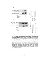
Figure 4 Determination of outliers in correlated variables FC_CTRL_FL and FC_CTRL_L. a) Plot of residuals RES_FL_L distributed over each
protein identified by Ratgene_sym. The thick line and the two thin straight lines above and below are average and +/-2 standard deviation
lines. Residual fold changes for each protein i were calculated using the linear regression model shown in Table 2 and calculated using the
following formula:Residue (i) = FC_CTRL_FL(i) - (a* FC_CTRL_L(i) + b), where FC_CTRL_FL(i) and FC_CTRL_L(i) refer to FC for FL_vs_CTRL and FC
for L_vs_CTRL for a given protein i, respectively. b) Normal probability plot of residual variable RES_FL_L over normal projection. The outliers are
indicated as blue solid dots in both panels. The normally distributed data points are indicated as red empty circles in both panels.
subset of the 76 candidate proteins from the FL_vs_L
condition that passed filter F2. These 42 proteins correspond to 21 genes, which we showed in Table 3.
In this table, we can further make several observations. First, protein ranks (indicator of confidence of
detection during search) derived from MS search software result as a default is not a reliable predictor for the
proteins’ biological significance. All significantly differentially expressed proteins in Table 3 have quite low
protein ranks, varying between 1500 and 2100. Second,
the patterns for differential expression changes are varied from one gene to another. For example, Capon,
Ddx21a, Rab40b (predicted), pdcd8, Serbinb13 (predicted) are all induced multiple folds from the resting
stage; Fbf1 (predicted), Pik4cb (predicted), Fcho2 (predicted), Slc1a3 (predicted) are all suppressed significantly from the resting stage; and Ddx18, Mrpl53
(predicted), and Mrpl45 (predicted) are all significantly
changed for FL_vs_CTRL conditions from L_vs_CTRL
conditions. Third, we have shown that at least in some
cases, a protein may be significantly differentially
expressed in the FL_vs_L condition for many reasons,
not necessarily due to a high FC_FL_L, e.g., Capon and
Rab40 (predicted)–both due to high FC_CTRL_L and
FC_CTRL_FL. Additional details of the protein quantification results for the proteins corresponding to the 21
genes are shown in Supplementary Table 1.
Activated protein signaling sub-network of molecular
response to fatigue loading
We mapped all significant Rat proteins to human proteins using gene homolog matching method describe in
the Experimental Procedures. 1,058 significantly changed
Rat IPI-identified proteins (using the F2 filter on all
comparative studies) out of 5,361 IPI-identified Rat proteins from the LC-MS/MS experiment were involved in
the mapping. These IPI-identified Rat proteins can be
mapped to 513 unique known Rat gene names (the
decrease was primarily due to aggregation of proteins
isoforms mapped to the same gene). 482 out of the original 513 Rat genes were successfully mapped to 484
human genes using the NCBI Homologene database.
The 484 human genes were mapped to 1,171 human
proteins identified with UniProt IDs. The slight increase
in total protein count from initial 1,058 Rat proteins to
1,171 human proteins suggest that there were a small
percentage of one-to-many homologous mapping relationships between Rat and human proteins.
Then, using the 42 Rat proteins representing 21 Rat
genes (as shown in Table 3) as seed proteins, we built a
protein interaction subnetwork. This network represented a coarse biological model that integrated prior
knowledge of the functional interaction relationships
among proteins and the latest acquired proteomics
knowledge on proteins quantitatively changed under
Li et al. BMC Systems Biology 2011, 5(Suppl 3):S7
/>
Page 9 of 14
Table 3 A list of 21 Rat genes whose proteins are found to be differentially expressed with statistical significance
between FL_vs_CTRL and L_vs_CTRL conditions
Rat Gene
Human Gene
FC (CTRL_L)
FC (CTRL_FL)
FC (FL_L)
Max Confidence
Peptide Evidence
Capon
NOS1AP
6.72884
6.00145
1.1212
0.98
≥6
Ddx18
DDX18
1.14716
2.13095
-1.85759
0.98
≥6
Ddx21a
Fbf1_predicted
DDX21
FBF1
3.28614
-3.10292
4.10949
-2.81444
-1.25055
-1.1025
0.96
0.98
≥6
≥6
Fcho2_predicted
FCHO2
-1.97277
-2.79227
1.41541
0.98
≥6
Klk14_predicted
KLK14
1.2212
1.88874
-1.54662
0.98
≥6
LOC301506
FSD1
-2.77612
-3.54757
1.27789
0.99
≥6
LOC306805
ASPN
1.83348
2.8254
-1.54101
0.99
≥12
Mrpl45_predicted
MRPL45
2.47117
3.98149
-1.61118
0.99
≥6
Mrpl53_predicted
MRPL53
3.70412
1.94325
1.90615
0.96
≥6
Pdcd8
Pik4cb
PDCD8
PIK4CB
2.91378
-2.77612
4.15437
-3.54757
-1.42577
1.27789
0.96
0.99
≥6
≥6
RGD1562139_predicted
RPL29
2.47771
3.28214
-1.32467
0.98
≥6
Rab40b_predicted
RAB40B
5.42109
4.99103
1.08617
0.98
≥6
Raf1
RAF1
-2.1328
-1.59117
-1.3404
0.97
≥6
Sema5b_predicted
SEMA5B
1.75998
2.60246
-1.47869
0.99
≥6
Serpinb13_predicted
SERPINB13
3.01946
3.82539
-1.26691
0.97
≥6
Slc1a3
SLC1A3
-1.97126
-2.78988
1.41528
0.98
≥6
Slc4a3
Tex101
SLC4A3
TEX101
2.15184
2.007
1.80834
1.60395
1.18995
1.25128
0.96
0.97
≥6
≥6
Upf2_predicted
UPF2
-1.72341
-2.54157
1.47474
0.98
≥6
* “Max Confidence” was calculated as 1- smallest q-value among all the comparison conditions (FL_L, CTRL_FL, and CTRL_L). “Peptide evidence” refers to total
number of peptides per group used to calculate Fold Change (FC) and q-value in groupwise comparisons for protein quantifications.
fatigue loading conditions compared with normal loading conditions. After the protein interaction network
expansion, the initial 42 seed proteins became expanded
into a set of 394 human protein interacting pairs covered by 297 human proteins. In Figure 5, we show a
visualization of the FL_vs_L expanded human protein
interaction sub-network (network with only one pair of
interactions are not shown). The largest connected component of this network consists of 9 genes (to be discussed in the next section), which can be used to reason
about molecular mechanisms why these proteins changed during mechanical stress conditions that ultimately
lead to microdamage in bones.
Pathway-protein association analysis
The 42 Rat proteins representing 21 Rat genes (as
shown in Table 3) were also used to perform pathwayprotein association analysis using the Kyoto Encyclopedia of Genes and Genomes ( />kegg/) [33]. Significance level for pathway comparisons
was set by represented number >3 due to results of
small counts. This allows avoiding any assumptions
about the shape of sampling distribution of population.
This pathway protein association matrix maps all the
biological pathways with pathway proteins. It enriches
the top frequent pathways in a given list of pathways,
which helps in discovering pathway markers. In Figure
6, 36 pathways and 21 proteins are associated with each
other for three comparisons (red for CTRL_L; green for
CTRL_FL; and blue for FL_L).
Discussions
Mechanical stimulation may cause bone cells to express
mechano-sensitive genes and proteins through membrane receptors and ion channels and downstream intracellualer signaling cascades [34-36]. These would lead to
differentiation of osteoblast progenitor cells and osteoblast prolifeRation [5]. Besides increase in bone formation, fatigue loading produce microdamage [9] in the
cortex which also leads to osteocyte apoptosis and
further activate bone remodeling through which the
damaged cortical bone is repaired [6,37].
In our study, we have found the enhanced expression
of proteins involved in receptor binding, RNA processing, cell division and etc. Cell division cycle 25 homolog B (CDC25B), DEAD (Asp-Glu-Ala-Asp) box
polypeptide 21 (DDX21), ribosomal protein L29 (RPL29)
(seed proteins) and the expanded proteins as shown in
Figure 5 were up-regulated. CDC25B that plays a role in
cell division seems to allow cell to go into cell division
during fatigue loading [38]. DDX21 and RPL29 all are
elevated in exercise conditions, and further elevated in
fatigue exercise conditions. DDX21 is putative RNA
helicase involved in RNA secondary structure alteRation,
Li et al. BMC Systems Biology 2011, 5(Suppl 3):S7
/>
Page 10 of 14
Figure 5 A protein interaction sub-network of FL_vs_L expanded differentially expressed proteins. Nodes colored in red or green are
FL_vs_L differentially expressed proteins (seeds) and nodes in light purple are non-seed expanded proteins recruited through human protein
interactions. Edges represent protein interactions recorded in the HAPPI database. Only HAPPI database protein interactions with quality ratings
at or above 3 are used. Proteins that are significantly differentially expressed in FL_vs_CTRL or L_vs_CTRL conditions are also shown using the
same color legend for FL_vs_L seed proteins, with the rectangle split into two half panels: the upper panel shows the gradient red (FC_CTRL_L
>0) or green (FC_CTRL_L <0) colors for the FC_CTRL_L value, while the lower panel shows the gradient red or green color using the same color
profile for the FC_CTRL_FL value. Standalone networks with only one pair of interactions are not shown.
and Ribosome reassembly [39]. RPL29 is ribosomal protein L29 involved in cell surface hairpin protein binding
[40].
NOS (Nitric Oxide Synthase) is increased under the
loading condition and further elevated by fatigue loading
in this study. NOS is the enzyme to produce Nitric
Oxide (NO) in cells [41]. NO has been shown to
increase in response to mechanical stimulation in osteoblastic cells [42]. It is also involved in mechanically
induced bone formation in vivo [43]. Our study further
verifies that NOS may mediate load induced bone formation at the periosteal surface in loading and fatigue
loading groups. In addition, the further elevated NOS
level under fatigue loading condition suggests NO may
Li et al. BMC Systems Biology 2011, 5(Suppl 3):S7
/>
also play a key role in mediating the repair of bone
damage, such as recruitment of osteoclast precursor,
because its actions include changes of the vascular permeability of the damaged area and stimulation of angiogenic activity [41].
Several apoptosis related proteins have been found to
change significantly in the current study. Raf1 human
(RAF proto-oncogene serine/threonine-protein kinase)
was down regulated in the present study. It has a role in
the transduction of mitogenic signals from the cell membrane to the nucleus [44]. Raf1 may promot cell survival
by antagonizing apoptosis signals-regulating kinase [45].
Our study indicates that loss of Raf1 coincide with
increased number of apoptotic osteocytes resulting from
fatigue loading, suggesting that Raf1 has a role in
Page 11 of 14
protection of osteocytes apoptosis. On the other hand,
PDCD8 (Programmed cell death 8) is up-regulated under
fatigue loading condition. Because PDCD8 is an apoptosis-inducing factor [46], it may induce osteocytes apoptosis following fatigue loading. Taken together, our study
shows that the combination of decreased anti-apoptotic
factor, Raf1, and increased pro-apoptotic factor, PDCD8,
results in significant increase in the number of apoptotic
osteocytes following fatigue loading. Several downstream
proteins of Raf1 and PDCD8 pathways, such as Bcl2 and
caspase proteins have previously been shown to be
involved in osteocyte apoptosis induced by fatigue loading [37,47]. Therefore, this study suggests that drugs targeting on Raf1 and PDCD8 may regulate bone
metabolism via prevention of osteocyte apoptosis.
Figure 6 A pathway-protein association matrix of differentially expressed proteins. The proteins in the first row from the fifth column to
25th column are differentially expressed with statistical significance between FL_vs_CTRL and L_vs_CTRL. The first column is KEGG pathway ID,
the second column is KEGG pathway name, the third column is number of represented proteins in a pathway, and the forth column is the total
number of molecular in a pathway. The up-arrow represents up-regulated expression, and the down-arrow represents down-regulated
expression. Three comparisons are shown (red for CTRL_L, green for CTRL_FL, and blue for FL_L).
Li et al. BMC Systems Biology 2011, 5(Suppl 3):S7
/>
In the pathway-protein association analysis, a list of 42
rat proteins differentially expressed with statistical significance between FL_vs_CTRL and L_vs_CTRL is used
to identify topmost frequent pathways. Of the 36 pathways in Figure 6, 13 are related to cancers; 18 to cellular
processes (6 immune system, 3 nervous system, 3 endocrine system,2 cell communication, 1 cell growth and
death, 1 cell motility, 1 circulatory system, 1 development); 4 to signal transduction; and 1 to carbohydrate
metabolism. The top eight pathway are Axon Guidance,
Inositol Phosphate Metabolism, Phosphatidylinositol Signaling System, Ribosome, MAPK Signaling Pathway,
Erbb Signaling Pathway, Chemokine Signaling Pathway,
and Apoptosis. Some of those pathways have been
reported to be related to bone metabolism. For example,
neural regulation of bone metabolism mediated in
osteoblastic and osteoclastic cells via Axon Guidance
pathway has been demonstrated in histochemical and
pharmacological studies [48] and Togari etc., in their
paper, suggested the extension of axons of peripheral
sensory and sympathetic neurons to osteoblastic and
osteoclastic cells and the possible neural regulation of
bone metabolism in these osteogenic cells. Inositol
phosphate metabolism and signal transduction pathways
was reported to regulate cytoplasmic Ca2+ concentrations in osteoblastic bone cells[49]. In addition, Kennea
etc. suggested that there would be robust and functional
intrinsic and extrinsic apoptotic pathways in human
fetal mesenchymal stem cells or or bone marrowderived stromal cells which could participate in the
repair of mesodermal tissues, such as bone in osteogenesis imperfecta and heart muscle in cardiac ischaemia
[50].
Of the 21 proteins, PDCD8 (A4QPB4_HUMAN;
AIFM1_HUMAN; B1ALN1_HUMAN) which is up regulated with statistical significance between FL_vs_CTRL
and L_vs_CTRL are involved in Apoptosis pathway,
RAF1 (C9J2U6_HUMAN; C9J3L4_HUMAN; RAF1_HUMAN) which is down regulated with statistical significance between FL_vs_CTRL and L_vs_CTRL is involved
in Cancers, Cellular Processes, and Signal Transduction
Pathways. This further indicates the effect of decreased
anti-apoptotic factor, Raf1, and increased pro-apoptotic
factor, PDCD8, on the increase in the number of apoptotic osteocytes following fatigue loading.
We also found other pathway-protein associations
such as PI4KB in Inositol phosphate metabolism and
Phosphatidylinositol signaling system pathways,
SEMA5B in Axon guidance, and RPL29 in Ribosome.
Some of them are linked to bone metabolism or bone
formation by previous reports. For example, Miller etc.
reported that the presence of HIP/RPL29 during early
chondrogenesis is essential for normal skeletal growth
and patterning. They designed a ribozyme-mediated
Page 12 of 14
knock-down approach to partially down-regulate HIP/
RPL29 expression in the multipotent mouse embryonic
skin fibroblast cell line C3H/10T To investigate the role
of HIP/RPL29 normal expression during cartilage formation [51]. And Mary showed that SEMA5B is a nerve
guidance factor which is involved in invasive growth,
vascular patterning, axon guidance, and bone development [52].
In addition, Rab40b is a member of Ras oncogene
family [53]. Ras oncogenes are small GTP-binding proteins [53]. Besides their role in cell prolifeRation, Ras
paradoxically induce both pro- and anti-apoptotic signaling [54]. It remains to be investigated whether Ras
plays any role in osteocyte apoptosis following fatigue
loading.
There is a possibility that other proteins, such as
MRPL45, SLC1A3, UPF2 and ASPN identified in this
study are involved in bone response to mechanical loading. ASPN has been found to be related to osteoarthritis
[32]. It is remained to be investigated if MRPL45,
SLC1A3 and UPF2 as intracellular transports could be
stimulated by mechanical stimulation.
In conclusion, using an integrated LC-MS/MS proteomics analysis for the first time in bone mechanical stimulation studies, we have identified several essential
proteins related to cell division, which can be linked to
osteoblast differentiation and proliferation and bone formation eventually in response to loading. More importantly, our study identified several new proteins
associated with osteocyte apoptosis induced by fatigue
loading. Our results suggest new insights for future
investigation of these proteins as candidate drug targets
to regulate bone metabolism and repair bone damage.
Additional material
Additional file 1: Protein quantification data for the 21 Rat genes
whose proteins levels are significantly changed in Loaded (L) or
Fully Loaded (FL) conditions compared with controls (CON). “CON_L”
refers to comparing L to CON. “CON_FL” refer to comparing FL to CON.
“FL_L” refers to comparing L to FL. q-value refers to adjusted p-values.
While p-value is an estimate of false positive rate, q-value is an estimate
of false discovery rate (FDR). FC refers to Fold Change. “Mean CON/L/FL”
refers to mean protein intensities. “%CV Injection” refers to % Coefficient
of Variation for injection variation, “%CV Inj + Sample %” refers to the
Coefficient of Variation for injection plus sample variation. “# of peptides/
group” refers to the number of distinct identified peptides for this
protein in any of the three groups: CON, L, or FL. “Mean Xcorr” refers to
the mean Xcorr of the peptides identified for this protein.
Acknowledgements
This work was partially supported by a Clinical Proteomic Technology
Assessment for Cancer (CPTAC) grant to Jake Chen (co-investigator) from
the National Cancer Institute (U24CA126480-01), seed funding to Indiana
Center for Systems Biology and Personalized Medicine from the Indiana
University - Purdue University Indianapolis, and NASA grant to Jiliang Li
(NNA04CD04G). We thank Dr. Mu Wang for consultation with us to develop
Li et al. BMC Systems Biology 2011, 5(Suppl 3):S7
/>
a proteomics service plan for this project and performing the LC-MS/MS
proteomics service. We also thank Ragini Pandey for helping us organize
materials during the manuscript writing process.
This article has been published as part of BMC Systems Biology Volume 5
Supplement 3, 2011: BIOCOMP 2010 - The 2010 International Conference on
Bioinformatics & Computational Biology: Systems Biology. The full contents
of the supplement are available online at />1752-0509/5?issue=S3.
Author details
Department of Biology, Purdue School of Science, Indiana University Purdue
University Indianapolis (IUPUI), Indianapolis, IN 46202, USA. 2Indiana
University School of Informatics, Indianapolis, IN 46202, USA. 3Indiana Center
for Systems Biology and Personalized Medicine, Indianapolis, IN 46202, USA.
4
Department of Computer and Information Science, Purdue University
School of Science, Indianapolis, IN 46202, USA.
1
Authors’ contributions
JYC conceived the initial work, designed the method for the data
construction. JL implemented the design of bone loading experiments and
generated the proteomics data using rat models. FZ collected and analyzed
the MS data, performed the statistical analyses. All authors are involved in
the drafting and revisions of the manuscript.
Competing interests
The authors declare that they have no competing interests.
Page 13 of 14
16.
17.
18.
19.
20.
21.
22.
Published: 23 December 2011
23.
References
1. Robling AG, Castillo AB, Turner CH: Biomechanical and molecular
regulation of bone remodeling. Annu Rev Biomed Eng 2006.
2. Ziegler R, Scheidt-Nave C, Scharla S: Pathophysiology of osteoporosis:
unresolved problems and new insights. J Nutr 1995, 125(7
Suppl):2033S-2037S.
3. Stromsoe K: Fracture fixation problems in osteoporosis. Injury 2004,
35(2):107-113.
4. Villemure I, Chung MA, Seck CS, Kimm MH, Matyas JR, Duncan NA: Static
compressive loading reduces the mRNA expression of type II and X
collagen in rat growth-plate chondrocytes during postnatal growth.
Connect Tissue Res 2005, 46(4-5):211-219.
5. Turner CH, Owan I, Alvey T, Hulman J, Hock JM: Recruitment and
proliferative responses of osteoblasts after mechanical loading in vivo
determined using sustained-release bromodeoxyuridine. Bone 1998,
22(5):463-469.
6. Bentolila V, Boyce TM, Fyhrie DP, Drumb R, Skerry TM, Schaffler MB:
Intracortical remodeling in adult rat long bones after fatigue loading.
Bone 1998, 23(3):275-281.
7. Burr DB, Martin RB, Schaffler MB, Radin EL: Bone remodeling in response
to in vivo fatigue microdamage. J Biomech 1985, 18(3):189-200.
8. Li J, Miller MA, Hutchins GD, Burr DB: Imaging bone microdamage in vivo
with positron emission tomography. Bone 2005, 37(6):819-824.
9. Li J, Waugh LJ, Hui SL, Burr DB, Warden SJ: Low-intensity pulsed
ultrasound and nonsteroidal anti-inflammatory drugs have opposing
effects during stress fracture repair. J Orthop Res 2007.
10. Tami AE, Nasser P, Schaffler MB, Knothe Tate ML: Noninvasive fatigue
fracture model of the rat ulna. J Orthop Res 2003, 21(6):1018-1024.
11. Verborgt O, Gibson GJ, Schaffler MB: Loss of osteocyte integrity in
association with microdamage and bone remodeling after fatigue in
vivo. J Bone Miner Res 2000, 15(1):60-67.
12. Pavalko FM, Norvell SM, Burr DB, Turner CH, Duncan RL, Bidwell JP: A
model for mechanotransduction in bone cells: the load-bearing
mechanosomes. J Cell Biochem 2003, 88(1):104-112.
13. Rubin J, Rubin C, Jacobs CR: Molecular pathways mediating mechanical
signaling in bone. Gene 2006, 367:1-16.
14. Armstrong VJ, Muzylak M, Sunters A, Zaman G, Saxon LK, Price JS,
Lanyon LE: Wnt/beta-catenin signaling is a component of osteoblastic
bone cell early responses to load-bearing and requires estrogen
receptor alpha. J Biol Chem 2007, 282(28):20715-20727.
15. Lau KH, Kapur S, Kesavan C, Baylink DJ: Up-regulation of the Wnt,
estrogen receptor, insulin-like growth factor-I, and bone morphogenetic
24.
25.
26.
27.
28.
29.
30.
31.
32.
33.
34.
35.
36.
37.
protein pathways in C57BL/6J osteoblasts as opposed to C3H/HeJ
osteoblasts in part contributes to the differential anabolic response to
fluid shear. J Biol Chem 2006, 281(14):9576-9588.
Ott LW, Resing KA, Sizemore AW, Heyen JW, Cocklin RR, Pedrick NM,
Woods HC, Chen JY, Goebl MG, Witzmann FA, et al: Tumor Necrosis
Factor-alpha- and interleukin-1-induced cellular responses: coupling
proteomic and genomic information. J Proteome Res 2007, 6(6):2176-2185.
Xie L, Pandey R, Xu B, Tsaprailis G, Chen QM: Genomic and proteomic
profiling of oxidative stress response in human diploid fibroblasts.
Biogerontology 2009, 10(2):125-151.
Wang M, You J, Bemis KG, Tegeler TJ, Brown DP: Label-free mass
spectrometry-based protein quantification technologies in proteomic
analysis. Brief Funct Genomic Proteomic 2008, 7(5):329-339.
Harezlak J, Wu MC, Wang M, Schwartzman A, Christiani DC, Lin X:
Biomarker discovery for arsenic exposure using functional data. Analysis
and feature learning of mass spectrometry proteomic data. J Proteome
Res 2008, 7(1):217-224.
Chen JY, Pinkerton SL, Shen C, Wang M: An integrated computational
proteomics method to extract protein targets for Fanconi anemia
studies. In 21st Annual ACM Symposium on Applied Computing. Volume 1.
Dijon, France; 2006:173-179.
McBride WJ, Schultz JA, Kimpel MW, McClintick JN, Wang M, You J,
Rodd ZA: Differential effects of ethanol in the nucleus accumbens shell
of alcohol-preferring (P), alcohol-non-preferring (NP) and Wistar rats: a
proteomics study. Pharmacol Biochem Behav 2009, 92(2):304-313.
Higgs RE, Knierman MD, Gelfanova V, Butler JP, Hale JE: Comprehensive
label-free method for the relative quantification of proteins from
biological samples. J Proteome Res 2005, 4(4):1442-1450.
Kersey PJ, Duarte J, Williams A, Karavidopoulou Y, Birney E, Apweiler R: The
International Protein Index: an integrated database for proteomics
experiments. Proteomics 2004, 4(7):1985-1988.
Link AJ, Eng J, Schieltz DM, Carmack E, Mize GJ, Morris DR, Garvik BM,
Yates JR: Direct analysis of protein complexes using mass spectrometry.
Nat Biotechnol 1999, 17(7):676-682.
Benjamini Y, Yekutieli D: The control of the false discovery rate in
multiple testing under dependency. Ann Statist 2001, 29(4):1165-1188.
Rice JA: Belmont, CA: Duxbury Press;, 2 1995.
Chen JY, Shen C, Sivachenko A: Mining alzheimer disease relevant
proteins from integrated protein interactome data. Pac Symp Biocomput
2006, 367-378.
Chen JY, Mamidipalli S, George B: HAPPI: A Database of Human
Annotated Protein-Protein Interactions. 2007, 2006.
Chen JY, Mamidipalli S, Huang T: HAPPI: an online database of
comprehensive human annotated and predicted protein interactions.
BMC Genomics 2009, 10(Suppl 1):S16.
Huan T, Sivachenko A, Harrison S, Chen JY: ProteoLens: a visual analytic
tool for multi-scale database-driven biological network data mining. BMC
Bioinformatics 2008, 9(Suppl 9):S5.
Kizawa H, Kou I, Iida A, Sudo A, Miyamoto Y, Fukuda A, Mabuchi A,
Kotani A, Kawakami A, Yamamoto S, et al: An aspartic acid repeat
polymorphism in asporin inhibits chondrogenesis and increases
susceptibility to osteoarthritis. Nat Genet 2005, 37(2):138-144.
Jiang Q, Shi D, Yi L, Ikegawa S, Wang Y, Nakamura T, Qiao D, Liu C, Dai J:
Replication of the association of the aspartic acid repeat polymorphism
in the asporin gene with knee-osteoarthritis susceptibility in Han
Chinese. J Hum Genet 2006, 51(12):1068-1072.
Kanehisa M, Araki M, Goto S, Hattori M, Hirakawa M, Itoh M, Katayama T,
Kawashima S, Okuda S, Tokimatsu T, et al: KEGG for linking genomes to
life and the environment. Nucleic Acids Res 2008, , 36 Database: D480-484.
Duncan RL, Turner CH: Mechanotransduction and the functional response
of bone to mechanical strain. Calcif Tissue Int 1995, 57(5):344-358.
Li J, Duncan RL, Burr DB, Gattone VH, Turner CH: Parathyroid hormone
enhances mechanically induced bone formation, possibly involving Ltype voltage-sensitive calcium channels. Endocrinology 2003,
144(4):1226-1233.
Li J, Duncan RL, Burr DB, Turner CH: L-type calcium channels mediate
mechanically induced bone formation in vivo. J Bone Miner Res 2002,
17(10):1795-1800.
Verborgt O, Tatton NA, Majeska RJ, Schaffler MB: Spatial distribution of Bax
and Bcl-2 in osteocytes after bone fatigue: complementary roles in bone
remodeling regulation? J Bone Miner Res 2002, 17(5):907-914.
Li et al. BMC Systems Biology 2011, 5(Suppl 3):S7
/>
Page 14 of 14
38. Lindqvist A, Kallstrom H, Karlsson Rosenthal C: Characterisation of Cdc25B
localisation and nuclear export during the cell cycle and in response to
stress. J Cell Sci 2004, 117(Pt 21):4979-4990.
39. Henning D, So RB, Jin R, Lau LF, Valdez BC: Silencing of RNA helicase II/
Gualpha inhibits mammalian ribosomal RNA production. J Biol Chem
2003, 278(52):52307-52314.
40. Liu JJ, Huang BH, Zhang J, Carson DD, Hooi SC: Repression of HIP/RPL29
expression induces differentiation in colon cancer cells. J Cell Physiol
2006, 207(2):287-292.
41. Garthwaite J, Charles SL, Chess-Williams R: Endothelium-derived relaxing
factor release on activation of NMDA receptors suggests role as
intercellular messenger in the brain. Nature 1988, 336(6197):385-388.
42. Pitsillides AA, Rawlinson SC, Suswillo RF, Bourrin S, Zaman G, Lanyon LE:
Mechanical strain-induced NO production by bone cells: a possible role
in adaptive bone (re)modeling? Faseb J 1995, 9(15):1614-1622.
43. Turner CH, Takano Y, Owan I, Murrell GA: Nitric oxide inhibitor L-NAME
suppresses mechanically induced bone formation in rats. Am J Physiol
1996, 270(4 Pt 1):E634-639.
44. Morrison DK: The Raf-1 kinase as a transducer of mitogenic signals.
Cancer Cells 1990, 2(12):377-382.
45. Chen J, Fujii K, Zhang L, Roberts T, Fu H: Raf-1 promotes cell survival by
antagonizing apoptosis signal-regulating kinase 1 through a MEK-ERK
independent mechanism. Proc Natl Acad Sci USA 2001, 98(14):7783-7788.
46. Susin SA, Lorenzo HK, Zamzami N, Marzo I, Snow BE, Brothers GM,
Mangion J, Jacotot E, Costantini P, Loeffler M, et al: Molecular
characterization of mitochondrial apoptosis-inducing factor. Nature 1999,
397(6718):441-446.
47. Follet H, Li J, Phipps RJ, Hui S, Condon K, Burr DB: Risedronate and
alendronate suppress osteocyte apoptosis following cyclic fatigue
loading. Bone 2007, 40(4):1172-1177.
48. Togari A, Mogi M, Arai M, Yamamoto S, Koshihara Y: Expression of mRNA
for axon guidance molecules, such as semaphorin-III, netrins and
neurotrophins, in human osteoblasts and osteoclasts. Brain Res 2000,
878(1-2):204-209.
49. Hughes AR, Horstman DA, Takemura H, Putney JW Jr: Inositol phosphate
metabolism and signal transduction. Am Rev Respir Dis 1990, 141(3 Pt 2):
S115-118.
50. Kennea NL, Stratou C, Naparus A, Fisk NM, Mehmet H: Functional intrinsic
and extrinsic apoptotic pathways in human fetal mesenchymal stem
cells. Cell Death Differ 2005, 12(11):1439-1441.
51. Miller SA, Brown AJ, Farach-Carson MC, Kirn-Safran CB: HIP/RPL29 downregulation accompanies terminal chondrocyte differentiation.
Differentiation 2003, 71(6):322-336.
52. Halloran MC, Severance SM, Yee CS, Gemza DL, Raper JA, Kuwada JY:
Analysis of a zebrafish semaphorin reveals potential functions in vivo.
Dev Dyn 1999, 214(1):13-25.
53. Pereira-Leal JB, Seabra MC: Evolution of the Rab family of small GTPbinding proteins. J Mol Biol 2001, 313(4):889-901.
54. Cox AD, Der CJ: The dark side of Ras: regulation of apoptosis. Oncogene
2003, 22(56):8999-9006.
doi:10.1186/1752-0509-5-S3-S7
Cite this article as: Li et al.: An integrated proteomics analysis of bone
tissues in response to mechanical stimulation. BMC Systems Biology 2011
5(Suppl 3):S7.
Submit your next manuscript to BioMed Central
and take full advantage of:
• Convenient online submission
• Thorough peer review
• No space constraints or color figure charges
• Immediate publication on acceptance
• Inclusion in PubMed, CAS, Scopus and Google Scholar
• Research which is freely available for redistribution
Submit your manuscript at
www.biomedcentral.com/submit