REGULATION OF CHOP TRANSLATION IN RESPONSE TO eIF2 PHOSPHORYLATION AND ITS ROLE IN CELL FATE
Bạn đang xem bản rút gọn của tài liệu. Xem và tải ngay bản đầy đủ của tài liệu tại đây (2.34 MB, 127 trang )
REGULATION OF CHOP TRANSLATION IN RESPONSE TO eIF2
PHOSPHORYLATION AND ITS ROLE IN CELL FATE
Lakshmi Reddy Palam
Submitted to the faculty of the University Graduate School
in partial fulfillment of the requirements
for the degree
Doctor of Philosophy
in the Department of Biochemistry and Molecular Biology
Indiana University
May 2012
ii
Accepted by the Faculty of Indiana University, in partial
fulfillment of the requirements for the degree of Doctor of Philosophy.
_____________________________________
Ronald C. Wek, Ph.D., Chair
_____________________________________
Robert A. Harris, Ph.D.
Doctoral Committee
_____________________________________
Brian P. Herring, Ph.D.
February 15, 2012
_____________________________________
David G. Skalnik, Ph.D.
iii
DEDICATION
I dedicate my thesis to my parents Sivarami Reddy Palam and Jayalakshmamma
Palam, who inspired me and gave me the courage to pursue my graduate school
aspirations. I am grateful for their tremendous support.
iv
ACKNOWLEDGEMENTS
I am greatly indebted to my mentor Dr. Ronald Wek for his valuable advice,
support, patience, and encouragement during my graduate career. I hope for similar
support in the future. I thank my committee members Dr. Robert Harris, Dr. David
Skalnik, and Dr. Paul Herring for their valuable time and advice over the years. I am
grateful to Sheree Wek for her help, and my fellow lab members and friends Souvik Dey,
Tom Baird, and Brian Teske for technical help, support, and suggestions from our many
conversations. I extend my thanks to Dr. Wek’s former students Dr. Kirk Staschke and
Dr. Dongui Zhou. Dr. Ivanov (University of Utah) kindly provided reagents which were
useful for my graduate studies. I also thank Dr. Howard Edenberg, Dr. Yunlong Liu, and
Dr. Jeanette McClintick for their assistance with my microarray analysis. Lastly, I
sincerely thank my wife Sreelatha Siripi for her support, encouragement, and
understanding.
v
ABSTRACT
Lakshmi Reddy Palam
REGULATION OF CHOP TRANSLATION IN RESPONE TO eIF2
PHOSPHORYLATION AND ITS ROLE IN CELL FATE
In response to different environmental stresses, phosphorylation of eukaryotic
initiation factor-2 (eIF2) rapidly reduces protein synthesis, which lowers energy
expenditure and facilitates reprogramming of gene expression to remediate stress
damage. Central to the changes in gene expression, eIF2 phosphorylation also enhances
translation of ATF4, a transcriptional activator of genes subject to the Integrated Stress
Response (ISR). The ISR increases the expression of genes important for alleviating
stress, or alternatively triggering apoptosis. One ISR target gene encodes the
transcriptional regulator CHOP whose accumulation is critical for stress-induced
apoptosis. In this dissertation research, I show that eIF2 phosphorylation induces
preferential translation of CHOP by a mechanism involving a single upstream ORF
(uORF) located in the 5’-leader of the CHOP mRNA. In the absence of stress and low
eIF2 phosphorylation, translation of the uORF serves as a barrier that prevents translation
of the downstream CHOP coding region. Enhanced eIF2 phosphorylation during stress
facilitates ribosome bypass of the uORF, and instead results in the translation of CHOP.
Stable cell lines were also constructed that express CHOP transcript containing the wild
type uORF or deleted for the uORF and each were analyzed for expression changes in
response to the different stress conditions. Increased CHOP levels due to the absence of
inhibitory uORF sensitized the cells to stress-induced apoptosis when compared to the
vi
cells that express CHOP mRNA containing the wild type uORF. This new mechanism of
translational control explains how expression of CHOP and the fate of cells are tightly
linked to the levels of phosphorylated eIF2 and stress damage.
Ronald C. Wek, Ph.D., Chair
vii
TABLE OF CONTENTS
LIST OF FIGURES x
ABBREVIATIONS xii
INTRODUCTION 1
1. Mechanisms regulating protein synthesis in response to environmental stresses 1
2. Multiple translation factors facilitate translation initiation 2
3. eIF2B facilitates eIF2-GTP exchange that is inhibited by phosphorylated eIF2 6
4. Feedback regulation by eIF2 dephosphorylation 7
5. Different mechanisms activate the eIF2 kinases 8
6. Mechanisms underlying gene-specific translation in response to eIF2~P 13
7. Additional regulators of the ISR are subject to translational control 21
8. PERK functions in conjunction with additional stress sensors during ER stress 23
9. The role of eIF2~P in disease 28
10. CHOP plays a critical role in eIF2~P-induced stress responses 30
11. Role of CHOP in apoptosis induced by ER stress 32
MATERIALS AND METHODS 34
1. Plasmid constructions 34
2. Cell culture and dual luciferase assays 36
3. Preparation of protein lysates and immunoblot analyses 37
4. Determining the transcriptional start site of CHOP mRNA 39
5. RNA isolation and real time PCR 40
6. Polysome analysis of CHOP translational control 41
7. Preparation of a CHOP
-/-
FRT recipient cell line 42
viii
8. Construction of the WT-uORF-CHOP/ FRT or ΔuORF-CHOP/FRT reporters 44
9. Stable expression of CHOP in FRT cells 45
10. Cell survival assays 45
11. Polysomal RNA preparation for micro array analysis 46
12. Microarray hybridization and normalization using spike-in controls 47
13. Genome-wide analysis of mRNA translational control in response to ER stress 48
RESULTS 50
1. Analysis of genome-wide mRNA association with polysomes in response to
ER stress 50
2. eIF2~P is required for CHOP transcription and translation 54
3. CHOP translational control is facilitated by an uORF in the 5’-leader of the
CHOP RNA 61
4. CHOP translational control is mediated by leaky scanning of ribosomes through
the inhibitory uORF 70
5. eIF1 facilitates ribosome bypass of inhibitory uORF and enhances CHOP
translation 72
6. The carboxy-terminal portion of the uORF is inhibitory to the downstream
CHOP ORF translation 76
7. Enhanced CHOP expression with deletion of the uORF 82
8. Enhanced expression of CHOP sensitizes cells to apoptosis 84
DISCUSSION 89
1. The uORF is central for regulation of CHOP translation in response to eIF2~P87 89
2. Translational control of CHOP and ATF4 differ in fundamental ways 91
ix
3. Role of CHOP translational control in stress responses 93
4. Multiple mechanisms regulate CHOP expression and activity in response to
stress 98
REFERENCES 100
CURRICULUM VITAE
x
LIST OF FIGURES
1. Diverse stress conditions activate family of eIF2 kinases and phosphorylate
eIF2α at serine 51 3
2. eIF2 in association with GTP and Met-tRNA
i
Met
participates in translation
initiation 4
3. eIF2 kinases, GCN2, HRI, PKR, and PERK regulate translation in
response to different stresses 11
4. Amino acid starvation induces eIF2 phosphorylation and GCN4 translation 15
5. Regulation of ATF4 and ATF5 mRNA translation in response stress and induced
eIF2 phosphorylation 18
6. eIF2~P contributes to the Unfolded Protein Response that is activated in
response to ER stress 24
7. Distribution of mRNA among polysomes in response to ER stress 52
8. Phosphorylation of eIF2 increases CHOP expression in response to ER stress 55
9. Both ATF4 and CHOP mRNAs are preferentially associated with large
polysomes during ER stress 58
10. Repression of translation initiation does not occur in A/A MEF cells in
response to ER stress 59
11. The 5’-leader of the CHOP mRNA contains an uORF that is required for
translational control in response to eIF2~P 64
12. The uORF is inhibitory to CHOP translation 66
13. CHOP-Luc mRNA is preferentially associated with large polysomes in
response to ER stress 68
xi
14. A strong start codon context for initiation of uORF translation thwarts bypass
of the inhibitory element in response to ER stress 73
15. Over-expression of eIF1 facilitates ribosome bypass of the inhibitory upstream
ORF and enhances CHOP expression 74
16. The carboxy terminal portion of the uORF is inhibitory to CHOP translation 78
17. The carboxy terminal region of the uORF-encoded peptide is inhibitory to CHOP
mRNA translation 80
18. Phosphorylation of eIF2 facilitates ribosome bypass of the inhibitory uORF,
enhancing translation of the CHOP coding region 81
19. Deletion of the uORF in the CHOP mRNA leads to elevated expression of
CHOP protein 83
20. Enhanced expression of CHOP sensitizes cells to apoptosis in response to
ER stress 88
21. Regulation of CHOP levels in response to stress and induced eIF2~P is
critical for cell fate 96
xii
ABBREVIATIONS
ASNS asparagine synthase
ATF activating transcription factor
ATF3 activating transcription factor 3
ATF4 activating transcription factor 4
ATF5 activating transcription factor 5
ATF6 activating transcription factor 6
bZIP basic zipper
C/EBP CCAAT enhancer binding portein
CHOP C/EBP homologous protein
CReP constitutive repressor of eIF2~P
C-terminus carboxy terminus
DsRBM double-stranded RNA-binding motif
DTT dithiothreitol
ERSE ER stress response element
EBER Epstein-Barr Virus Small RNA
eIF eukaryotic initiation factor
eIF2 eukaryotic initiation factor-2
eIF2B eukaryotic initiation factor B
ER endoplasmic reticulum
GAAC general amino acid control
GADD34 growth arrest and DNA damage-inducible protein 34
GCN general control nonderepressible
xiii
GCN2 general control nonderepressible 2
GDI guanosine diphosphate dissociation inhibitor
GEF guanine nucleotide exchange factor
HisRS histidyl-tRNA synthetase
HIV-1 human immunodeficiency virus type 1
HRI heme-regulated inhibitor
IFN interferon
IPTG isopropyl β-D-1-thiogalactopyranoside
ISR integrated stress response
Met-tRNAi methione- initiator methionyl-tRNA
Min minute(s)
mRNA messenger RNA
mTOR mammalian target-of-rapamycin
NaF sodium fluoride
NF-κB nuclear factor kappa B
PCR polymerase chain reaction
PEK pancreatic eIF2 kinase
PERK PKR-like ER kinase
PKR double-stranded RNA-activated kinase
PMSF phenylmethylsulfonyl fluoride
PP1 protein phosphatase 1
PP1c catalytic subunit of protein phosphatase 1
qRT quantitative reverse transcription
xiv
RT reverse transcriptase
S.D. standard deviation
S.E. standard error
SLIC sequence and ligase independent cloning
SS signal sequence
TC ternary complex
TF transcription factor
TOR target-of-rapamycin
TM transmembrane
uORF upstream open reading frame
UPR unfolded protein response
UTR untranslated region
WRS Wolcott-Rallison Syndrome
1
INTRODUCTION
1. Mechanisms regulating protein synthesis in response to environmental stresses
Rapid changes in global and gene-specific translation occur in response to many
different environmental stresses. For example, translation is repressed when there is
accumulation of misfolded protein in the endoplasmic reticulum, which prevents further
overload of the secretory pathway and provides time for reconfiguration of gene
expression with a focus on stress alleviation (1, 2). A central mechanism for this
translational control involves phosphorylation of eukaryotic initiation factor 2 (eIF2~P)
by the double-stranded RNA activated protein kinase (PKR) like ER kinase (PERK) or
pancreatic eIF2 kinase (PEK) (3, 4). eIF2 is a translation initiation factor that combines
with initiator Met-tRNAi
Met
and GTP and participates in the selection of the start codon.
Phosphorylation of the α subunit of eIF2 at Ser-51 in response to endoplasmic reticulum
(ER) stress blocks the exchange of eIF2-GDP to eIF2-GTP, thus reducing global
translation initiation and subsequent protein synthesis (5, 6).
In addition to PERK, three other eIF2 kinases respond to other stress conditions,
including general control nonderepressible 2 (GCN2) induced by nutritional deprivation,
heme-regulated inhibitor (HRI) activated by heme deficiency in erythroid cells, and PKR
which functions in an anti-viral defense pathway (4, 5). Accompanying this repression of
global translational initiation, eIF2~P selectively enhances the translation of ATF4
mRNA, encoding a basic zipper (bZIP) transcriptional activator of stress-related genes
involved in metabolism, protection against oxidative damage, and regulation of apoptosis
(1, 3, 7-9). The idea that ATF4 is a common downstream target that integrates signaling
from PERK and other eIF2 kinases has led to the eIF2~P/ATF4 pathway being
2
collectively referred to as the Integrated Stress Response (ISR) (10). Elevated ATF4
levels induce additional bZIP transcriptiona l regulators, such as CHOP and ATF3, which
together direct a program of gene expression important for cellular remediation, or
alternatively apoptosis (9-11). Deregulation of eIF2 kinase pathways may lead to disease
complications (1-3, 5, 12-14).
2. Multiple translation factors facilitate translation initiation
The eIF2 consists of three subunits (α,β, and γ) and binds with GTP and initiator
Met-tRNA
i
Met
during translation initiation (5, 6). The so-called eIF2 ternary complex
associates with the 40S ribosomal subunit, resulting in a 43S pre-initiation complex that
is also joined with additional translation initiation factors, eIF1, eIF1A, eIF3 and eIF5 (5,
6). The 43S complex then localizes to the cap structure and associated eIF4F proteins
situated at the 5'-end of target mRNAs. Upon binding to the cap structure, the 43S
ribosome scans in 5’ to 3’ direction along the 5’-leader of the mRNA, searching for an
initiation codon. This is typically the first AUG codon, and selection can be enhanced by
an optimum sequence context -GCC(A/G
-3
)CCAAUGG
+4
-, with the initiation codon in
underline and bold and a flanking purine at the -3 and a G at the +4 positions (15).
Together the eIF1 and eIF1A facilitate the recognition and selection of initiating codons.
eIF1 plays a key role in the fidelity of AUG selection by preventing translation initiation
at non-AUG codons (16, 17). Conformational changes in 43S complex accelerate the
GTPase activity of eIF5 that facilitates the eIF2-GTP hydrolysis to eIF2-GDP and
inorganic phosphate (18). The irreversible eIF2-GTP hydrolysis occurs only when Pi
3
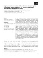
Figure 1. Diverse stress conditions activate family of eIF2 kinases and phosphorylate
eIF2α at serine 51. Protein kinases PKR, HRI, GCN2, and PERK, each respond to
different stress conditions and phosphorylate eIF2. Phosphorylated eIF2 acts as
competitive inhibitor to eIF2B, a guanine nucleotide exchange factor that is required for
conversion of eIF2-GDP to eIF2-GTP. The resulting lowered levels of eIF2-GTP repress
global translation initiation.
4
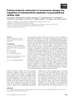
Figure 2. eIF2 in association with GTP and Met-tRNA
i
Met
participates in translation
initiation. eIF2 forms a ternary complex with GTP and Met-tRNA
i
Met
, and facilitates
joining of the initiator tRNA to the 40S ribosomal subunit. The 40S ribosomal subunit
with the ternary complex forms a 43S pre-initiation complex together with other initiation
factors eIF1, eIF1A, and eIF5. eIF4F facilitates loading of the 43S complex to 5’-cap of
mRNAs consisting of a 7’-methyl guanosine. With the help of eIF3 and the RNA helicase
eIF4A, the 43S complex progressively scans in 5’ to 3’ direction along the 5’ leader of
the mRNA in search of an initiation codon. The GTPase function of eIF5 facilitates the
eIF2-GTP hydrolysis to eIF2-GDP and Pi. Upon recognition of the initiator AUG in the P
site of the 43S complex, the Pi is released from eIF2. Following the dissociation of eIF2-
GDP, the 60S ribosomal subunit combines with the 40S ribosomal subunit to form the
5
80S complex, and translation elongation begins. A family of eIF2 kinases phosphorylates
the α subunit of eIF2 at serine 51 in response to various stress stimuli. Phosphorylated
eIF2 itself becomes a competitive inhibitor to the guanine nucleotide exchange factor,
eIF2B, which is required for recycling of eIF2-GDP to eIF2-GTP. The decrease in eIF2-
GTP levels during eIF2 phosphorylation results in reduced translation initiation. Lowered
protein synthesis allows cells sufficient time to remedy the stress damage. A program of
gene expression is also initiated in response to stress induced eIF2~P, which allows cells
to adapt to the stress conditions.
6
releases from eIF2, which occurs upon base pairing between the anticodon of tRNA
i
Met
and the initiation codon of the mRNA. The release of Pi from eIF2 is regulated by
dissociation of eIF1 from the 43S/mRNA complex (19, 20). With release of eIF2-GDP,
eIF5B facilitates the 60S ribosomal subunit joining to the 40S subunit to form the 80S
ribosomal complex. Translation elongation then begins by accepting aminoacyl-tRNAs
into the A (aminoacyl) site of the ribosome for subsequent formation of peptide bonds
(6).
3. eIF2B facilitates eIF2-GTP exchange that is inhibited by phosphorylated eIF2
Eukaryotic initiation factor 2B (eIF2B) is the guanine nucleotide exchange factor
(GEF) for eIF2 that recycles eIF2 associated with GDP to eIF2-GTP. eIF2B is a
heteropentameric complex that consists of 5 subunits, designated α, β, γ, δ and ε in
mammals, and the yeast counterparts Gcn3p, Gcd7p, Gcd1p, Gcd2p, and Gcd6p,
respectively (6, 21, 22). The γ and ε subunits facilitate the catalytic function of eIF2B,
while the α, β, and δ subunits serve a regulatory function (23-26). Phosphorylation of the
α subunit of eIF2 at serine 51 in response to various stresses alters the initiation factor
from a substrate to a competitive inhibitor of eIF2B, associating with the regulatory
portion of eIF2B and blocking exchange of eIF2-GDP to eIF2-GTP (5, 6).
Recent studies indicate that the yeast eIF5 can control the eIF2-GTP levels
through its novel GDP dissociation inhibitor (GDI) function (27). eIF5 binds to eIF2-
GDP by its carboxyl terminal domain, and sequesters available eIF2 from eIF2B, thus
reducing the exchange to eIF2-GTP. Furthermore, eIF5 was reported to have a high
7
affinity for eIF2 when its α subunit is phosphorylated, suggesting that eIF5 (GDI) can
assist in the regulation of eIF2 in response to stress conditions (27, 28).
4. Feedback regulation by eIF2 dephosphorylation
Cells reduce translation and conserve energy resources through eIF2~P during
diverse stress conditions. Dephosphorylation of eIF2 is required for resumption of
general protein synthesis. The first identified phosphatase complex dephosphorylating
eIF2~P consisted of cellular catalytic subunit protein phosphatase-1 (PP1c) and a viral
regulatory subunit encoded by the herpes simplex virus gene γ
1
34.5 (29). By
dephosphorylating eIF2, herpes virus escapes the antiviral effects of PKR. Growth arrest
and DNA damage -34 (GADD34) is a cellular homolog of γ
1
34.5 that recruits type 1
serine/threonine protein phosphatase 1 specifically to eIF2 (30-33). GADD34 is not
readily detectable in normal cells but is transcriptionally up regulated by ATF4 in
response to stress (34, 35). This feedback mechanism facilitates resumption of general
protein synthesis. Constitutive Repressor of eIF2~P (CReP) is another well-studied
protein that specifically recruits PP1 to phosphorylated eIF2 (36). Unlike GADD34,
CReP is constitutively expressed in cells. Mice deleted for CReP survive gestation, but
exhibit severe growth retardation and impaired erythropoiesis (37). Deletion of both
CReP and GADD34 in mice leads to embryonic lethality, indicating that proper
regulation of eIF2~P is important for developmental processes (37).
8
5. Different mechanisms activate the eIF2 kinases
Each of the eIF2 kinases are activated by different stresses. PERK is induced in
response to accumulation of unfolded protein in the ER (1-3). PERK is a transmembrane
protein with its regulatory region in the lumen of the ER and a cytosolic protein kinase
domain. BiP is a molecular chaperone present in the ER that is reported to bind to the
PERK luminal domain in the absence of stress. Upon stress induction, BiP dissociates
from PERK, allowing PERK dimerization (38, 39). PERK dimerization is suggested to
lead to a conformational change that contributes to autophosphorylation in the kinase
activation loop of PERK, which leads to enhanced eIF2~P. PERK phosphorylation of
eIF2 increases the expression of ATF4, which then contributes to activation of a cascade
of transcription factors, including ATF3 and CHOP (4, 11, 40, 41). This model is
supported by studies showing that the fusion of a dimerization domain to the PERK
kinase domain leads to activation of this eIF2 kinase in the absence of stress (42).
Furthermore, PERK inactivation occurs by deletion of a dimerization region from amino
acid residues 102 to 407 in PERK (38).
In addition to nutrient deprivation, GCN2 can be activated by UV irradiation and
proteasome inhibition (43-45). Central to the regulation of GCN2 is a region homologous
to histidyl-tRNA synthetase enzymes, referred to as the HisRS-related domain. The
mechanism of GCN2 activation involves binding of uncharged tRNA that accumulates
during amino acid limitation to the HisRS-related regulatory domain (22, 46-50).
Uncharged tRNAs binding to the HisRS region is suggested to cause conformational
changes in GCN2, which facilitates GCN2 autophosphorylation at sequences in the
protein kinase activation loop (47, 49). In response to UV irradiation, GCN2
9
phosphorylates eIF2 and reduces protein synthesis. Lowered protein synthesis diminishes
the levels of the labile IκBα protein, which functions as an inhibitor of the transcription
factor NF-κB (43). Thus GCN2 confers resistance to apoptosis in response to UV
irradiation through activation of NF-κB and induced expression of its target genes (43).
ATF4 is differentially regulated in response to various stresses. Even though
robust eIF2~P occurs in response to UV irradiation, ATF4 synthesis is hampered. The
underlying reason for the uncoupling between eIF2~P and induced ATF4 synthesis is that
ATF4 transcription is repressed during UV irradiation (51). Therefore, there are only low
levels of ATF4 mRNA, which cannot be readily translated in response to eIF2~P. Forced
expression of ATF4 by salubrinal pretreatment followed by UV irradiation suggests that
elevated levels of ATF4 during UV stress is detrimental to cell survival (51).
The eIF2 kinase PKR participates in an anti-viral defense mechanism that is
mediated by interferon (IFN) (52-54). PKR contains two double-stranded RNA-binding
motifs (dsRBMs) upstream of its protein kinase domain, which are central for induced
eIF2~P (52-54). Double-stranded RNAs which can accumulate during many different
viral infections is suggested to bind to the dsRBMs, facilitating a bridge between PKR
polypeptides, which triggers PKR autophosphorylation and an activated eIF2 kinase (55).
Interferons α and β that are produced during viral infection further induce this mode of
translational control by increasing the transcription of PKR. The eIF2~P in turn reduces
cellular mRNA and viral mRNA translation, thus limiting viral proliferation (55).
Viruses can mitigate the PKR-defense system by producing RNAs or proteins
that directly or indirectly alter PKR activity (55-57). For example, the NS5A protein from
hepatitis virus was reported to directly bind to PKR and inactivate the eIF2 kinase (58).
10
Vaccinia virus protein K3L mimics the substrate eIF2α, thus acting as substrate decoy
that binds to and blocks the PKR catalytic pocket (59). The E3L from vaccinia virus and
NS1 from influenza virus are proteins with dsRBMs that are proposed to bind and
sequester the dsRNA, thus precluding PKR activation (60). In the case of herpes virus, as
discussed above, the protein γ
1
34.5 recruits PP1c to dephosphorylate eIF2~P, and thereby
avoid PKR repression of translation (61). Along with eIF2~P regulation, PKR was shown
to function in a variety of signal transduction pathways, including those involving
interleukin-3, NF-κB, p53, interferon regulatory factor-1, platelet-derived growth factor,
IFN-β, STAT1, and mitogen-activated protein kinases (62). These pathways can affect
cell survival, with PKR being suggested to trigger apoptosis as part of the strategy to
thwart viral infection and proliferation.
HRI is expressed predominantly in erythroid cells. HRI is regulated by heme
through the two heme-binding regions in HRI: an N-terminal domain of HRI and in an
insert region in the protein kinase domain of HRI (63). Heme, in the presence of iron,
binds to α and β globin chains in ratio of 1:2:2, respectively. In response to iron
deficiency, conformational changes in heme cause a release from the kinase insert portion
of HRI, allowing for HRI autophosphorylation and activation to occur (63, 64). The
activated HRI then phosphorylates eIF2 and inhibits protein synthesis. Therefore, globin
protein synthesis is reduced during heme deprivation and the balance between the levels
of globin protein are retained with respect to available iron and heme content. HRI
-/-
mice
show high globin content despite iron depletion, resulting in globin aggregation and
11
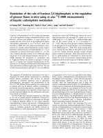
Figure 3. eIF2 Kinases, GCN2, HRI, PKR, and PERK regulate translation in
response to different stresses. Each eIF2 kinase has a conserved protein kinase domain
and distinct regulatory domains that serve to recognize different stress conditions. In
response to amino acid starvation, accumulated uncharged tRNAs bind to the HisRS-
related domain of GCN2 causing conformational changes that facilitates activation of the
protein kinases. The carboxyl terminal region allows for GCN2 dimerization and also for
this eIF2 kinase to associate with ribosomes. Heme binds to amino-terminal sequences of
HRI, along with a kinase insert region, leading to inhibition of eIF2 kinase activity. In
response to iron deficiency in erythrocytes, heme is released from HRI, facilitating
phosphorylation of eIF2. During viral infection, accumulated double-stranded RNA binds
to two dsRBMs, facilitating a conformation change that enhance PKR
autophosphorylation and increase the phosphorylation of eIF2. PERK exists as a