Modern food microbiology 7th ed phần 120
Bạn đang xem bản rút gọn của tài liệu. Xem và tải ngay bản đầy đủ của tài liệu tại đây (87.16 KB, 5 trang )
Foodborne Listeriosis
601
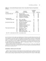
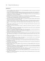
Table 25–4 Summary of Some Findings on the Thermal Destruction of L. monocytogenes
Strains Tested/State
Scott A, free suspension
Scott A, intracellular
Scott A, free suspension
F5069, intracellular
F5069, free suspension
Scott A, free suspension
Ten strains
Chicken/meat isolate
Number
of Cells
(ml)
Heating Menstrum
∼ 105
∼ 105
∼ 105
∼ 105
∼ 105
∼ 106
∼ 106
∼ 105
∼ 108
∼ 108
∼ 107
∼ 107
∼ 105
∼ 105
Sterile skim milk
Sterile skim milk
Sterile skim milk
Whole raw milk
Whole raw milk
Sterile whole milk
Sterile whole milk
Ice cream mix
pH 7.2, phosphate buffer
pH 5.9, meat slurry
Liquid whole egg
Irradiated ground meats
Beef
Minced chicken
Heating
D
z
Temperature Value Value
(◦ C)
(sec) (◦ C) Reference
71.7
71.7
71.7
71.7
71.7
71.7
71.7
79.4
70.0
70.0
72.0
62.0
70.0
70.0
1.7
2.0
0.9
1.9
1.6
5.0
3.1
2.6
9.0
13.8
36.0
61.0
6.5
6.5
6.3
6.0
6.1
8.0
7.3
7.0
—
—
7.1
4.92
7.2
6.7
9
109
11
15
15
14
14
9
8
8
41
36
88
88
been reported on its thermal destruction in dairy products. D values have been determined on many
strains of L. monocytogenes in whole and skim milk, cream, ice cream, and various meat products.
As this organism is an intracellular pathogen, several studies were undertaken to determine its relative
heat resistance inside and outside phagocytes. Overall, standard pasteurization protocols for milk are
adequate for destroying L. monocytogenes at levels of 105 –106 /ml, whether freely suspended or in
intracellular state. Some of the specific findings are presented below. For a more extensive review, see
Doyle et al.29
Dairy Products
A summary of thermal D and z values for some L. monocytogenes strains is presented in Table 25–4.
The D values indicate that the high-temperature, short-time (HTST) protocol for milk (71.7◦ C for 15
seconds) is adequate to reduce normally existing numbers of this organism below detectable levels.
The vat or low-temperature, long-time (LTLT) pasteurization protocol (62.8◦ C for 30 minutes) is even
more destructive (see Chapter 17). Employing the Scott A strain (serovar 4b from the Massachusetts
outbreak), D values ranged from 0.9 to 2.0 seconds with z values of 6.0–6.5◦ C. The F5069 strain
(serovar 4b) appeared to be a bit more heat resistant than Scott A from these results, although Scott A
was the most heat resistant of three other strains evaluated, not including F5069.11
The thermal resistance of L. monocytogenes is not affected by its intracellular position. With Scott
A freely suspended in whole raw milk at mean levels of 2.6 × 10⁵ cfu/ml and heating at 71.7◦ C for
15 seconds, no survivors could be found after five heating trials.82 In seven heating trials with Scott A
engulfed in vitro by bovine phagocytes, no survivors could be detected with a mean number of 5 × 10⁴
cfu/ml. Further, these investigators experimentally infected cows with Scott A and were still unable
to find survivors following 11 pasteurization trials at 71.7◦ C for 15 seconds with numbers of Scott A
that ranged from 1.4 × 10³ to 9.5 × 10³ cfu/ml I90I employing five strains of L. monocytogenes in
602
Modern Food Microbiology
whole milk, skim milk, and 11% nonfat milk solids, Donnelly and Briggs27 found that composition
did not affect heat destruction and that at 62.7◦ C, the D values were 60 seconds or less. The five
strains employed included serotypes 1, 3, and 4. When milk that was naturally contaminated with a
serotype 1 strain at around 104 /ml was subjected to an HTST protocol at temperatures ranging from
60◦ to 78◦ C, no viable cells could be detected at processing temperatures of 69◦ C or above.37 In
their review of the early studies on the thermal resistance of L. monocytogenes in milk, Mackey and
Bratchell89 concluded that normal pasteurization procedures will inactivate this organism but that the
margin of safety is greater for the vat protocol (LTLT) than the HTST protocol. Their mathematical
model predicted a 39 D reduction for vat and a 5.2 D reduction for HTST.
Nondairy Products
For liquid whole egg and meat products, D values are generally higher than for milk, a fact not
unpredicted, considering the effect of proteins and lipids on the thermal resistance of microorganisms
(discussed in Chapter 17). For one strain of L. monocytogenes isolated from a chicken product, D
values at 70◦ C were 6.6–8.4 seconds; they were essentially the same in beef and two poultry meats.88
In one study, viable cells could be recovered by enrichments from eight of nine samples following
heating in ground beef to 70◦ C.8 In a study of blue crabmeat, strain Scott A at levels of about 107 had
a D value of 2.61 minutes with a z of 8.4◦ C, indicating that the crabmeat pasteurization protocol of
30 minutes at 85◦ C was adequate to render the product safe from this organism.55 Processing frankfurters to an internal temperature of 160◦ F (71.1◦ C) has been shown to effect at least a 3-log cycle
reduction of strain Scott A.133 The cooking of meat products to an internal temperature of 70◦ C for 2
minutes will destroy L. monocytogenes.43,83,89
In liquid whole egg (LWE) exposed to 60◦ C for 3.5 minutes, the calculated D value for strain Scott
A was 2.1 minutes.5 However, the same strain in LWE + 10% NaCl heated at 63◦ C for 3.5 minutes
had a D of 13.7 minutes, whereas LWE + 10% sucrose gave a D of 1.9 minutes under the same
conditions. The 10% NaCl lowered the aw from 0.98 to 0.915, which could account in part for the
higher D value. Higher D values were found for seven serovars incubated at 4◦ C for 5 days followed
by 37◦ C incubation for 7 days.119 In saline, D60 values were 0.72–3.1 and D62 were 0.30–1.3 minutes.
In the sausage-type meat employed by Farber,36 the D value at 62◦ C was 61 seconds, but when
cure ingredients were added, the D value increased to 7.1 minutes, indicating some heat-protective
effects of the cure compounds, which consisted of nitrite, dextrose, lactose, corn syrup, and 3% (w/v)
NaCl. An approximate doubling in D value in ground beef containing 30% fat, 3.5% NaCl, 200 ppm
nitrite, and 300 ppm nitrate was found by Mackey et al.88 who attributed the increased heat resistance
to the 3.5% NaCl. The destruction of strain Scott A by microwave cooking was investigated by Lund
et al.,86 where more than 107 cells/g were placed in chicken stuffing and 106 –107 /g on chicken skin. By
use of a home-type microwave unit, the adequacy of heating to an internal temperature of 70◦ C for 1
minute was shown to give a 6-log reduction in numbers. The thermal destruction of L. monocytogenes
is similar to that of most other bacteria relative to pH of suspending menstrum where resistance is
higher at pH values closer to 7.0 than values in the acid range. This was demonstrated in cabbage
juice, where D values were higher at a pH of 5.6 than at 4.6.7
In a study of rainbow trout from retail markets in east Tennessee, 51% of the 74 samples were
positive for L. monocytogenes.31 The log10 means for aerobic plate counts (APC) and coliforms were
6.2 and 3.2, respectively, and the higher percentage of L. monocytogenes was associated with samples
that had the highest APC and coliform numbers. See Chapters 4, 5 and 9 for numbers of listeriae in a
variety of foods.
Foodborne Listeriosis
603
Effect of Sublethal Heating on Thermotolerance
It is unclear whether sublethal heating of L. monocytogenes cells renders them more resistant
to subsequent thermal treatments. Some investigators have reported no effect,11,13 and others have
reported increased resistance.35,38,79 In one study, the heat shocking of strain Scott A at 48◦ C for 20
minutes resulted in a 2.3-fold increase in D values at 55◦ C.79 In another study employing Scott A
in broth and ultrahigh temperature (UHT)-treated milk, an increase in heat resistance was observed
following exposure to 48◦ C for 60 minutes and subsequent exposure to 60◦ C.38 Finally, in a study
employing 10 strains at a level of about 107 /g in a sausage mix and heat shocking at 48◦ C for
30 or 60 minutes, no significant increase in thermotolerance was observed at 62◦ or 64◦ C, but those
shocked for 120 minutes did show an average 2.4-fold increase in D values at 64◦ C.35 In this study, the
thermotolerance was maintained for at least 24 hours when the cells were stored at 4◦ C. If sublethal
heating does lead to greater thermoresistance, it would not pose a problem for milk that contains fewer
than 10 cells/ml assuming that a twofold to threefold increase in D value occurs.
VIRULENCE PROPERTIES
Of listerial species, L. monocytogenes is the pathogen of concern for humans. Although L. ivanovii
can multiply in the mouse model, it does so to a much less degree than L. monocytogenes, and up to 106
cells caused no infection in the mouse.59 L. innocua, L. welshimeri, and L. seeligeri are nonpathogens,
although the last produces a hemolysin. The most significant virulence factor associated with
L. monocytogenes is listeriolysin O (LLO).
Listeriolysin O and Ivanolysin O
In general, the pathogenic/virulent strains of L. monocytogenes produce beta-hemolysis on blood
agar and acid from rhamnose but not from xylose. Strains whose hemolysis can be enhanced with
either the prepurified exo-substance or by direct use of the culture are potentially pathogenic.117
Regarding hemolysis, the evidence is overwhelming that all virulent strains of this species produce
a specific substance that is responsible for beta-hemolysis on erythrocytes and the destruction of
phagocytic cells that engulf them. The substance and perfringolysin O (PFO) have been shown to
be highly homologous to streptolysin O (SLO) and pneumolysin O (PLO). It has been purified and
shown to have a molecular weight of 60,000 Da and to consist of 504 amino acids.44,95 It is produced
mainly during the exponential growth phase, with maximum levels after 8–10 hours of growth.43 Less
LLO is synthesized at 26◦ C than at 37◦ C with high glucose, and synthesis was found to be best with
0.2% glucose at 37◦ C.24 Sorbate at a level of 2% inhibited LLO synthesis at 35◦ C under aerobic or
anaerobic conditions.72 LLO has been detected in all strains of L. monocytogenes, including some
that were nonhemolytic, but not in L. welshimeri or L. grayi. The gene that encodes its production is
chromosomal and it is designated hly. Its role in virulence is discussed below.
L. ivanovii and L. seeligeri produce thiol-dependent exotoxins that are similar but not identical to
LLO. Large quantities are produced by L. ivanovii but only small quantities by L. seeligeri.43 The L.
ivanovii thiol-activated cylolysin is ivanolysin O (ILO). Antiserum raised to the L. ivanovii product
cross-reacts with that from L. monocytogenes and SLO.73 ILO-deficient mutants have been shown to
be avirulent in mice and chick embryos.1
604
Modern Food Microbiology
Purified LLO has been shown to share in common with SLO and PLO the following properties:
activated by SH-compounds such as cysteine, inhibited by low quantities of cholesterol, and common antigenic sites as evidenced by immunological cross-reactivity. Unlike SLO, LLO is active at
a pH of 5.5 but not at pH 7.0, suggesting the possibility of its activity in macrophage phagosomes
(phagolysosomes). Its LD50 for mice is about 0.8 µg, and it induces an inflammatory response when
injected intradermally.44 It appears that LLO and the other poreforming toxins evolved from a single
progenitor gene.
Intracellular Invasion
When L. monocytogenes is contracted via the oral route, it apparently colonizes the intestinal tract
by mechanisms that are poorly understood. From the intestinal tract, the organism invades tissues,
including the placenta in pregnant women, and enters the blood stream, from which it reaches other
susceptible body cells. As an intracellular pathogen, it must first enter susceptible cells, and then it
must possess means of replicating within these cells. In the case of phagocytes, entry occurs in two
steps: directly into phagosomes and from the phagosomes into the phagocyte’s cytoplasm.
Entry or uptake into nonphagocytic cells is different. In nonphagocytic cell lines, uptake requires
surface-bound proteins of the bacterium designated In1A and In1B.78 The former has a molecular
weight of 88 kDa and the latter 65 kDa. They are involved in aiding the entry of L. monocytogenes cells into host cells. The In1A protein, i.e., internalin, and its mammalian surface receptor is
E-cadherin. It is required for entry into cultured epithelial cells, whereas In1B is required for invasion
of cultured mouse hepatocytes.32 Another invasion-associated protein of Listeria is p60, a 60-kDa
protein encoded by the iap gene. It is secreted by all species of Listeria. Another surface protein, ActA
(90 kDa), is required for actin polymerization and it allows for intracytoplasmic movement of cells.61
Ami (ca. 90 kDa) is located on the surface of L. monocytogenes and it is a bacteriolysin. From a study
of 150 food isolates, Ami was present in 149 while 283 of 300 human isolates contained this protein.61
All of the positive strains contained LLO, InlB, and ActA.
L. monocytogenes survives inside macrophages by escaping from phagolysosomal membranes into
the cytoplasm (cytosol), and this process is facilitated in part by LLO. Once inside the cytosol, the surface protein ActA (encoded by actA) aids in the formation of actin tails that propel the organism toward
the cytoplasmic membrane. At the membrane, double membrane vacuoles form. With LLO and the two
bacterial phospholipases, the phosphatidylinositol-specific phospholipase C (encoded by plcA) and the
broad-range phospholipase C (encoded by plcB), the bacteria are freed and the process is repeated upon
entry of bacteria into adjacent host cells. The latter occurs following the pushing out of the membrane
to form a filopodium (a projection), which is absorbed by an adjacent cell and the invasion process is
repeated. Thus, the spread of L. monocytogenes from cell to cell occurs without the bacterium having
to leave the inner parts of host cells. For more information, see reference 97 and Chapter 22.
Monocytosis-Producing Activity
An interesting yet incompletely understood part of the L. monocytogenes cell is a lipid-containing
component of the cell envelope that shares at least one property with the lipopolysaccharide (LPS) that
is typical of Gram-negative bacteria. In Gram-negative bacteria, LPS is located in the outer membrane,
but listeriae and other Gram-positive bacteria do not possess outer membranes. The L. monocytogenes
substance is lipoteichoic acid (LTA). It was shown several decades ago that phenol–water extracts of
L. monocytogenes cells induce the production of monocytes, and it was because of this monocytosisproducing activity (MPA) factor that the organism was given the species name monocytogenes. This
Foodborne Listeriosis
605
LTA fraction accounts for about 6% of the dry weight of cells and is associated with the plasma membrane. It has a molecular weight of about 1,000 Da, contains no amino acids or carbohydrates, and
stimulates only mononuclear cells.42 It possesses low tissue toxicity and is serologically inactive121 but
it kills macrophages in vitro.42 It has been shown to share the following properties with LPS: it is pyrogenic and lethal in rabbits, produces a localized Schwartzman reaction, contains acylated hydroxy fatty
acids, produces a positive reaction with the Limulus amoebocyte lysate (LAL) reagent, contains 2-keto3-deoxyoctonic acid (KDO), and contains heptose. Regarding its LAL reactivity, 1 µg/ml was required
to produce a positive reaction115 whereas the same can be achieved with picogram quantities of LPS.
Sphingomyelinase
L. ivanovii is known to be infectious for sheep, in which it causes abortions, and to be a prolific
producer of hemolysin on sheep erythrocytes. It has been shown to possess an LLO-like hemolysin
(ILO), sphingomyelinase, and lecithinase.73 Sphingomyelinase has a molecular weight of 27,000
Da.127 Whereas the LLO-like agent is responsible for the inner complete zone of hemolysis on sheep
erythrocytes, the halo of incomplete hemolysis that is enhanced by Rhodococcus equi appears to be
caused by the two enzymes noted. In one study, a mutant defective in sphingomyelinase and another
protein exhibited lower virulence than wild-type strains.1
ANIMAL MODELS AND INFECTIOUS DOSE
The first animal model employed to test the virulence of L. monocytogenes was the administration
of a suspension of cells into the eye of a rabbit or guinea pig (Anton’s test), where 106 cells produced
conjunctivitis.2 Chicken embryos have been studied by a large number of investigators. Inocula of
100 cells of L. monocytogenes into the allantoic sac of 10-day-old embryos led to death within 2–5
days, and the LD50 was less than 6 × 10² cells for virulent strains. L. ivanovii is also lethal by this
method. Injections of 100–30,000 cells/egg into the chorioallantoic membrane of 10-day-old chick
embryos resulted in death within 72 hours compared to about 5 days for mice.123 Although Anton’s
test and chick embryos may be used to assess the relative virulence of strains of listeriae, the mouse
is the model of choice for the additional information that it gives relative to cellular immunity.
Not only the mouse is the most widely used laboratory animal for virulence studies of listeriae,
but also it is widely used in studies of T cell immunity in general. This model is employed by use of
normal, baby, juvenile, or adult mice, as well as a variety of specially bred strains such as athymic
(T cell-deficient) nude mice. Listerial cells have been administered intraperitoneally (IP), intravenously
(IV), and intragastrically (IG). When normal adult mice are used, all smooth and hemolytic strains
of L. monocytogenes at levels of 103 –104 /mouse multiply in the spleen.59 With many strains, inocula
of 105 –106 are lethal to normal adult mice, although numbers as high as 7 × 10⁹ have been found
necessary to produce an LD50 . A low of 50 cells for 15-g mice has been reported (see below).
Whereas the IP route of injection is often used for mice, IG administration is employed to assess
gastrointestinal behavior of listeriae. The administration of L. monocytogenes to 15-g mice by the
IG route produced more rapid infection and more deaths in the first 3 days of the 6-day test than
by IP.105 By this method, the approximate 50% lethal dose (ALD50 ) ranged from 50 to 4.4 × 10⁵
cells for 15 food and clinical isolates of L. monocytogenes.105 Six- to eight-week-old mice were given
IP and oral challenges of a serovar 4b strain by one group to study their effect under normal and
compromised states. Cells were suspended in 11% nonfat milk solids and administered to four groups
of mice: normal, hydrocortisone treated, pregnant, and cimetidine treated. Minimum numbers of cells