Modern food microbiology 7th ed phần 21
Bạn đang xem bản rút gọn của tài liệu. Xem và tải ngay bản đầy đủ của tài liệu tại đây (89.72 KB, 5 trang )
Fresh Meats and Poultry
89
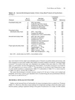
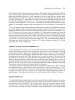
Table 4–14 General Microbiological Quality of Some Turkey Meat Products (All log Numbers
are log10 )
Products
Precooked turkey rolls
No. of
Samples
Microbial
Group/Target
% Samples
Meeting
Target
Reference
100
67
83
4
0
126
126
126
126
126
6
6
6
48
48
APC: log 3.00/g
Coliforms: log 2.00 or less/g
Enterococci: log 2.00 or less/g
Presence of salmonellae
Presence of C. perfringens
30
29
29
APC:
Presence of E. coli or salmonellae
20
21
0
187
187
187
Ground fresh turkey meat
74
75
75
75
75
75
APC: log 7.00 or less/g
Presence of coliforms
Presence of E. coli
Presence of “fecal streptococci”
Presence of S. aureus
Presence of salmonellae
51
99
41
95
69
28
77
77
77
77
77
77
Frozen ground turkey meat
50
50
50
50
50
APC 32◦ C: <106 /g
Psychrotrophs: <106 /g
MPN E. coli: <10/g
MPN S. aureus: <10/g
MPN “fecal streptococci”: <10/g
54
32
80
94
54
78
78
78
78
78
Precooked turkey
rolls/sliced turkey meat
Note: APC = Aerobic plate count; MPN = most probable number.
spp. were found to be the single most abundant genus of bacteria on poultry during processing, with
more organisms on neck-skin samples than feather-associated samples for both pre- and postscalded
carcasses.68 Corynebacterium spp. were abundant in air samples in the same study. In a 1994–1995
study of 1,297 broiler carcasses thoughout the United States, Clostridium perfringens was found in
43% and Staphylococcus aureus in 64%.177 The prevalence of Arcobacter and Campylobacter spp.
on some poultry products is summarized in Table 4–4; salmonellae in Table 4–5; L. monocytogenes
in Table 4–6; and E. coli 0157:H7 in Table 4–7.
The yeasts that developed on broiler carcasses stored at 4◦ C for up to 14 days were isolated and
identified, and at least 7 genera were identified with a Candida being the most predominant followed
by Cryptococcus and Yarrowia.86
MICROBIAL SPOILAGE OF POULTRY
Studies on the bacterial biota of fresh poultry by many investigators have revealed over 25 genera
(Table 4–1). However, when these meats undergo low-temperature spoilage, almost all workers agree
that the primary spoilage organisms belong to the genus Pseudomonas. In a study of 5,920 isolates
90
Modern Food Microbiology
from chicken carcasses,104 pseudomonads were found to constitute 30.5%, Acinetobacter 22.7%,
Flavobacterium 13.9%, and Corynebacterium 12.7%, with yeasts, Enterobacteriaceae, and others
in lower numbers. Of the pseudomonads, these investigators found that 61.8% were fluorescent on
King’s medium and that 95.2% of all pseudomonads oxidized glucose. A previous characterization of
pseudomonads on poultry undergoing spoilage was made by Barnes and Impey,11 who showed that the
pigmented pseudomonads decreased from 34% to 16% from initial storage to the development of strong
off-odors, whereas the nonpigmented actually increased from 11% to 58%. Acinetobacter and other
species of bacteria decreased along with the pseudomonads. A similar process occurs in spoiling fish.
Fungi are of considerably less importance in poultry spoilage except when antibiotics are employed
to suppress bacterial growth. When antibiotics are employed, however, molds become the primary
agents of spoilage. The genera Candida, Rhodotorula, Debaryomyces, and Yarrowia are the most
important yeasts found on poultry (Table 4–2). The essential feature of poultry spoilage is sliminess
at the outer surfaces of the carcass or cuts. The visceral cavity often displays sour odors or what is
commonly called visceral taint. This is especially true of the spoilage of New York-dressed poultry,
where the viscera are left inside. The causative organisms here are also bacteria of the type noted
above in addition to enterococci.
In a study of yeasts on fresh and spoiling poultry carcasses in South Africa, Candida and Debaryomyces spp. were the two most dominant genera on both fresh and spoiled carcasses, while Rhodotorula
was not found on spoiled carcasses.180 Trichosporon spp. were not found on fresh poultry but they were
on 5% of spoiled while 3% of fresh and 11% of spoiled contained Yarrowia. The two most abundant
species found on fresh and spoiled were Candida zeylanoides and Debaryomyces hansenii.180
S. putrefaciens grows well at 5◦ C and produces potent off-odors in 7 days when growing on chicken
muscle.124 Among odor producers in general, there is a selection of types that produce strong odors
among the varied biota that exists on fresh poultry.122 The study noted was conducted with chicken
breast muscle, which spoils differently than leg muscles because the latter have a higher pH. With
chicken leg muscle stored at 2◦ C for 16 days, 79% of the biota consisted of pseudomonads, 17% of
Acinetobacter-Moraxella, and 4% of S. putrefaciens.123 All isolates of the latter produced sulfidelike odors and this organism produces H2 S, methyl mercaptan, and dimethyl sulfide. It was not of
significance in the spoilage of chicken breast muscles.
When New York-dressed poultry undergoes microbial spoilage, the organisms make their way
through the gut walls and invade inner tissues of the intestinal cavity. The characteristic sharpness
associated with the spoilage of this type of poultry is referred to as “visceral taint.”
As poultry undergoes spoilage, off-odors are generally noted before sliminess, with the former being
first detected when log10 numbers/cm2 are about 7.2 to 8.0. Sliminess generally occurs shortly after the
appearance of off-odors, with the log10 counts/cm2 about 8.6 Total aerobic plate counts/cm2 of slimy
surface rarely go higher than log10 9.5. With the initial growth first confined to poultry surfaces, the
tissue below the skin remains essentially free of bacteria for some time. Gradually, however, bacteria
begin to enter the deep tissues, bringing about increased hydration of muscle proteins, much as occurs
with beef. Whether autolysis plays an important role in the spoilage of inner poultry tissues is not clear.
The primary reasons that poultry spoilage is mainly restricted to the surfaces are as follows. The inner
portions of poultry tissue are generally sterile, or contain relatively few organisms, which generally
do not grow at low temperatures. The spoilage biota, therefore, is restricted to the surfaces and hide
where it is deposited from water, processing, and handling. The surfaces of fresh poultry stored in an
environment of high humidity are susceptible to the growth of aerobic bacteria such as pseudomonads.
These organisms grow well on the surfaces, where they form minute colonies that later coalesce to
produce the sliminess (biofilm) characteristic of spoiled poultry. May et al.121 showed that poultry skin
Fresh Meats and Poultry
91
supports the growth of the poultry spoilage biota better than even the muscle tissue. In the advanced
stages of poultry spoilage, the surfaces will often fluoresce when illuminated with ultraviolet light
because of the presence of large numbers of fluorescent pseudomonads. Surface spoilage organisms
can be recovered directly from the slime for plating, or one can prepare slides for viewing by smearing
with portions of slime. Upon Gram staining, one may note the uniform appearance of organisms
indistinguishable from those listed. Tetrazolium (2,3,5-triphenyltetrazolium chloride) can be used
also to assess microbial activity on poultry surfaces. When an eviscerated carcass is sprayed with this
compound, a red pigment develops in areas of high microbial activity. These areas generally consist
of cut muscle surfaces and other damaged areas such as feather follicles.140 When the skin is removed
from a fresh chicken carcass, leg muscles are more likely to spoil faster than breast muscles since the
pH of the former is typically in the pH range of 6.3–6.6 while the latter is lower, i.e., 5.7–5.9.
Pseudomonads are favored at the lowest growth temperature. When poultry was spoiled at 1◦ C,
these organisms dominated while at 10◦ C and 15◦ C, enteric and other bacteria became significant.12
More information on poultry spoilage can be found in reference 28. The spoilage of poultry and other
meats under vacuum and modified atmosphere packaging is covered in Chapter 14.
CARCASS SANITIZING/WASHING
Just prior to slaughter, the outer surfaces of meat animals are laden with dust, dirt, and fecal matter.
It is inevitable that some of the microorganisms from these sources will be found on the carcasses of
slaughtered animals, and although most are nonpathogens, pathogens may be present. In an effort to
reduce the number and types of pathogens on dressed carcasses and finished products, a number of
methods have emerged:
1. Trimming—the excising of skin or outer tissue
2. Washing—the use of plain water at varying temperatures and hose pressures
3. Organic acids—the addition to wash water of acetic, citric, or lactic acid at concentrations of 2%
to 5%
4. Other chemicals—the addition to wash water of hydrogen peroxide, chlorine dioxide, or
chlorhexidine
5. Steam vacuum treatments—the application of steam for 5 to 10 seconds at 80◦ C or higher as the
final carcass preparation step
6. Combinations—the use of two or more of the above
In the USDA’s pathogen reduction program for beef carcasses, 1 out of every 300 carcasses is to
be examined by sponging 100-cm2 sections from three carcass areas (rump, flank, and brisket) for E.
coli, which should be <5 cfu/cm2 .175 The sponging method is one of six that were compared for beef
carcasses.37
Overall, a large number of studies have been conducted on most of the methods noted above for
removing microorganisms from slaughter carcasses, and reduction of APCs on the order of 1 to 3 log
cycles is common. Many studies have employed laboratory and genetically modified strains of certain
pathogens that were mixed with fresh animal feces and then rubbed onto meat cuts. The removal of
biota applied in this way may be expected to be different from that acquired naturally, but comparative
studies are wanting. The long-term effect of acid and steam treatments on meat biota is unknown
because these procedures are relatively new for commercial use. The emergence of acid-resistant
92
Modern Food Microbiology
organisms after prolonged use is a likely outcome based on the long-term and widespread use of
antimicrobials in general. It has been noted that multiple treatments are better than any one method
alone23 and this approach could reduce the emergence of resistant organisms. For catfish, the shelf
life of fillets was extended by spraying with 4% lactic or 2–4% propionic acid.52
The combined use of a hot water wash followed by organic acid rinse was more effective for hog
carcasses than either alone, and they effected about a 2-log cycle reduction.48 These investigators
suggested using water at 80◦ C. Using cold beef carcass sufaces spiked with E. coli 0157:H7 and S.
Typhimurium, a 30 s 4% lactic acid spray effected a 5.2-log reduction of the two pathogens.24 Using
a postchill 30 s lactic acid (4%) spray at 55◦ C effected an additional reduction of E. coli 0157:H7 of
2 to 2.4 log cycles and of 1.6–1.9 for S. Typhimurium.
The stages of microbial contamination of pig carcasses was investigated in an Iberian slaughterhouse.
Microbial numbers of E. coli were decreased by scalding and singing but increased by dehairing.151
The E. coli count was decreased significantly by closure of the anus and evisceration. A final carcass
wash with high pressure potable water failed to decrease microbial surface number.151
REFERENCES
1. Acuff, G.R., C. Vanderzant, M.O. Hanna, J.G. Ehlers, F.A. Golan, and F.A. Gardner. 1986. Prevalence of Campylobacter
jejuni in turkey carcass processing and further processing of turkey products. J. Food Protect. 49:712–717.
2. Acuff, G.R., C. Vanderzant, F.A. Gardner, and F.A. Golan. 1982. Examination of turkey eggs, poults, and brooder house
facilities for Campylobacter jejuni. J. Food Protect. 45:1279–1281.
3. Antoniollo, P.G., F. da Silva Bandeira, M.M. Jantzen, E.H. Duval, and W.P. da Silva. 2003. Prevalence of Listeria spp. in
feces and carcasses of a lamb packing plant in Brazil. J. Food Protect. 66:328–330.
4. Atabay, H.I., J.E.L. Corry, and S.L.W. On. 1998. Diversity and prevalence of Arcobacter spp. in broiler chickens. J. Appl.
Microbiol. 84:1007–1016.
5. Ayres, J.C. 1960. The relationship of organisms of the genus Pseudomonas to the spoilage of meat, poultry and eggs. J.
Appl. Bacteriol. 23:471–486.
6. Ayres, J.C., W.S. Ogilvy, and G.F. Stewart. 1950. Post mortem changes in stored meats. I. Microorganisms associated
with development of slime on eviscerated cut-up poultry. Food Technol. 4:199–205.
7. Baek, S.-Y., S.-Y. Lim, D.-H. Lee, K.-H. Min, and C.-M. Kim. 2000. Incidence and characterization of Listeria monocytogenes from domestic and imported foods in Korea. J. Food Protect. 63:186–189.
8. Bailey, J.S., N.A. Cox, S.E. Craven, and D.E. Cosby 2002. Serotype tracking of Salmonella through integrated broiler
chicken operations. J. Food Protect. 65:742–745.
9. Barbe, C.D., R.W. Mandigo, and R.L. Henrickson. 1966. Bacterial flora associated with rapid-processed ham. J. Food
Sci. 31:988–993.
10. Barber, D.A., P.B. Bahnson, R. Isaacson, C.J. Jones, and R.M. Weigei. 2002. Distributioon of Salmonella in swine
production ecosystems. J. Food Protect. 65:1861–1868.
11. Barnes, E.M., and C.S. Impey. 1968. Psychrophilic spoilage bacteria of poultry. J. Appl. Bacteriol. 31:97–107.
12. Barnes, E.M., and M.J. Thornley. 1966. The spoilage flora of eviscerated chickens stored at different temperatures.
J. Food Technol. 1:113–119.
13. Bauer, F.T., J.A. Carpenter, and J.O. Reagan. 1981. Prevalence of Clostridium perfringens in pork during processing.
J. Food Protect. 44:279–283.
14. Bell, W.N., and L.A. Shelef. 1978. Availability and microbial stability of retail beef-soy blends. J. Food Sci. 43:315–318,
333.
15. Borton, R.J., J. Bratzler, and J.F. Price. 1970. Effects of four species of bacteria on porcine muscle. 2. Electrophoretic
patterns of extracts of salt-soluble protein. J. Food Sci. 35:783–786.
16. Brooks, H.J.L., B.D. Mollison, K.A. Bettelheim, K. Matejka, K.A. Patterson, and V.K. Ward. 2001. Occurrence and
virulence factors of non-0157 Shiga toxin-producing Escherichia coli in retail meat in Dunedin, New Zealand. Lett. Appl.
Microbiol. 32:118–122.
Fresh Meats and Poultry
93
17. Brown, M.H., C.O. Gill, J. Hollingsworth, R. Nickelson, II, S. Seward, J.J. Sheridan, T. Stevenson, J.L. Sumner, D.M.
Theno, W.R. Usborne, and D. Zink. 2000. The role of microbiological testing in systems for assuring the safety of beef.
Int. J. Food Microbiol. 62:7–16.
18. Bryan, F.L., J.C. Ayres, and A.A. Kraft. 1968. Salmonellae associated with further-processed turkey products. Appl.
Microbiol. 16:1–9.
19. Byrne, C.M., I. Erol, J.E. Call, C.W. Kasper, D.R. Buege, C.J. Hiemke, P.J. Fedorka-Cray, A.K. Benson, F.M. Wallace,
and J.B. Luchansky. 2003. Characterization of Escherichia coli 0157:H7 from downer and healthy dairy cattle in the upper
midwest region of the United States. Appl. Environ. Microbiol. 69:4683–4688.
20. Byun, J.-S., J.S. Min, I.S. Kim, J.-W. Kim, M.-S. Chung, and M. Lee. 2003. Comparison of indicators of microbial quality
of meat during aerobic cold storage. J. Food Protect. 66:1733–1737.
21. Carl, K.E. 1975. Oregon’s experience with microbial standards for meat. J. Milk Food Technol. 38:483–486.
22. Carrami˜nana, J.J., J. Vangăuella, D. Blanco, C. Rota, A.I. Agustin, A. Ari˜no, and A. Herrera. 1997. Salmonella incidence
and distribution of serotypes throughout processing in a Spanish poultry slaughterhouse. J. Food Protect. 60:1312–1317.
23. Castillo, A.L., M. Lucia, K.J. Goodson, J.W. Savell, and G.R. Acuff. 1998. Comparison of water wash, trimming, and
combined hot water and lactic acid treatments for reducing bacteria of fecal origin on beef carcasses. J. Food Protect.
61:823–828.
24. Castillo, A., L.M. Lucia, D.B. Roberson, T.H. Stevenson, L. Mercado, and G.R. Acuff. 2001. Lactic acid sprays reduce
bacterial pathogens on cold beef carcass surfaces and in subsequently produced ground beef. J. Food Protect. 64:58–62.
25. Ceylan, E., and D.Y.C. Fung. 2000. Destruction of Yersinia enterocolitica by Lactobacillus sake and Pediococcus acidilactici duing low-temperature fermentation of Turkish dry sausage (sucuk). J. Food Sci. 65:876–879.
26. Chang, Y.H. 2000. Prevalence of Salmonella spp. in poultry broilers and shell eggs in Korea. J. Food Protect. 63:655–658.
27. Chapman, P.A., A.T.C. Malo, M. Ellin, R. Ashton, and M.A. Harkin. 2001. Escherichia coli 0157:H7 in cattle and sheep
at slaughter, on beef and lamb carcasses and in raw beef and lamb products in South Yorkshire, UK. Int. J. Food Microbiol.
64:139–150.
28. Cox, N.A., S.M. Russell, and J.S. Bailey. 1998. The microbiology of stored poultry. In The Microbiology of Meat and
Poultry, ed. A. Davies and R. Board, 266–287. New York: Kluwer Academic Publishers, Inc.
29. Cox, N.A., A.J. Mercuri, and J.E. Thompson. 1975. Enterobacteriaceae at various stages of poultry chilling. J. Food Sci.
40:44–46.
30. Craven, S.E., and A.J. Mercuri. 1977. Total aerobic and coliform counts in beef-soy and chicken-soy patties during
refrigerated storage. J. Food Protect. 40:112–115.
31. Dainty, R.H., B.G. Shaw, K.A. DeBoer, and E.S.J. Scheps. 1975. Protein changes caused by bacterial growth on beef.
J. Appl. Bacteriol. 39:73–81.
32. Davidson, C.M., M.J. Dowdell, and R.G. Board. 1973, Properties of Gram negative aerobes isolated from meats. J. Food
Sci. 38:303–305.
33. Delmore, R.J., J.N. Sofos, K.E. Belk, W.R. Lloyd, G.L. Bellinger, G.R. Schmidt, and G.C. Smith. 1999. Good manufacturing practices for improving the microbiological quality of beef variety meats. Dairy Fd. Environ. Sanit. 19:742–
752.
34. Dillon, V.M., and R.G. Board. 1991. Yeasts associated with red meats. J. Appl. Bacteriol. 71:93–108.
35. Dillon, V.M. 1998. Yeasts and moulds associated with meat and meat products. In The Microbiology of Meat and Poultry,
ed. A. Davies and R. Board, 85–117. New York: Kluwer Academic Publishers, Inc.
36. Dodd, C.C., M.W. Sanderson, J.M. Sargeant, T.G. Nagaraja, R.D. Oberst, R.A. Smith, and D.D. Griffin. 2003. Prevalence
of Escherichia coli 0157 in cattle feeds in midwestern feedlots. Appl. Environ. Microbiol. 69:5243–5247.
37. Dorsa, W.J., E.N. Cutter, and G.R. Siragusa. 1996. Evaluation of six sampling methods for recovery of bacteria from beef
carcass surfaces. Lett. Appl. Microbiol. 22:39–41.
38. Doyle, M.P., M.B. Hugdahl, and S.L. Taylor. 1981. Isolation of virulent Yersinia enterocolitica from porcine tongues.
Appl. Environ. Microbiol. 42:661–666.
39. Draughon, F.A. 1980. Effect of plant-derived extenders on microbiological stability of foods. Food Technol. 34:69–74.
40. Drosinos, E.H., and R.G. Board. 1994. Metabolic activities of pseudomonads in batch cultures in extract of minced lamb.
J. Appl. Bacteriol. 77:613–620.
41. Duffy, E.A., K.E. Belk, J.N. Sofos, G.R. Bellinger, A. Pape, and G.C. Smith. 2001. Extent of microbial contamination in
United States pork retail products. J. Food. Protect. 64:172–178.