Andersons pediatric cardiology 1171
Bạn đang xem bản rút gọn của tài liệu. Xem và tải ngay bản đầy đủ của tài liệu tại đây (187.61 KB, 3 trang )
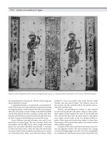
FIG.44.3 Closednormalaorticvalvephotographedfromitsarterial
aspect.Notethatthezonesofapposition(arrows)betweenthethree
leafletsextendfromtheirattachmentsatthesinutubularjunction(stars)to
thecenterofthevalvarorifice(circle).Theentiretyofthesezones
representsthe“commissures,”althoughtraditionallyitisonlytheperipheral
attachmentsthatarecreditedwiththistitle.
Furthermore,allpartsofthesezonesneedtoopenwithouthindranceifthe
valveistofunctionproperly.Thereisoneadditionalpotentialringwithinthe
outflowtract.However,thisringisavirtualentity.Itismadebycreatingaline
thatjoinstogetherthemostproximalattachmentsofthethreevalvarleaflets
withintheleftventricle.Itisthisvirtualbasalringthatisusuallyidentifiedby
echocardiographersasthevalvarannulus.Theentiretyoftheroot,consideredin
threedimensions,takestheformofacrown(Fig.44.4).
FIG.44.4 Crownlikearrangementofthesemilunarattachmentsofthe
leafletsoftheaorticvalve(redline)andhowtheseextendfromthe
sinutubularjunction(bluering)tothevirtualbasalring(greenring)
constructedbyjoiningtogetherthemostproximalattachmentsofthe
leafletswithintheleftventricle.Notethat,intheleftventricularoutflowtract,
thesemilunarattachmentscrosstheanatomicventriculoarterialjunction
onlyatthebasesofthetwovalvarsinusesthatgiverisetothecoronary
arteries,incorporatingmyocardiumatthebasesofthesetwosinuses(see
Fig.44.2).
Thereforeitfollowsthat“annulus”doesnotseemthemostobviouswordwith
whichtodescribedthesupportprovidedforthevalvarleafletswithintheroot.
Thisisthemoresobecauseamajorityofsurgeonsdescribethesemilunar
remnantsoftheleafletssubsequenttotheremovaloftheirgreaterpartsduring
operativeproceduresasthesurgical“annulus.”4Theoverallanatomic
arrangementshouldbetakenintoaccountwhenmeasurementsaremadeofthe
outflowtract.Whendiagramsaremadetoillustratetheconceptofmeasurement
ofthe“annulus,”theyoftenshowalinedrawnbetweenproximalpointsof
attachmentoftheleaflets(Fig.44.5,right).
FIG.44.5 Right,Idealizedarrangementoftheaorticroot,asfrequently
illustratedwhendemonstratingmeasurementsfortheaorticannulus.The
dimensionusuallytakenisthebasalone,betweentheattachmentsof
opposingleaflets,whichrepresentsthevirtualbasalring.Left,These
dimensions(redarrows)donotrepresentthewidestdiameteroftheroot,
whichextendsfromthenadirofoneleaflettothezoneofapposition
betweentheoppositeleaflets(greenarrow).Itisalsoimportanttotake
noteofthedimensionsasshownatrightatmid-sinusallevelandatthe
sinutubularjunction.
Suchdiagramsmustinvolveadegreeofpoeticlicenseonbehalfofthe
observerbecausethesectionillustratedcannevercutthefulldiameterofthe
arterialroot(seeFig.44.5,left).Allofthisnormalanatomyisofrelevancewhen
consideringthestructureofstenoticlesionswithintheoutflowtract,particularly
thefactthatso-calledsupravalvarstenosisinvolvestetheringofthevalvar
leafletsatthelevelofthesinutubularjunction.
ValvarStenosis
Thestenoticaorticvalveistraditionallyconsideredasshowingunicuspid,
bicuspid,ortricuspidpatterns.Strictlyspeaking,a“cusp”isapointorelevation.
Despiteitspopularity,itisnottheidealadjectivetousewhenaccountingfor
lesionsoftheabnormalvalve.Ourpreferenceistodescribeunifoliate,bifoliate,
ortrifoliatevalves,accordingtothenumberofleafletspresent.Nonetheless,we
recognizethattheabnormalvalveswillcontinuetobedescribedintermsof
cusps.However,whenmakingsuchdescriptions,itisnecessarytotakeaccount
alsoofthemorphologyofthevalvarsinuses.Thisisbecause,whenthecurtain