Pediatric emergency medicine trisk 42
Bạn đang xem bản rút gọn của tài liệu. Xem và tải ngay bản đầy đủ của tài liệu tại đây (129.29 KB, 4 trang )
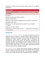
TABLE 8.1
ANATOMIC AND PHYSIOLOGIC FEATURES IN CHILDREN
PERTINENT TO LARYNGOSCOPY AND INTUBATION
Anatomy
• Size —airway structures are smaller and field of vision is narrower.
• Adenoidal hypertrophy is common in young children.
• Developing teeth —while young infants are edentulous, the underlying
alveolar ridge contains developing tooth buds that are susceptible to
disruption.
• Primary teeth in young children can be easily avulsed and/or aspirated.
• Tongue is large relative to size of oropharynx.
• Superior larynx —often referred to as “anterior,” the laryngeal opening in
infants and young children is actually located in a superior position (in
infants, the larynx is opposite C3–C4 as opposed to C4–C5 in adults). This
makes the angle of the laryngeal opening with respect to the base of the
tongue more acute and visualization more difficult.
• The hyoepiglottic ligament (connects base of tongue to epiglottis) has less
strength in young children—thus, a laryngoscope blade in the vallecula will
not elevate the epiglottis as efficiently as in an adult.
• The epiglottis of children is narrow and angled acutely with respect to the
tracheal axis; thus the epiglottis covers the tracheal opening to a greater
extent and can be more difficult to mobilize.
• The narrowest point occurs at the level of the cricoid cartilage.
Physiology
• Lung —smaller and fewer alveoli, decreased gas exchange surface area,
absent collateral channels of ventilation.
• Respiratory mechanics —the cartilaginous chest wall in children has poor
elastic recoil and leads to increased compliance. The closing volume (CV),
the volume at which terminal bronchioles collapse as a result of extrinsic
pressure exceeding intrabronchial pressure + elastic recoil forces is
frequently higher than functional residual capacity (FRC), leading to a
greater tendency for atelectasis and collapse.
• Cellular physiology —increased oxygen consumption in infants; prone to
significant increase with physiologic perturbation (e.g., fever, hypothermia).
• Cardiovascular —high vagal tone, greater tendency for bradycardia with
hypoxia, laryngeal stimulation.
High-Flow Nasal Cannula
High-flow nasal cannula (HFNC) devices deliver oxygen at rates that match or
exceed patients’ inspiratory flow rates (up to as high as 60 L/min in adults),
resulting in a higher concentration of oxygen (FiO2 ) due to limited entrainment
of room air. HFNC can deliver an FiO2 from 21% to 100%, with heated and
humidified air being well tolerated. Nasal cannulas come in sizes for neonatal,
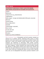
pediatric, and adult use. The flow should be adjusted based on patient age ( Table
8.2 ). If adequate respiratory support can be achieved with HFNC devices, the
need for sedation and risk of ventilator-associated pneumonia associated with
endotracheal intubation and mechanical ventilation can be avoided. In addition,
HFNC devices may be better tolerated than face masks by pediatric patients
because they are less constricting and permit speaking and feeding. HFNC has an
excellent safety profile, with only case-reportable complications related to
barotrauma (pneumothorax, pneumomediastinum, pneumocephalus).
TABLE 8.2
SUGGESTED FLOW RATES FOR HIGH-FLOW NASAL CANNULA
(HFNC)
Patient weight (kg)
Starting flow (L/min)
Maximum flow (L/min)
<5
5–10
10–20
20–40
>40
6
8
15–20
25–30
25–30
8
15
20
40
40–60
Noninvasive Ventilation
NIV encompasses mechanical respiratory support without endotracheal intubation
through either continuous positive airway pressure (CPAP) or bilevel positive
airway support (BPAP). CPAP provides a constant distending airway pressure
throughout the respiratory cycle. BPAP provides two levels of pressure referred to
as inspiratory positive airway pressure (IPAP) and expiratory positive airway
pressure (EPAP). Breaths can be synchronized to spontaneous respiratory effort
or delivered independently. Both CPAP and BPAP can be delivered through a
range of interfaces, including nasal cannula, nasal mask, full-face mask, or
helmet. Choosing an appropriate interface is often the greatest challenge in
pediatrics. Interfaces should be chosen balancing the desire to maximize comfort
and compliance while ensuring minimal leak. In addition to pressure, NIV can
also deliver supplemental oxygen and inhaled therapies such as albuterol or
racemic epinephrine.
Similar to HFNC, NIV can be used for either acute hypoxic or hypercarbic
respiratory failure. CPAP may be appropriate when hypoxemia is the primary
indication. Because it delivers higher mean airway pressures while offloading
inspiratory effort, BPAP can be used for more severe hypoxemia and to address
hypercapnia. Multiple parameters can be titrated with NIV, including CPAP
(typically 5- to 10-cm H2 O), EPAP and IPAP (typically 5 to 10 cm H2 0 and 8 to
22 cm H2 O, respectively), FiO2 , and backup ventilation rate for patients
experiencing intermittent apnea or hypopnea. If successful, NIV eliminates some
complications related to intubation, such as laryngeal or tracheal injury or
ventilator-associated pneumonia, as well the risks associated with sedation and
neuromuscular blockade. NIV should not be used in patients requiring immediate
endotracheal intubation, or those with impaired mental status or requiring airway
protection. Relative contraindications include facial injury, upper gastrointestinal
bleeding, untreated pneumothorax, and significant or escalating vasopressor
support. Most children, with appropriate coaching and provider patience during
initiation, will tolerate NIV though some require anxiolysis or sedation. Beyond
the potential for failure of NIV, the significant complications include barotrauma,
aspiration, and hemodynamic instability due to decreased venous return. Minor
complications include skin breakdown, eye irritation, nasal mucosal trauma, and
gastric distention.
APPROACH TO ENDOTRACHEAL INTUBATION
Rapid sequence intubation (RSI) is the favored approach when advanced airway
management is required in pediatrics. RSI can optimize intubating conditions and
results in higher intubation success rates than sedation alone approaches. In brief,
RSI involves the near simultaneous administration of a sedative and
neuromuscular-blocking agent (NMBA) to render a patient unconscious and
immobile, with blunted natural airway reflexes. In pure RSI, bag mask ventilation
is not performed to avoid gastric insufflation and increased risk of aspiration in
patients presumed not to be fasted. Modified or controlled RSI differs in that it
includes the delivery of gentle positive pressure breaths following medication
administration to prevent hypoxia or hypercarbia during apnea. Sedation-only
intubation may be preferred in cases of upper airway obstruction or in cases
where a difficult airway is predicted, and maintaining spontaneous respiration and
airway patency is deemed prudent ( Fig. 8.1 ).
RSI is generally considered to be a controlled series of steps starting with
preparing for the procedure and ending with management following placement.
The steps are often described as the 7 Ps: preparation, preoxygnenation,
pretreatment/preoptimization (i.e., adjunctive therapies), paralysis with induction,
positioning, placement of the endotracheal tube, and finally postintubation
management. The key components of RSI are reviewed in the sections that
follow.
EQUIPMENT
Anticipating the need for increasing airway support and having necessary
advanced airway equipment available are critical. An oxygen supply source,
devices for passive oxygen delivery, and a resuscitation bag and mask are needed
for preparation as well as during advanced airway management procedures.
Monitoring equipment including capnography should be available. For advanced
airway management, oral and nasal airways, endotracheal tubes (ETTs), stylets,
and traditional laryngoscope blades and handles and/or a videolaryngoscope in
the appropriate size for the patient should be available. To facilitate preparation,
the mnemonic “SOAP ME” (suction, oxygen, airway equipment, positioning,
monitors and meds, end-tidal CO2 monitor and equipment) can be used.
Alternatively, centers are increasingly using preintubation checklists to assure
appropriate equipment, personnel resources, and medications are available.
Endotracheal Tubes
Both cuffed and uncuffed ETTs are available for use in pediatrics. Historically,
uncuffed tubes were preferentially used in young children to allow use of the
maximal tube size that would be accommodated by the anatomic narrowing at the
level of the subglottis. More recent bronchoscopic and radiolographic data
(airway CT and MRI) suggest that the pediatric airway may by more elliptically
shaped at this level rather than circumferentially narrowed. In addition, newly
designed cuffed pediatric ETTs are manufactured with balloons that are low
profile and moved distally on the tube to avoid laryngeal structures when
appropriately positioned. Use of these new cuffed tubes has been shown to
decrease the need for tube exchange secondary to inappropriate sizing, with no
increase in postextubation stridor, need for racemic epinephrine, or long-term
complications. Pediatric Advanced Life Support (PALS) guidelines as well as the
anesthesia literature now support that, beyond the newborn period, cuffed ETTs
are equally as safe as uncuffed tubes. In addition, cuffed tubes are favored in