Anatomy for anaesthetists - 8th edition
Bạn đang xem bản rút gọn của tài liệu. Xem và tải ngay bản đầy đủ của tài liệu tại đây (7.76 MB, 366 trang )
Anatomy for Anaesthetists
AFAA01 11/11/03 13:38 Page i
This page intentionally left blank
ANATOMY FOR
ANAESTHETISTS
HAROLD ELLIS
CBE, MA, DM, FRCS, FACS(Hon)
Clinical Anatomist,
Guy’s King’s and St Thomas’s School of Biomedical Sciences, London
Emeritus Professor of Surgery,
University of London
STANLEY FELDMAN
BSc, MB, FRCA
Emeritus Professor of Anaesthetics,
Charing Cross and Westminster Medical School
WILLIAM HARROP-GRIFFITHS
MA, MB, BS, FRCA
Consultant Anaesthetist,
St Mary’s Hospital, Paddington, London
with a chapter on the
Anatomy of Pain
contributed by
ANDREW LAWSON
FFARCSI, FANZCA, FRCA, MSc
Consultant in Anaesthesia and Pain Management,
Royal Berkshire Hospital, Reading
Eighth edition
AFAA01 11/11/03 13:38 Page iii
© 1963, 1969, 1977, 1983, 1988, 1993, 1997, 2004
by Blackwell Science Ltd
a Blackwell Publishing company
Blackwell Science, Inc., 350 Main Street, Malden,
Massachusetts 02148-5020, USA
Blackwell Publishing Ltd, 9600 Garsington Road,
Oxford OX4 2DQ, UK
Blackwell Science Asia Pty Ltd, 550 Swanston Street,
Carlton, Victoria 3053, Australia
The right of the Author to be identified
as the Author of this Work has been
asserted in accordance with the
Copyright, Designs and Patents Act 1988.
All rights reserved. No part of
this publication may be reproduced,
stored in a retrieval system, or
transmitted, in any form or by any
means, electronic, mechanical,
photocopying, recording or otherwise,
except as permitted by the UK
Copyright, Designs and Patents Act
1988, without the prior permission
of the publisher.
First published 1963
Second edition 1969
Third edition 1977
Reprinted 1979
Fourth edition 1983
Fifth edition 1988
Reprinted 1990
Sixth edition 1993
Reprinted 1995
Seventh edition 1997
Reprinted 1998
Italian first edition 1972
Japanese fourth edition 1989
German fifth edition 1992
Library of Congress Cataloging-in-Publication Data
Ellis, Harold, 1926 –
Anatomy for anaesthetists / Harold Ellis, Stanley
Feldman, William Harrop-Griffiths; with a chapter
on the Anatomy of pain contributed by Andrew
Lawson.—8th ed.
p. ; cm.
Includes bibliographical references and index.
ISBN 1-4051-0663-8
1. Human anatomy. 2. Anesthesiology.
[DNLM: 1. Anatomy. 2. Anesthesia.
QS 4 E47a 2003] I. Feldman, Stanley A.
II. Harrop-Griffiths, William. III. Title.
QM23.2.E42 2003
611′.0024617—dc22
2003020753
ISBN 1405 1066 38
A catalogue record for this title is available from the
British Library
Set in 10/13.5pt Sabon by Graphicraft Limited,
Hong Kong
Printed and bound in Denmark, by Narayana Press,
Odder
Commissioning Editor: Stuart Tayler
Editorial Assistant: Katrina Chandler
Production Editor: Rebecca Huxley
Production Controller: Kate Charman
For further information on Blackwell Publishing,
visit our website:
AFAA01 11/17/03 10:26 Page iv
Contents
Part 1: The Respiratory Pathway 1
The Mouth 3
The Nose 7
The Pharynx 16
The Larynx 26
The Trachea 42
The Main Bronchi 48
The Pleura 50
The Lungs 53
Part 2: The Heart 71
The Pericardium 73
The Heart 75
Developmental Anatomy 86
Part 3: The Vertebral Canal and its Contents 95
The Vertebrae and Sacrum 97
The Spinal Meninges 119
The Spinal Cord 125
Part 4: The Peripheral Nerves 137
The Spinal Nerves 139
The Cervical Plexus 146
The Brachial Plexus 153
The Thoracic Nerves 180
The Lumbar Plexus 183
The Sacral and Coccygeal Plexuses 192
Part 5: The Autonomic Nervous System 213
Introduction 215
The Sympathetic System 218
The Parasympathetic System 228
Part 6: The Cranial Nerves 233
Introduction 235
The Olfactory Nerve (I) 238
The Optic Nerve (II) 239 v
AFAA01 11/11/03 13:38 Page v
vi Contents
The Oculomotor Nerve (III) 242
The Trochlear Nerve (IV) 244
The Trigeminal Nerve (V) 245
The Abducent Nerve (VI) 266
The Facial Nerve (VII) 267
The Auditory (Vestibulocochlear) Nerve (VIII) 272
The Glossopharyngeal Nerve (IX) 273
The Vagus Nerve (X) 276
The Accessory Nerve (XI) 282
The Hypoglossal Nerve (XII) 283
Part 7: The Anatomy of Pain 285
Introduction 287
Classification of Pain 288
Peripheral Receptors and Afferent fibres 288
The Spinal Cord and Central Projections 290
Modulation of Pain 294
The Gate Control Theory of Pain 295
The Sympathetic Nervous System and Pain 296
Part 8: Zones of Anaesthetic Interest 297
The Thoracic inlet 299
The Diaphragm 305
The Intercostal Spaces 311
The Abdominal Wall 318
The Antecubital Fossa 324
The Great Veins of the Neck 330
The Orbit and its Contents 336
Index 349
AFAA01 11/11/03 13:38 Page vi
Acknowledgements
The first two editions of this textbook were prepared in collaboration with that
skilled medical artist Miss Margaret McLarty. The illustrations for the sixth
edition were almost all drawn or redrafted by Rachel Chesterton; we thank her
for the excellent way in which they have been executed. Further illustrations for
the seventh and this edition were prepared by Jane Fallows with great skill.
vii
AFAA01 11/11/03 13:38 Page vii
Introduction
The anaesthetist requires a particularly specialized knowledge of anatomy. Some
regions of the body, for example the respiratory passages, the major veins and
the peripheral nerves, the anaesthetist must know with an intimacy of detail that
rivals or even exceeds that of the surgeon; other areas can be all but ignored.
Although formal anatomy teaching is no longer part of the syllabus of the FRCA
in the UK, its importance for the safe practice of anaesthesia is recognized by
the examiners, who always include questions on anatomy related to anaesthesia
in this examination. The role of anatomy in anaesthetic teaching is often con-
sidered merely as a prerequisite for the safe practice of local anaesthetic blocks.
However, it is also important in understanding the anatomy of the airway, the
function of the lungs, the circulation, venous access, monitoring neuromuscular
block and many other aspects of practical anaesthesia. For this reason, this book
is not intended to be a textbook for regional anaesthetic techniques; there are
many excellent books in this field. It is an anatomy book written for anaesthetists,
keeping in mind the special requirements of their daily practice.
In this eighth edition, we have revised much of the text, we have taken the
opportunity to expand and update the sections of special interest to anaesthetists
and we have included new and improved illustrations. William Harrop-Griffiths
of St Mary’s Hospital, London, joins us as our new co-author. He brings with
him special expertise in modern anaesthetic technology and has greatly assisted
us in updating the text and illustrations. Dr Andrew Lawson has fully updated
his important section on the Anatomy of Pain and has given valuable advice on
procedures relevant to the practice of pain medicine.
viii
AFAA01 11/11/03 13:38 Page viii
Part 1
The Respiratory Pathway
AFAC01 11/11/03 13:39 Page 1
This page intentionally left blank
3
The Mouth
The mouth is made up of the vestibule and the mouth cavity, the former commun-
icating with the latter through the aperture of the mouth.
The vestibule is formed by the lips and cheeks without and by the gums and
teeth within. An important feature is the opening of the parotid duct on a small
papilla opposite the 2nd upper molar tooth. Normally the walls of the vestibule
are kept together by the tone of the facial muscles; a characteristic feature of a
facial (VII) nerve paralysis is that the cheek falls away from the teeth and gums,
enabling food and drink to collect in, and dribble out of, the now patulous
vestibule.
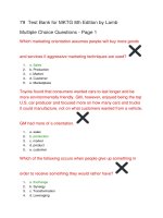
The mouth cavity (Fig. 1) is bounded by the alveolar arch of the maxilla and
the mandible, and teeth in front, the hard and soft palate above, the anterior
two-thirds of the tongue and the reflection of its mucosa forward onto the
mandible below, and the oropharyngeal isthmus behind.
The mucosa of the floor of the mouth between the tongue and mandible
bears the median frenulum linguae, on either side of which are the orifices of the
Uvula
Palatopharyngeal
arch
Palatine tonsil
Palatoglossal
arch
Fig. 1 View of the open mouth with the tongue depressed.
AFAC01 11/11/03 13:39 Page 3
4 The Respiratory Pathway
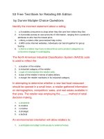
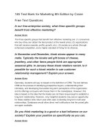
submandibular salivary glands (Fig. 2). Backwards and outwards from these
ducts extend the sublingual folds that cover the sublingual glands on each side
(Fig. 3); the majority of the ducts of these glands open as a series of tiny orifices
along the overlying fold, but some drain into the duct of the submandibular gland
(Wharton’s duct).
The palate
The hard palate is made up of the palatine processes of the maxillae and the
horizontal plates of the palatine bones. The mucous membrane covering the
hard palate is peculiar in that the stratified squamous mucosa is closely con-
nected to the underlying periosteum, so that the two dissect away at operation
as a single sheet termed the mucoperiosteum. This is thin in the midline, but
thicker more laterally due to the presence of numerous small palatine salivary
glands, an uncommon but well-recognized site for the development of mixed
salivary tumours.
The soft palate hangs like a curtain suspended from the posterior edge of the
hard palate. Its free border bears the uvula centrally and blends on either side with
the pharyngeal wall. The anterior aspect of this curtain faces the mouth cavity
and is covered by a stratified squamous epithelium. The posterior aspect is part
Frenulum linguae
Sublingual fold
Orifice of
submandibular
duct
Fig. 2 View of the open mouth with the tongue elevated.
AFAC01 11/11/03 13:39 Page 4
The Mouth 5
of the nasopharynx and is lined by a ciliated columnar epithelium under which
is a thick stratum of mucous and serous glands embedded in lymphoid tissue.
The ‘skeleton’ of the soft palate is a tough fibrous sheet termed the palatine
aponeurosis, which is attached to the posterior edge of the hard palate. The
aponeurosis is continuous on each side with the tendon of tensor palati and may,
in fact, represent an expansion of this tendon.
The muscles of the soft palate are five in number: the tensor palati, the levator
palati, the palatoglossus, the palatopharyngeus and the musculus uvulae (see
Fig. 13).
The tensor palati (tensor veli palatini) arises from the scaphoid fossa at the root
of the medial pterygoid plate, from the lateral side of the Eustachian cartilage
and the medial side of the spine of the sphenoid. Its fibres descend laterally to the
superior constrictor and the medial pterygoid plate to end in a tendon that pierces
the pharynx, loops medially around the hook of the hamulus to be inserted into
the palatine aponeurosis. Its action is to tighten and flatten the soft palate.
The levator palati (levator veli palatini) arises from the undersurface of the
petrous temporal bone and from the medial side of the Eustachian tube, enters
the upper surface of the soft palate and meets its fellow of the opposite side.
It elevates the soft palate.
The palatoglossus arises in the soft palate, descends in the palatoglossal fold
and blends with the side of the tongue. It approximates the palatoglossal folds.
Tongue
Hyoglossus
Submandibular
duct and gland
Geniohyoid
Mylohyoid
Anterior belly
of digastric
Lingual A.
Genioglossus
Lingual N.
Sublingual gland
Fig. 3 Coronal section through the floor of the mouth.
AFAC01 11/11/03 13:39 Page 5
6 The Respiratory Pathway
The palatopharyngeus descends from the soft palate in the palatopharyngeal
fold to merge into the side wall of the pharynx: some fibres become inserted along
the posterior border of the thyroid cartilage. It approximates the palatopharyngeal
folds.
The musculus uvulae takes origin from the palatine aponeurosis at the posterior
nasal spine of the palatine bone and is inserted into the uvula. Injury to the cranial
root of the accessory nerve, which supplies this muscle via the vagus nerve, results
in the uvula becoming drawn across and upwards towards the opposite side.
The tensor palati is innervated by the mandibular branch of the trigeminal
nerve via the otic ganglion (see p. 265). The other palatine muscles are supplied
by the pharyngeal plexus, which transmits cranial fibres of the accessory nerve
via the vagus.
The palatine muscles help to close off the nasopharynx from the mouth in
deglutition and phonation. In this, they are aided by contraction of the upper
part of the superior constrictor, which produces a transverse ridge on the back
and side walls of the pharynx at the level of the 2nd cervical vertebra termed
the ridge of Passavant.
Partial clefts of palate
Premaxilla
Vomer
Unilateral complete
cleft palate
Bilateral complete
cleft palate
Fig. 4 Types of cleft palate deformity.
AFAC01 11/11/03 13:39 Page 6
10 The Respiratory Pathway
The nasolacrimal duct drains tears into the anterior end of the inferior meatus
in solitary splendour.
The paranasal sinuses
The paranasal air sinuses comprise the maxillary, sphenoid, frontal and eth-
moidal sinuses. They are, in effect, the out-pouchings from the lateral wall of
the nasal cavity into which they drain; they all differ considerably from subject
to subject in their size and extent, and they are rarely symmetrical. There are
traces of the maxillary and sphenoid sinuses in the newborn; the rest become
evident about the age of 7 or 8 years in association with the eruption of the
second dentition and lengthening of the face. They only become fully developed
at adolescence.
The maxillary sinus (the antrum of Highmore) is the largest of the sinuses.
It is pyramid-shaped, and occupies the body of the maxilla (Fig. 8). The base of
this pyramid is the lateral wall of the nasal cavity and its apex points laterally
towards the zygomatic process.
Orbit
Crista galli
Ethmoidal
air cells
Superior
concha
Antral
ostium
Middle and
inferior
conchae
Maxillary
antrum
Nasal
septum
Hard palate
(maxilla)
Fig. 8 The maxillary sinus in coronal section.
AFAC01 11/11/03 13:39 Page 10
The Nose 11
The floor of the sinus extends into the alveolar process of the maxilla, which
lies about 1.25 cm below the level of the floor of the nose. Bulges in the floor
are produced by the roots of at least the 1st and 2nd molars; the number of such
projections is variable and may include all the teeth derived from the maxillary
process, i.e. the canine, premolars and molars. The floor may actually be per-
forated by one or more of the roots.
The roof is formed by the orbital plate of the maxilla, which bears the canal
of the infra-orbital branch of the maxillary nerve. Medially, the antrum drains
into the middle meatus; the ostium is situated high up on this wall and is thus
inefficiently placed from the mechanical point of view. Drainage from this sinus
is therefore dependent on the effectiveness of the cilia lining its wall. There may
be one or more accessory openings from the antrum into the middle meatus.
The sphenoid sinuses lie side by side in the body of the sphenoid. Occasionally,
they extend into the basisphenoid and the clinoid processes. They are seldom
equal in size, and the septum between them is usually incomplete. They open
into the spheno-ethmoidal recess.
The frontal sinuses occupy the frontal bone above the orbits and the root
of the nose. They are usually unequal, and their dividing septum may be incom-
plete. It is interesting that their extent is in no way related to the size of the
superciliary ridges. They drain through the frontonasal duct into the middle
meatus.
The ethmoidal sinuses or air cells are made up of some 8–10 loculi suspended
from the outer extremity of the cribriform plate of the ethmoid and bounded
laterally by its orbital plate. They thus occupy the upper lateral wall of the nasal
cavity. The cells are divided into anterior, middle and posterior groups by bony
septa; their openings have already been described above.
Blood supply
The upper part of the nasal cavity receives its arterial supply from the anterior
and posterior ethmoidal branches of the ophthalmic artery, a branch of the
internal carotid artery. The sphenopalatine branch of the maxillary artery is dis-
tributed to the lower part of the cavity and links up with the septal branch of
the superior labial branch of the facial artery on the antero-inferior part of the
septum. It is from this zone, just within the vestibule of the nose, that epistaxis
occurs in some 90% of cases (Little’s area).
A rich submucous venous plexus drains into the sphenopalatine, facial and
ophthalmic veins, and through the latter links up with the cavernous sinus. Small
tributaries also pass through the cribriform plate to veins on the undersurface of
the orbital lobe of the brain. These connections account for the potential danger
of boils and other infections within and adjacent to the nose.
AFAC01 11/11/03 13:39 Page 11
12 The Respiratory Pathway
Nerve supply
The olfactory nerve (I) supplies the specialized olfactory zone of the nose, which
occupies an area of some 2 cm
2
in the uppermost parts of the septum and lateral
walls of the nasal cavity (see p. 238).
The nerves of common sensation are derived from the nasociliary branch
of the 1st division of trigeminal nerve (V′) and also from the 2nd, or maxillary,
division (V″). These nerves are considered fully in the chapter on the cranial
nerves, but may conveniently be summarized here.
1 The septum (Fig. 9) is supplied, in the main, by the nasopalatine nerve, derived
from V″ via the pterygopalatine ganglion. The postero-superior corner receives
branches of the medial postero-superior nasal nerves from the same source, and
the anterior part of the septum is supplied by the septal branches of the anterior
ethmoidal nerve (a branch of the nasociliary branch of V′).
2 The lateral wall (Fig. 10) is innervated in its upper part, in the region of the
superior and middle conchae, by the lateral posterior superior nasal nerve. The
inferior concha receives branches from the anterior superior alveolar nerve (arising
from the maxillary nerve in the infra-orbital canal) and from the anterior (greater)
palatine nerve (derived from the pterygopalatine ganglion). The anterior part
of the lateral wall, in front of the conchae, is supplied by the anterior ethmoidal
branch of the nasociliary nerve. This branch then leaves the nasal cavity between
Septal branch of
anterior ethmoidal N.
Medial posterior
superior nasal NN.
Nasopalatine N.
Fig. 9 The nerve supply of the nasal septum.
AFAC01 11/11/03 13:39 Page 12
The Nose 13
the nasal bone and the upper nasal cartilage to become the external nasal nerve,
which supplies the outer aspect of the nose; the anterior ethmoidal nerve thus
innervates the cartilaginous tip of the nose on both its inner and outer aspects.
3 The floor is supplied in its anterior part by the antero-superior alveolar nerve
and posteriorly by the anterior (greater) palatine nerve.
4 The vestibule receives terminal twigs of the infra-orbital branch of the maxillary
nerve, which also supplies the skin immediately lateral to, and beneath, the nose.
5 The paranasal sinuses are innervated by V′ and V″. The maxillary sinus is
supplied entirely by the maxillary nerve; its roof by the infra-orbital nerve, floor
by the anterior palatine nerve, medial wall by the medial postero-superior nasal
and the anterior (greater) palatine nerves, and the anterior, posterior and lateral
walls by the superior alveolar branches. The other sinuses are supplied by the
ophthalmic division of V: the ethmoidal and sphenoidal sinuses by the anterior
and posterior ethmoidal nerves, and the frontal sinus by the supra-orbital and
supratrochlear nerves.
Structure
The vestibule is lined by a stratified squamous epithelium bearing stiff straight
hairs, sebaceous glands and sweat glands. The remainder of the nasal cavity, apart
Termination of
nasopalatine N.
Septal branch of
anterior ethmoidal N.
Lateral posterior
superior nasal NN.
Nasopalatine N.
Anterior (Greater)
palatine N.
Posterior (Lesser)
palatine NN.
Fig. 10 The nerve supply of the lateral wall of the nose.
AFAC01 11/11/03 13:39 Page 13
14 The Respiratory Pathway
from the small olfactory area, bears tall columnar ciliated cells interspersed with
mucus-secreting goblet cells, and forms a continuous epithelial sheet with the
mucosa of the nasal sinuses. Beneath the epithelium is a highly vascular connective
tissue containing copious lymphoid aggregates and carrying mucous and serous
glands. The mucous membrane is thick and velvety over the greater part of the
nasal septum and over the conchae. However, it is thin over the septum immedi-
ately within the vestibule (where the blood vessels of Little’s area show through
the mucosa) and also over the meati and the floor of the nose.
The mucosa of the nose and its accessory sinuses is closely adherent to the
underlying periosteum or perichondrium; surgically, the two layers strip away
together and are termed the mucoperiosteum.
The functions of the nose
The nose acts as a respiratory pathway, through which air becomes warmed,
humidified and filtered, as the organ of olfaction and as a resonator in speech.
There is a strong inborn reflex to breathe through the nose. This is natural to
the survival of babies during suckling. As a result, nasal obstruction may cause
gross discomfort; thus, packing the nose after surgery may cause restlessness
upon emergence from on anaesthetic, and choanal atresia may cause cyanosis
in the newborn. The natural expiratory resistance of the upper airways is in the
order of 1–2 cmH
2
O and can be increased subconsciously to provide a natural
form of continuous positive airway pressure (CPAP). Intubation of the trachea
decreases this natural expiratory resistance.
Air passes through the nose, not directly along the inferior meatus, but in
a curve through the upper reaches of the nasal cavity. The vascular cavernous
plexuses, arranged longitudinally like so many radiator pipes, increase the
temperature of the air to that of the body by the time it reaches the nasopharynx.
Water, derived partly from the mucous and serous glands, partly from the goblet
cells, but mainly by exudation from the mucous surfaces, produces nearly 100%
saturation of the inhaled air. Filtration is effected by the blanket of mucus covering
the nasal cavity and its related sinuses. The mucus is swept towards the pharynx
like a sticky conveyor belt by the action of the cilia and then swallowed. Reflex
sneezing also helps rid the nose of irritants.
The blood supply to the nasal mucosa is under reflex control. General warm-
ing of the subject produces reflex hyperaemia whereas general cooling results in
vasoconstriction. Hence the well-known observation that one’s stuffy nose in a
hot room clears on going out into the cold air.
A part of the Horner’s syndrome produced in a cervical sympathetic block
(see p. 304) is blockage of the nasal passage on that side as a result of paralysis of
sympathetic vasoconstrictor fibres to the nasal mucosa.
AFAC01 11/11/03 13:39 Page 14
The Nose 15
Nasal intubation
The major nasal air passage lies beneath the inferior concha, and a nasotracheal
tube should be encouraged to use this passage by passing it directly backwards
along the floor of the nose. Occasionally, the posterior end of the inferior turbinate
may be hypertrophied and may offer resistance to the easy passage of the tube.
The delicate mucosa of the nose and the posterior pharyngeal wall may easily
be torn, and force must never be used in this manoeuvre. Cases are on record
of nasal tubes being passed through the mucosa of the posterior pharyngeal wall
into the retropharyngeal space and of serious haemorrhage from injury to the
posterior ethmoidal vessels, which are branches of the internal carotid artery via
the ophthalmic artery and therefore impossible to control by proximal ligation.
It can be seen from Fig. 11 that a nasotracheal tube must curve anteriorly as it
passes through the nasopharynx. It may be possible to pass a well-curved tube
in a ‘blind’ manner, but more flexible tubes will need assistance if they are to
be passed through the vocal cords. Magill’s intubating forceps are commonly
Hyoid
Dens
Fig. 11 Nasal intubation; note the curvatures of the tracheal tube.
AFAC01 11/11/03 13:39 Page 15
16 The Respiratory Pathway
used for this purpose. A well-curved and rigid tube may increase the chances of
success of attempts at blind nasal intubation, but may also increase the chances
of trauma to the anterior tracheal wall.
The Pharynx
The pharynx is a wide muscular tube that forms the common upper pathway
of the respiratory and alimentary tracts. Anteriorly, it is in free communication
with the nasal cavity, the mouth and the larynx, which conveniently divide
Nasopharynx and
opening of
Eustachian tube
Oropharynx
Laryngopharynx
Fig. 12 A sagittal section through the head and neck to show the subdivisions of the pharynx.
AFAC01 11/11/03 13:39 Page 16
The Pharynx 17
it into three parts, termed the nasopharynx, oropharynx and laryngopharynx,
respectively (Figs 12 & 13). In extent, it reaches from the skull (the basilar part
of the occipital bone) to the origin of the oesophagus at the level of the 6th
cervical vertebra (C6). Posteriorly, it rests against the cervical vertebrae and the
prevertebral fascia.
The nasopharynx
The nasopharynx lies behind the nasal cavity and above the soft palate. It com-
municates with the oropharynx through the pharyngeal isthmus, which becomes
closed off during the act of swallowing (see p. 22). On the lateral wall of the
nasopharynx, 1 cm behind and just below the inferior nasal concha, lies the
pharyngeal opening of the pharyngotympanic (Eustachian) tube. The underlying
cartilage of the tube produces a bulge immediately behind its opening, termed the
tubal elevation, and behind this, in turn, is a small depression, the pharyngeal
recessafossa of Rosenmüller (see Fig. 7).
Levator veli palatini
Tensor veli palatini
Salpingo-pharyngeus
Tip of hamulus
Stylopharyngeus
Palatopharyngeus
Tip of hyoid
Superior and
inferior cornua of
thyroid cartilage
Piriform fossa
Inferior constrictor
Uvula of soft palate
Fig. 13 The pharynx: the posterior wall has been removed and the interior is viewed from behind.
On the left side the palatal muscles have been exposed.
AFAC01 11/11/03 13:39 Page 17
18 The Respiratory Pathway
The nasopharyngeal tonsil (‘adenoids’) lies on the roof and posterior wall
of the nasopharynx. It consists of a collection of lymphoid tissue covered by
ciliated epithelium and lies directly against the superior constrictor muscle; it
has no well-defined fibrous capsule. The lymphoid tissue begins to atrophy at
puberty and has all but disappeared by early adult life.
Postero-superiorly to the nasopharynx lies the sphenoid sinus that separates
the pharynx from the sella turcica containing the pituitary gland. This is the
basis of the transnasal approach to the pituitary.
The oropharynx
The mouth cavity leads into the oropharynx through the oropharyngeal isthmus,
which is bounded by the palatoglossal arches, the soft palate and the dorsum
of the tongue (see Fig. 1). The oropharynx itself extends in height from the soft
palate to the tip of the epiglottis. Its most important features are the tonsils.
The palatine tonsils are the collections of lymphoid tissue that lie on each side
in the triangle formed by the palatoglossal and palatopharyngeal arches (the
pillars of the fauces), connected across the base by the dorsum of the tongue (see
Fig. 1). The free surface of each palatine tonsil presents about 12–20 tonsillar
pits, and its upper part bears the intratonsillar cleft. This free surface is covered
by a stratified squamous epithelium: the unique combination of squamous
epithelium with underlying lymphoid tissue renders a section through the tonsil
unmistakable under the microscope.
The deep surface of the palatine tonsil may send processes of lymphoid tissue
into the dorsum of the tongue, into the soft palate and into the faucal pillars.
The palatine tonsil is bounded on this deep aspect by a dense fibrous capsule
of thickened pharyngeal aponeurosis, which is separated by a film of lax con-
nective tissue from the underlying superior constrictor muscle (Fig. 14). In the
absence of inflammation, this capsule enables complete enucleation of the tonsil
to be effected. However, after repeated quinsies, the capsule becomes adherent
to the underlying muscle and tonsillectomy then requires sharp dissection.
Vascular, lymphatic and nerve supply
The principal blood supply of the palatine tonsil is the tonsillar branch of
the facial artery which, accompanied by its two venae comitantes, pierces the
superior constrictor muscle to enter the inferior pole of the tonsil. In addition,
twigs from the lingual, ascending palatine, ascending pharyngeal and maxillary
arteries all add their contributions. Venous drainage passes into the venae com-
itantes of the tonsillar branch of the facial artery and also into a paratonsillar
vein that descends from the soft palate across the outer aspect of the tonsillar
AFAC01 11/11/03 13:39 Page 18
The Pharynx 19
capsule to pierce the pharyngeal wall into the pharyngeal venous plexus. It
is this vein which is the cause of occasional unpleasant venous bleeding after
tonsillectomy. The internal carotid artery, it should be noted, is a precious 2.5 cm
away from the tonsillar capsule, and is out of harm’s way during tonsillectomy
(see Fig. 14).
Lymph drainage is to the upper deep cervical nodes, particularly to the jugulo-
digastric node (or tonsillar node) at the point where the common facial joins the
internal jugular vein.
There is a threefold sensory nerve supply:
1 the glossopharyngeal nerve via the pharyngeal plexus;
2 the posterior palatine branch of the maxillary nerve;
3 twigs from the lingual branch of the mandibular nerve.
For this reason, infiltration anaesthesia of the tonsil is more practicable than
attempts at nerve blockade.
The palatine and pharyngeal tonsils, together with lymph collections on the
posterior part of the tongue and in relation to the Eustachian orifice, form a
more or less continuous ring of lymphoid tissue around the pharyngeal entrance,
which is termed Waldeyer’s ring.
The laryngopharynx
The third part of the pharynx extends from the tip of the epiglottis to the lower
border of the cricoid at the level of C6 (see Fig. 13). Its anterior aspect faces
Ext. and int.
carotid arteries
Ascending
pharyngeal A.
Ascending
palatine A.
Facial A.
+ tonsillar
branch
Posterior pillar
+ palatopharyngeus
Palatine tonsil
Tonsillar capsule
Anterior pillar
+ palatoglossus
Superior constrictor
muscle
Fig. 14 Diagram of the tonsil and its surroundings in horizontal section.
AFAC01 11/11/03 13:39 Page 19