Biochemistry (lippincotts illustrated reviews series)
Bạn đang xem bản rút gọn của tài liệu. Xem và tải ngay bản đầy đủ của tài liệu tại đây (24.54 MB, 1,108 trang )
XC
hange E
O
W
U
B
www.pdfgrip.com
ac
.c
tr
om
to
k
lic
C
.c
om
k
lic
C
t
Y
N
Y
U
B
to
re
.
.
k e r- s o ft w a
w
w
ac
ww
ww
tr
di
!
F-
or
O
W
t
N
di
PD
hange E
!
XC
or
PD
F-
k e r- s o ft w a
re
!
O
W
t
N
PD
di
U
B
ac
k e r- s o ft w a
Sixth Edition
www.pdfgrip.com
.c
k
tr
om
to
.c
om
k
lic
C
hange E
Y
N
Y
U
B
to
re
.
.
k e r- s o ft w a
w
w
ac
XC
Lippincott’s
Illustrated Reviews:
Biochemistr
ww
ww
tr
F-
or
O
W
t
lic
di
C
hange E
!
XC
or
PD
F-
re
O
W
t
ac
k e r- s o ft w a
Sixth Edition
Denise R. Ferrier, PhD
Professor
Department of Biochemistry and Molecular Biology
Drexel University College of Medicine
Philadelphia, Pennsylvania
Health
Philadelphia • Baltimore • New York • London
Buenos Aires • Hong Kong • Sydney • Tokyo
www.pdfgrip.com
.c
om
.c
tr
om
to
B
U
Y
N
PD
di
!
hange E
k
O
W
N
Y
U
B
to
k
lic
C
XC
or
re
.
.
k e r- s o ft w a
w
w
ac
ww
ww
tr
F-
Lippincott’s
Illustrated Reviews:
Biochemistr
t
lic
di
C
hange E
!
XC
or
PD
F-
re
hange E
N
Y
U
B
Marketing Manager: Joy Fisher-Williams
ac
om
k
.
.
tr
.c
.c
lic
k
lic
C
w
w
r
C
om
ww
ww
Development
Editor: Kelly Horvath
tr
e
k e r- s o ft w a
t
to
to
B
U
Y
N
Product Manager: Angela Collins
ac
di
!
PD
!
O
W
XC
or
or
PD
F-
O
W
ang
h
eE
XC
di
Acquisitions
Editor: Susan Rhyner
Ft
k e r- s o ft w a
re
Production Manager: David Saltzberg
Art and Page Design: Debbie McQuade
Cover Design: Holly McLaughlin
Sixth Edition
Copyright © 2014 (2011, 2008, 2005, 1994, 1987) Lippincott Williams & Wilkins, a Wolters Kluwer business
351 West Camden Street
Baltimore, MD 21201
Two Commerce Square; 2001 Market Street
Philadelphia, PA 19103
Printed in China
All rights reserved. This book is protected by copyright. No part of this book may be reproduced or transmitted in any
form or by any means, including as photocopies or scanned-in or other electronic copies, or utilized by any information
storage and retrieval system without written permission from the copyright owner, except for brief quotations embodied in
critical articles and reviews. Materials appearing in this book prepared by individuals as part of their official duties as U.S.
government employees are not covered by the above-mentioned copyright. To request permission, please contact
Lippincott Williams & Wilkins at Two Commerce Square, 2001 Market Street, Philadelphia, PA 19103, via email at
, or via website at lww.com (products and services).
987654321
Library of Congress Cataloging-in-Publication Data
Ferrier, Denise R.
Biochemistry / Denise R. Ferrier. -- 6th ed.
p. ; cm. -- (Lippincott’s illustrated reviews)
Rev. ed. of: Biochemistry / Richard A. Harvey, Denise R. Ferrier. 5th ed. c2011.
Includes bibliographical references and index.
ISBN 978-1-4511-7562-2 (alk. paper)
I. Title. II. Series: Lippincott’s illustrated reviews.
[DNLM: 1. Biochemistry--Examination Questions. QU 18.2]
612.3′9--dc23
2012025941
DISCLAIMER
Care has been taken to confirm the accuracy of the information presented and to describe generally accepted practices.
However, the authors, editors, and publisher are not responsible for errors or omissions or for any consequences from
application of the information in this book and make no warranty, expressed or implied, with respect to the currency,
completeness, or accuracy of the contents of the publication. Application of this information in a particular situation remains
www.pdfgrip.com
O
W
!
PD
!
O
W
N
Y
U
B
to
to
B
U
Y
N
considered absolute and universal recommendations.
k
lic
tr
ac
.c
C
om
k
lic
C
.c
re
.
.
wa
w
w
k
om
The authors, editors, and publisher have exerted every effort to ensure that drug selection and dosage set forth in this
ac
ww
ww
tr
or
or
PD
ange E
hange E
di
di be
theF-XCprofessional
responsibility of the practitioner; the clinical treatments described and recommended mayF-XChnot
t
t
k
e r- s o ft
e r- s o ft
text are
in accordance with the current recommendations and practice at the time of publication. However, in view of
wa
re
ongoing research, changes in government regulations, and the constant flow of information relating to drug therapy and
drug reactions, the reader is urged to check the package insert for each drug for any change in indications and dosage and
for added warnings and precautions. This is particularly important when the recommended agent is a new or infrequently
employed drug.
Some drugs and medical devices presented in this publication have Food and Drug Administration (FDA) clearance for
limited use in restricted research settings. It is the responsibility of the health care provider to ascertain the FDA status of
each drug or device planned for use in their clinical practice.
The publishers have made every effort to trace the copyright holders for borrowed material. If they have inadvertently
overlooked any, they will be pleased to make the necessary arrangements at the first opportunity.
To purchase additional copies of this book, call our customer service department at (800) 638-3030 or fax orders to
(301) 223-2320. International customers should call (301) 223-2300.
Visit Lippincott Williams & Wilkins on the Internet: . Lippincott Williams & Wilkins customer service
representatives are available from 8:30 am to 6:00 pm, EST.
www.pdfgrip.com
XC
O
W
t
k
lic
om
om
to
B
U
Y
N
Y
U
B
to
k
ww
ww
lic
di
!
F-
or
O
W
hange E
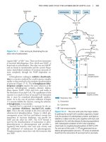
Acknowledgments
t
N
di
PD
hange E
!
XC
or
PD
F-
k e r- s o ft w a
tr
ac
.c
re
C
.c
C
ac
.
.
tr
w
w
I am grateful to my colleagues at Drexel University College of Medicine who generously
shared their expertise to help make this book as accurate and as useful to medical
students as possible. I am particularly appreciative of the many helpful comments of Dr.
Åke Rökaeus of the Karolinska Institute as they have enhanced the accuracy of this work.
In addition, the author thanks Dr. Susan K. Fried and Dr. Richard B. Horenstein for their
valuable contributions to the Obesity chapter in previous editions of this text. A special
thank you to Dr. Alan Katz for his helpful comments on the clinical aspects of the cases in
the Appendix. Ms. Barbara Engle was an invaluable sounding board throughout the
process.
k e r- s o ft w a
re
The editors and production staff of Lippincott Williams & Wilkins were an important
source of encouragement. I particularly want to acknowledge the contributions of Susan
Ryner, the Acquisitions Editor, and Angela Collins, the Managing Editor. Many thanks are
due to Kelly Horvath, Development Editor, for her assistance in the final editing of this
book. I also want to thank Deborah McQuade for her work in the assembly of the 6th
edition.
www.pdfgrip.com
www.pdfgrip.com
di
O
W
!
hange E
t
tr
ac
om
to
B
U
Y
N
PD
XC
.c
.c
om
k
lic
C
F-
k
N
Y
U
B
to
re
.
.
k e r- s o ft w a
w
w
ac
ww
ww
tr
This book is dedicated to my
husband John, whose loving
support made the task possible;
to my students, who have taught
me so much over the last 20
years; and to Richard Harvey and
the late Pamela Champe, who
helped me develop as an author.
or
O
W
t
lic
di
C
hange E
!
XC
or
PD
F-
k e r- s o ft w a
re
O
W
Y
U
to
1:
2:
3:
4:
5:
k
lic
tr
ac
.c
.c
Chapter
Chapter
Chapter
Chapter
Chapter
C
om
k
lic
C
t
B
U
B
to
re
.
.
k e r- s o ft w a
w
w
ac
ww
ww
UNIT I: Protein Structure and Function
tr
di
!
hange E
k e r- s o ft w a
Amino Acids
Structure of Proteins
Globular Proteins
Fibrous Proteins
Enzymes
UNIT II: Bioenergetics and Carbohydrate Metabolism
Chapter
Chapter
Chapter
Chapter
Chapter
Chapter
Chapter
Chapter
6:
7:
8:
9:
10:
11:
12:
13:
Bioenergetics and Oxidative Phosphorylation
Introduction to Carbohydrates
Introduction to Metabolism and Glycolysis
Tricarboxylic Acid Cycle and Pyruvate Dehydrogenase Complex
Gluconeogenesis
Glycogen Metabolism
Metabolism of Monosaccharides and Disaccharides
Pentose Phosphate Pathway and Nicotinamide Adenine Dinucleotide
Phosphate
Chapter 14: Glycosaminoglycans, Proteoglycans, and Glycoproteins
UNIT III: Lipid Metabolism
Chapter
Chapter
Chapter
Chapter
15:
16:
17:
18:
Dietary Lipids Metabolism
Fatty Acid, Ketone Body, and Triacylglycerol Metabolism
Phospholipid, Glycosphingolipid, and Eicosanoid Metabolism
Cholesterol, Lipoprotein, and Steroid Metabolism
UNIT IV: Nitrogen Metabolism
Chapter
Chapter
Chapter
Chapter
19:
20:
21:
22:
Amino Acids: Disposal of Nitrogen
Amino Acid Degradation and Synthesis
Conversion of Amino Acids to Specialized Products
Nucleotide Metabolism
UNIT V: Integration of Metabolism
Chapter
Chapter
Chapter
Chapter
23:
24:
25:
26:
Metabolic Effects of Insulin and Glucagon
The Feed–Fast Cycle
Diabetes Mellitus
Obesity
www.pdfgrip.com
om
O
W
XC
Y
N
F-
or
Contents
t
N
di
PD
hange E
!
XC
or
PD
F-
re
XC
hange E
O
W
N
Y
B
UNIT VI: Storage and Expression of Genetic Information
29:
30:
31:
32:
33:
om
to
k
lic
tr
ac
.c
C
om
.c
re
Chapter
Chapter
Chapter
Chapter
Chapter
t
U
Y
U
B
to
k
lic
k e r- s o ft w a
.
.
ac
w
w
C
ww
ww
tr
di
!
F-
N
O
W
t
PD
di
!
hange E
or
Chapter 27: Nutrition
Chapter 28: Vitamins
XC
or
PD
F-
k e r- s o ft w a
re
DNA Structure, Replication, and Repair
RNA Structure, Synthesis, and Processing
Protein Synthesis
Regulation of Gene Expression
Biotechnology and Human Disease
Appendix: Clinical Cases
Index
Bonus chapter online! Chapter 34: Blood Clotting (Use your scratch-off code
provided in the front of this book for access to this and other free online resources on
the point.)
www.pdfgrip.com
!
O
W
t
N
PD
di
U
B
Amino Acids
k
om
to
tr
ac
.c
.c
om
k
lic
C
XC
Y
N
Y
U
B
to
re
.
.
k e r- s o ft w a
w
w
ac
ww
ww
tr
hange E
UNIT I:
Protein Structure and Function
F-
or
O
W
t
lic
di
C
hange E
!
XC
or
PD
F-
k e r- s o ft w a
re
1
www.pdfgrip.com
XC
hange E
di
!
F-
N
Y
U
B
B
U
Y
N
t
or
O
W
t
PD
di
!
XC
or
PD
F-
O
W
hange E
I. OVERVIEW
ac
.c
tr
k e r- s o ft w a
Figure 1.1 Structural features of amino acids (shown in their fully protonated form).
www.pdfgrip.com
om
k
lic
C
om
k
lic
C
.c
re
.
.
k e r- s o ft w a
w
w
ac
ww
ww
tr
to
to
Proteins are the most abundant and functionally diverse molecules in living systems.
Virtually every life process depends on this class of macromolecules. For example,
enzymes and polypeptide hormones direct and regulate metabolism in the body, whereas
contractile proteins in muscle permit movement. In bone, the protein collagen forms a
framework for the deposition of calcium phosphate crystals, acting like the steel cables in
reinforced concrete. In the bloodstream, proteins, such as hemoglobin and plasma
albumin, shuttle molecules essential to life, whereas immunoglobulins fight infectious
bacteria and viruses. In short, proteins display an incredible diversity of functions, yet all
share the common structural feature of being linear polymers of amino acids. This
chapter describes the properties of amino acids. Chapter 2 explores how these simple
building blocks are joined to form proteins that have unique three-dimensional structures,
making them capable of performing specific biologic functions.
re
XC
hange E
di
!
F-
N
Y
U
B
B
U
Y
N
t
or
O
W
t
PD
di
!
XC
or
PD
F-
O
W
hange E
II. STRUCTURE
ac
om
k
lic
tr
.c
re
C
.c
om
k
lic
k e r- s o ft w a
.
.
ac
w
w
C
ww
ww
tr
to
to
Although more than 300 different amino acids have been described in nature, only 20 are
commonly found as constituents of mammalian proteins. [Note: These are the only amino
acids that are coded for by DNA, the genetic material in the cell (see p. 395).] Each
amino acid has a carboxyl group, a primary amino group (except for proline, which has a
secondary amino group), and a distinctive side chain (“R group”) bonded to the α-carbon
atom (Figure 1.1A). At physiologic pH (approximately 7.4), the carboxyl group is
dissociated, forming the negatively charged carboxylate ion (–COO–), and the amino
group is protonated (–NH3+). In proteins, almost all of these carboxyl and amino groups
are combined through peptide linkage and, in general, are not available for chemical
reaction except for hydrogen bond formation (Figure 1.1B). Thus, it is the nature of the
side chains that ultimately dictates the role an amino acid plays in a protein. It is,
therefore, useful to classify the amino acids according to the properties of their side
chains, that is, whether they are nonpolar (have an even distribution of electrons) or
polar (have an uneven distribution of electrons, such as acids and bases) as shown in
Figures 1.2 and 1.3.
k e r- s o ft w a
re
A. Amino acids with nonpolar side chains
Each of these amino acids has a nonpolar side chain that does not gain or lose protons
or participate in hydrogen or ionic bonds (see Figure 1.2). The side chains of these
amino acids can be thought of as “oily” or lipid-like, a property that promotes
hydrophobic inter-actions (see Figure 2.10, p. 19).
1. Location of nonpolar amino acids in proteins: In proteins found in aqueous
solutions (a polar environment) the side chains of the nonpolar amino acids tend to
cluster together in the interior of the protein (Figure 1.4). This phenomenon, known
as the hydrophobic effect, is the result of the hydrophobicity of the nonpolar R
groups, which act much like droplets of oil that coalesce in an aqueous environment.
The nonpolar R groups, thus, fill up the interior of the folded protein and help give it
its three-dimensional shape. However, for proteins that are located in a hydrophobic
environment, such as a membrane, the nonpolar R groups are found on the outside
surface of the protein, interacting with the lipid environment see Figure 1.4. The
importance of these hydrophobic interactions in stabilizing protein structure is
discussed on p. 19.
Figure 1.2 Classification of the 20 amino acids commonly found in proteins, according to
the charge and polarity of their side chains at acidic pH is shown here and continues in
Figure 1.3. Each amino acid is shown in its fully protonated form, with dissociable
hydrogen ions represented in red print. The pK values for the α-carboxyl and α-amino
groups of the nonpolar amino acids are similar to those shown for glycine.
www.pdfgrip.com
XC
hange E
O
W
U
B
ac
.c
tr
om
to
k
lic
C
.c
om
k
lic
C
t
Y
N
Y
U
B
to
re
.
.
k e r- s o ft w a
w
w
ac
ww
ww
tr
di
!
F-
or
O
W
t
N
di
PD
hange E
!
XC
or
PD
F-
k e r- s o ft w a
re
Figure 1.3 Classification of the 20 amino acids commonly found in proteins, according to
the charge and polarity of their side chains at acidic pH (continued from Figure 1.2).
www.pdfgrip.com
t
Y
to
B
U
Y
U
B
to
k
lic
tr
ac
.c
re
C
.c
om
k
lic
k e r- s o ft w a
.
.
ac
w
w
C
ww
ww
tr
di
!
hange E
N
!
XC
N
O
W
F-
or
or
PD
t
O
W
di
om
hange E
PD
XC
Figure 1.4 Location of nonpolar amino acids in soluble and membrane proteins.
F-
k e r- s o ft w a
re
Sickle cell anemia, a sickling disease of red blood cells, results from the
replacement of polar glutamate with nonpolar valine at the sixth position in the
β subunit of hemoglobin (see p. 36).
Figure 1.5 Comparison of the secondary amino group found in proline with the primary
amino group found in other amino acids such as alanine.
2. Proline: Proline differs from other amino acids in that its side chain and α-amino N
form a rigid, five-membered ring structure (Figure 1.5). Proline, then, has a
secondary (rather than a primary) amino group. It is frequently referred to as an
“imino acid.” The unique geometry of proline contributes to the formation of the
fibrous structure of collagen (see p. 45) and often interrupts the α-helices found in
globular proteins (see p. 26).
B. Amino acids with uncharged polar side chains
These amino acids have zero net charge at physiologic pH, although the side chains of
cysteine and tyrosine can lose a proton at an alkaline pH (see Figure 1.3). Serine,
threonine, and tyrosine each contain a polar hydroxyl group that can participate in
hydrogen bond formation (Figure 1.6). The side chains of asparagine and glutamine
each contain a carbonyl group and an amide group, both of which can also participate
in hydrogen bonds.
1. Disulfide bond: The side chain of cysteine contains a sulfhydryl (thiol) group (–
SH), which is an important component of the active site of many enzymes. In
proteins, the –SH groups of two cysteines can be oxidized to form a covalent crosswww.pdfgrip.com
link called a disulfide bond (–S–S–). Two disulfide-linked cysteines are referred to as
“cystine.” (See p. 19 for a further discussion of disulfide bond formation.)
XC
hange E
O
W
t
k
lic
tr
ac
.c
C
om
.c
re
om
to
B
U
Y
N
Y
U
B
to
k
lic
k e r- s o ft w a
.
.
ac
w
w
C
ww
ww
tr
di
!
F-
or
O
W
t
N
di
PD
hange E
!
XC
or
PD
F-
k e r- s o ft w a
re
Many extracellular proteins are stabilized by disulfide bonds. Albumin, a blood
protein that functions as a transporter for a variety of molecules, is an
example.
Figure 1.6 Hydrogen bond between the phenolic hydroxyl group of tyrosine and another
molecule containing a carbonyl group.
2. Side chains as sites of attachment for other compounds: The polar hydroxyl
group of serine; threonine; and, rarely, tyrosine, can serve as a site of attachment
for structures such as a phosphate group. In addition, the amide group of
asparagine, as well as the hydroxyl group of serine or threonine, can serve as a site
of attachment for oligosaccharide chains in glycoproteins (see p. 165).
C. Amino acids with acidic side chains
The amino acids aspartic and glutamic acid are proton donors. At physiologic pH, the
side chains of these amino acids are fully ionized, containing a negatively charged
carboxylate group (–COO–). They are, therefore, called aspartate or glutamate to
emphasize that these amino acids are negatively charged at physiologic pH (see
Figure 1.3).
D. Amino acids with basic side chains
The side chains of the basic amino acids accept protons (see Figure 1.3). At
physiologic pH, the R groups of lysine and arginine are fully ionized and positively
charged. In contrast, histidine is weakly basic, and the free amino acid is largely
uncharged at physiologic pH. However, when histidine is incorporated into a protein,
its R group can be either positively charged (protonated) or neutral, depending on the
ionic environment provided by the protein. This is an important property of histidine
that contributes to the buffering role it plays in the functioning of proteins such as
hemoglobin (see p. 31). [Note: Histidine is the only amino acid with a side chain that
can ionize within the physiologic pH range.]
www.pdfgrip.com
t
Y
to
B
U
Y
U
B
to
k
lic
tr
ac
.c
re
C
.c
om
k
lic
k e r- s o ft w a
.
.
ac
w
w
C
ww
ww
tr
di
!
hange E
N
!
XC
N
O
W
F-
or
or
PD
t
O
W
di
om
hange E
PD
XC
Figure 1.7 Abbreviations and symbols for the commonly occurring amino acids.
F-
k e r- s o ft w a
re
E. Abbreviations and symbols for commonly occurring amino acids
Each amino acid name has an associated three-letter abbreviation and a one-letter
symbol (Figure 1.7). The one-letter codes are determined by the following rules.
1. Unique first letter: If only one amino acid begins with a given letter, then that
letter is used as its symbol. For example, V = valine.
2. Most commonly occurring amino acids have priority: If more than one amino
acid begins with a particular letter, the most common of these amino acids receives
this letter as its symbol. For example, glycine is more common than glutamate, so G
= glycine.
3. Similar sounding names: Some one-letter symbols sound like the amino acid they
represent. For example, F = phenylalanine, or W = tryptophan (“twyptophan” as
Elmer Fudd would say).
4. Letter close to initial letter: For the remaining amino acids, a one-letter symbol is
assigned that is as close in the alphabet as possible to the initial letter of the amino
acid, for example, K = lysine. Furthermore, B is assigned to Asx, signifying either
aspartic acid or asparagine, Z is assigned to Glx, signifying either glutamic acid or
glutamine, and X is assigned to an unidentified amino acid.
Figure 1.8
D
and L forms of alanine are mirror images.
www.pdfgrip.com
XC
hange E
O
W
t
k
lic
tr
ac
.c
C
om
.c
re
om
to
B
U
Y
N
Y
U
B
to
k
lic
k e r- s o ft w a
.
.
ac
w
w
C
ww
ww
tr
di
!
F-
or
O
W
t
N
di
PD
hange E
!
XC
or
PD
F-
k e r- s o ft w a
re
F. Optical properties of amino acids
The α-carbon of an amino acid is attached to four different chemical groups
(asymmetric) and is, therefore, a chiral, or optically active carbon atom. Glycine is the
exception because its α-carbon has two hydrogen substituents. Amino acids with an
asymmetric center at the α-carbon can exist in two forms, designated D and L, that
are mirror images of each other (Figure 1.8). The two forms in each pair are termed
stereoisomers, optical isomers, or enantiomers. All amino acids found in proteins are
of the L configuration. However, D-amino acids are found in some antibiotics and in
bacterial cell walls. (See p. 252 for a discussion of D-amino acids.)
www.pdfgrip.com
XC
hange E
di
t
Y
U
B
B
U
Y
N
O
W
!
F-
or
O
W
t
N
di
!
XC
or
PD
F-
PD
hange E
III. ACIDIC AND BASIC PROPERTIES OF AMINO ACIDS
ac
om
k
lic
tr
.c
re
C
.c
om
k
lic
k e r- s o ft w a
.
.
ac
w
w
C
ww
ww
tr
to
to
Amino acids in aqueous solution contain weakly acidic α-carboxyl groups and weakly basic
α-amino groups. In addition, each of the acidic and basic amino acids contains an
ionizable group in its side chain. Thus, both free amino acids and some amino acids
combined in peptide linkages can act as buffers. Recall that acids may be defined as
proton donors and bases as proton acceptors. Acids (or bases) described as “weak” ionize
to only a limited extent. The concentration of protons in aqueous solution is expressed as
pH, where pH = log 1/[H+] or –log [H+]. The quantitative relationship between the pH of
the solution and concentration of a weak acid (HA) and its conjugate base (A–) is
described by the Henderson-Hasselbalch equation.
k e r- s o ft w a
re
Figure 1.9 Titration curve of acetic acid.
A. Derivation of the equation
Consider the release of a proton by a weak acid represented by HA:
The “salt” or conjugate base, A–, is the ionized form of a weak acid. By definition, the
dissociation constant of the acid, Ka, is
[Note: The larger the Ka, the stronger the acid, because most of the HA has
dissociated into H+ and A–. Conversely, the smaller the K a, the less acid has
dissociated and, therefore, the weaker the acid.] By solving for the [H +] in the above
equation, taking the logarithm of both sides of the equation, multiplying both sides of
the equation by –1, and substituting pH = –log [H+] and pKa = –log Ka, we obtain the
Henderson-Hasselbalch equation:
www.pdfgrip.com
XC
hange E
O
W
U
B
B. Buffers
ac
.c
tr
om
to
k
lic
C
.c
om
k
lic
C
t
Y
N
Y
U
B
to
re
.
.
k e r- s o ft w a
w
w
ac
ww
ww
tr
di
!
F-
or
O
W
t
N
di
PD
hange E
!
XC
or
PD
F-
k e r- s o ft w a
re
A buffer is a solution that resists change in pH following the addition of an acid or base. A
buffer can be created by mixing a weak acid (HA) with its conjugate base (A–). If an acid
such as HCl is added to a buffer, A – can neutralize it, being converted to HA in the
process. If a base is added, HA can neutralize it, being converted to A– in the process.
Maximum buffering capacity occurs at a pH equal to the pKa, but a conjugate acid–base
pair can still serve as an effective buffer when the pH of a solution is within
approximately ±1 pH unit of the pKa. If the amounts of HA and A– are equal, the pH is
equal to the pKa. As shown in Figure 1.9, a solution containing acetic acid (HA = CH3 –
COOH) and acetate (A– = CH3 –COO–) with a pKa of 4.8 resists a change in pH from pH
3.8 to 5.8, with maximum buffering at pH 4.8. At pH values less than the pKa, the
protonated acid form (CH3 – COOH) is the predominant species in solution. At pH values
greater than the pKa, the deprotonated base form (CH3 – COO–) is the predominant
species.
Figure 1.10 Ionic forms of alanine in acidic, neutral, and basic solutions.
C. Titration of an amino acid
1. Dissociation of the carboxyl group: The titration curve of an amino acid can be
analyzed in the same way as described for acetic acid. Consider alanine, for
example, which contains an ionizable α-carboxyl and α-amino group. [Note: Its –CH3
R group is nonionizable.] At a low (acidic) pH, both of these groups are protonated
(shown in Figure 1.10). As the pH of the solution is raised, the – COOH group of form
I can dissociate by donating a proton to the medium. The release of a proton results
in the formation of the carboxylate group, – COO–. This structure is shown as form
II, which is the dipolar form of the molecule (see Figure 1.10). This form, also called
a zwitterion, is the isoelectric form of alanine, that is, it has an overall (net) charge
of zero.
2. Application of the Henderson-Hasselbalch equation: The dissociation constant
of the carboxyl group of an amino acid is called K1, rather than Ka, because the
www.pdfgrip.com
molecule contains a second titratable group. The Henderson-Hasselbalch equation
can be used to analyze the dissociation of the carboxyl group of alanine in the same
way as described for acetic acid:
XC
hange E
O
W
U
B
ac
.c
tr
om
to
k
lic
C
.c
om
k
lic
C
t
Y
N
Y
to
B
U
re
.
.
k e r- s o ft w a
w
w
ac
ww
ww
tr
di
!
F-
or
O
W
t
N
di
PD
hange E
!
XC
or
PD
F-
k e r- s o ft w a
re
where I is the fully protonated form of alanine, and II is the isoelectric form of
alanine (see Figure 1.10). This equation can be rearranged and converted to its
logarithmic form to yield:
3. Dissociation of the amino group: The second titratable group of alanine is the
amino (– NH3+) group shown in Figure 1.10. This is a much weaker acid than the –
COOH group and, therefore, has a much smaller dissociation constant, K2. [Note: Its
pKa is, therefore, larger.] Release of a proton from the protonated amino group of
form II results in the fully deprotonated form of alanine, form III (see Figure 1.10).
Figure 1.11 The titration curve of alanine.
4. pKs of alanine: The sequential dissociation of protons from the carboxyl and amino
groups of alanine is summarized in Figure 1.10. Each titratable group has a pKa that
is numerically equal to the pH at which exactly one half of the protons have been
removed from that group. The pKa for the most acidic group (–COOH) is pK1,
whereas the pKa for the next most acidic group (– NH3+) is pK2. [Note: The pKa of
the α-carboxyl group of amino acids is approximately 2, whereas that of the α-amino
is approximately 9.]
www.pdfgrip.com
XC
hange E
t
k
lic
ac
.c
C
om
k
lic
C
.c
tr
om
to
B
U
Y
N
Y
U
B
to
re
.
.
k e r- s o ft w a
w
w
ac
ww
ww
tr
di
!
F-
or
O
W
t
O
W
di
N
hange E
PD
XC
!
F-
or
PD
5. Titration curve of alanine: By applying the Henderson-Hasselbalch equation to
each dissociable acidic group, it is possible to calculate the complete titration curve
of a weak acid. Figure 1.11 shows the change in pH that occurs during the addition
of base to the fully protonated form of alanine (I) to produce the completely
deprotonated form (III). Note the following:
k e r- s o ft w a
re
a. Buffer pairs: The – COOH/– COO– pair can serve as a buffer in the pH region
around pK1, and the – NH3+/– NH2 pair can buffer in the region around pK2.
b. When pH = pK: When the pH is equal to pK1 (2.3), equal amounts of forms I
and II of alanine exist in solution. When the pH is equal to pK2 (9.1), equal
amounts of forms II and III are present in solution.
c. Isoelectric point: At neutral pH, alanine exists predominantly as the dipolar
form II in which the amino and carboxyl groups are ionized, but the net charge is
zero. The isoelectric point (pI) is the pH at which an amino acid is electrically
neutral, that is, in which the sum of the positive charges equals the sum of the
negative charges. For an amino acid, such as alanine, that has only two
dissociable hydrogens (one from the α-carboxyl and one from the α-amino
group), the pI is the average of pK1 and pK2 (pI = [2.3 + 9.1]/2 = 5.7) as shown
i n Figure 1.11. The pI is, thus, midway between pK1 (2.3) and pK2 (9.1). pI
corresponds to the pH at which the form II (with a net charge of zero)
predominates and at which there are also equal amounts of forms I (net charge
of +1) and III (net charge of –1).
Separation of plasma proteins by charge typically is done at a pH above the pI
of the major proteins. Thus, the charge on the proteins is negative. In an
electric field, the proteins will move toward the positive electrode at a rate
determined by their net negative charge. Variations in the mobility pattern are
suggestive of certain diseases.
6. Net charge of amino acids at neutral pH: At physiologic pH, amino acids have a
negatively charged group (– COO–) and a positively charged group (– NH3+), both
attached to the α-carbon. [Note: Glutamate, aspartate, histidine, arginine, and lysine
have additional potentially charged groups in their side chains.] Substances such as
amino acids that can act either as an acid or a base are defined as amphoteric and
are referred to as ampholytes (amphoteric electrolytes).
Figure 1.12 The Henderson-Hasselbalch equation is used to predict: A, changes in pH as
the concentrations of HCO3– or CO2 are altered, or B, the ionic forms of drugs.
www.pdfgrip.com
XC
hange E
O
W
t
k
lic
tr
ac
.c
C
om
.c
re
om
to
B
U
Y
N
Y
U
B
to
k
lic
k e r- s o ft w a
.
.
ac
w
w
C
ww
ww
tr
di
!
F-
or
O
W
t
N
di
PD
hange E
!
XC
or
PD
F-
k e r- s o ft w a
re
D. Other applications of the Henderson-Hasselbalch equation
The Henderson-Hasselbalch equation can be used to calculate how the pH of a
physiologic solution responds to changes in the concentration of a weak acid and/or its
corresponding “salt” form. For example, in the bicarbonate buffer system, the
Henderson-Hasselbalch equation predicts how shifts in the bicarbonate ion
concentration, [HCO3–], and CO2 influence pH (Figure 1.12A). The equation is also
useful for calculating the abundance of ionic forms of acidic and basic drugs. For
example, most drugs are either weak acids or weak bases (Figure 1.12B). Acidic drugs
(HA) release a proton (H+), causing a charged anion (A–) to form.
HA
H+ + A-
Weak bases (BH+) can also release a H+. However, the protonated form of basic drugs
is usually charged, and the loss of a proton produces the uncharged base (B).
BH+
B + H+
A drug passes through membranes more readily if it is uncharged. Thus, for a weak
acid, such as aspirin, the uncharged HA can permeate through membranes, but A–
www.pdfgrip.com
cannot. For a weak base, such as morphine, the uncharged form, B, penetrates
through the cell membrane, but BH+ does not. Therefore, the effective concentration
of the permeable form of each drug at its absorption site is determined by the relative
concentrations of the charged (impermeant) and uncharged (permeant) forms. The
ratio between the two forms is determined by the pH at the site of absorption, and by
the strength of the weak acid or base, which is represented by the pKa of the ionizable
group. The Henderson-Hasselbalch equation is useful in determining how much drug is
found on either side of a membrane that separates two compartments that differ in
pH, for example, the stomach (pH 1.0–1.5) and blood plasma (pH 7.4).
XC
hange E
O
W
U
B
www.pdfgrip.com
ac
.c
tr
om
to
k
lic
C
.c
om
k
lic
C
t
Y
N
Y
U
B
to
re
.
.
k e r- s o ft w a
w
w
ac
ww
ww
tr
di
!
F-
or
O
W
t
N
di
PD
hange E
!
XC
or
PD
F-
k e r- s o ft w a
re
XC
hange E
di
!
F-
N
Y
U
B
B
U
Y
N
t
or
O
W
t
PD
di
!
XC
or
PD
F-
O
W
hange E
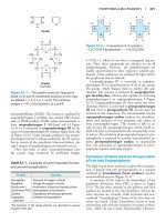
IV. CONCEPT MAPS
Figure 1.13 Symbols used in concept maps.
www.pdfgrip.com
ac
.c
tr
om
k
lic
C
om
k
lic
C
.c
re
.
.
k e r- s o ft w a
w
w
ac
ww
ww
tr
to
to
Students sometimes view biochemistry as a list of facts or equations to be memorized,
rather than a body of concepts to be understood. Details provided to enrich
understanding of these concepts inadvertently turn into distractions. What seems to be
missing is a road map—a guide that provides the student with an understanding of how
various topics fit together to make sense. Therefore, a series of biochemical concept
maps have been created to graphically illustrate relationships between ideas presented in
a chapter and to show how the information can be grouped or organized. A concept map
is, thus, a tool for visualizing the connections between concepts. Material is represented
in a hierarchic fashion, with the most inclusive, most general concepts at the top of the
map and the more specific, less general concepts arranged beneath. The concept maps
ideally function as templates or guides for organizing information, so the student can
readily find the best ways to integrate new information into knowledge they already
possess.
k e r- s o ft w a
re
XC
hange E
di
O
W
!
F-
t
Y
U
U
Y
N
O
W
t
N
di
PD
hange E
!
XC
or
PD
F-
or
A. How is a concept map constructed?
B
ac
.c
tr
om
k
lic
C
om
k
lic
.c
re
.
.
k e r- s o ft w a
w
w
C
ww
ww
ac
to
to
B
1. Concept boxes and links: Educators define concepts as “perceived regularities in
events or objects.” In the biochemical maps, concepts include abstractions (for
example, free energy), processes (for example, oxidative phosphorylation), and
compounds (for example, glucose 6-phosphate). These broadly defined concepts are
prioritized with the central idea positioned at the top of the page. The concepts that
follow from this central idea are then drawn in boxes (Figure 1.13A). The size of the
type indicates the relative importance of each idea. Lines are drawn between
concept boxes to show which are related. The label on the line defines the
relationship between two concepts, so that it reads as a valid statement, that is, the
connection creates meaning. The lines with arrowheads indicate in which direction
the connection should be read (Figure 1.14).
tr
k e r- s o ft w a
re
2. Cross-links: Unlike linear flow charts or outlines, concept maps may contain crosslinks that allow the reader to visualize complex relationships between ideas
represented in different parts of the map (Figure 1.13B), or between the map and
other chapters in this book (Figure 1.13C). Cross-links can, thus, identify concepts
that are central to more than one topic in biochemistry, empowering students to be
effective in clinical situations and on the United States Medical Licensure
Examination (USMLE) or other examinations that require integration of material.
Students learn to visually perceive nonlinear relationships between facts, in contrast
to cross-referencing within linear text.
www.pdfgrip.com