Identifying specific matrix metalloproteinase-2-inhibiting peptides through phage display-based subtractive screening
Bạn đang xem bản rút gọn của tài liệu. Xem và tải ngay bản đầy đủ của tài liệu tại đây (10.71 MB, 9 trang )
Turkish Journal of Biology
Turk J Biol
(2021) 45: 674-682
© TÜBİTAK
doi:10.3906/biy-2105-6
/>
Research Article
Identifying specific matrix metalloproteinase-2-inhibiting peptides through phage
display-based subtractive screening
1,
1
1
2
3
Aylin ÖZDEMİR BAHADIR *, Bertan Koray BALCIOĞLU , Müge SERHATLI , Şeyma IŞIK , Berrin ERDAĞ
1
Genetic Engineering and Biotechnology Institute, Marmara Research Center, TÜBİTAK, Kocaeli, Turkey
2
Department of Medical Biotechnology Institute of Health Sciences Acıbadem Mehmet Ali Aydınlar University, İstanbul, Turkey
3
Department of Medical Biology, Basic Medical Sciences, İstanbul Aydın University, İs-tanbul, Turkey
Received: 04.05.2021
Accepted/Published Online: 27.09.2021
Final Version: 14.12.2021
Abstract: Gelatinases A and B, which are members of the matrix metalloproteinase (MMP) family, play essential roles in cancer
development and metastasis, as they can break down basal membranes. Therefore, the determination and inhibition of gelatinases
is essential for cancer treatment. Peptides that can specifically block each gelatinase may, therefore, be useful for cancer treatment.
In this study, subtractive panning was carried out using a 12-mer peptide library to identify peptides that block gelatinase A activity
(MMP-2), which is a key pharmacological target. Using this method, 17 unique peptide sequences were determined. MMP-2 inhibition
by these peptides was evaluated through zymogram analyses, which revealed that four peptides inhibited MMP-2 activity by at least
65%. These four peptides were synthesized and used for in vitro wound healing using human umbilical vein endothelial cells, and two
peptides, AOMP12 and AOMP29, were found to inhibit wound healing by 40%. These peptides are, thus, potential candidates for MMP2 inhibition for cancer treatment. Furthermore, our findings suggest that our substractive biopanning screening method is a suitable
strategy for identifying peptides that selectively inhibit MMP-2.
Key words: Phage display, peptide, matrix metalloproteinases, MMP-2, subtractive panning
1. Introduction
The extracellular matrix (ECM), also referred to as
the connective tissue, is a complex structure that
surrounds and supports cells in mammalian tissues.
The remodeling of tissues and the regulation of cellular
migration are linked to the controlled destruction of
the ECM by a special group of endopeptidase enzymes.
Matrix metalloproteinases (MMPs), collectively called
matrixins, are involved in ECM degradation. The MMP
family of enzymes consists of more than 20 types (Pilcher
et al., 1999; Vihinen and Kähäri, 2002; Dufour and
Overall, 2013). MMPs are secreted primarily from the
mesenchyme and fibroblasts during tissue development
and regeneration. Normal physiological events, such as
embryonic development, inflammatory cell migration,
wound healing, and angiogenesis are modulated by the
activity of extracellular enzymes and natural inhibitors
of MMPs. In particular, MMPs are expressed at low levels
in normal tissue but are expressed at high levels in cases
of inflammation and physiological remodeling alongside
other biomarkers, such as cytokines, developmental
factors, and ECM components. Disrupted expression and
activation of MMPs have been linked to diseases such as
cancer, autoimmune diseases, tissue ulcers, atherosclerosis,
and aneurysm (Nagaset and Woessner, 1999).
Gelatinases A and B (MMP-2 and MMP-9, respectively)
are the most important members of the MMP family.
Unlike other MMPs, MMP-2 and MMP-9 break up
gelatins, one of the main elements of the basal membrane.
They are secreted as zymogens and then cleaved into the
active form (Cieplak and Strongin, 2017). In metastatic
tumors, type IV collagen activity is high as the metastatic
cells need to replace the basal membrane and blood vessels.
MMPs secreted by tumor cells, stromal fibroblasts, and
infiltrating inflammatory cells play key roles in various
stages of tumor cell invasion and metastatic progression
(Deryugina and Quigley, 2006). MMP-2 and -9 are known
to be essential to malignant cancer invasion by disrupting
the surrounding ECM and accelerating cancer metastasis
and angiogenesis (Giannopoulos et al., 2008; Kumar et
al., 2010). Therefore, both gelatinases, primarily MMP-2,
are good targets for anticancer drugs. MMPs also play a
role in angiogenesis by disrupting the vascular basement
membrane and by remodeling the ECM (Overall and
*Correspondence:
674
This work is licensed under a Creative Commons Attribution 4.0 International License.
ƯZDEMİR BAHADIR et al. / Turk J Biol
López-Otín, 2002; Chakraborti et al., 2003). Therefore,
these enzymes are important targets for the development
of antitumor drugs. Furthermore, studies have shown
that these enzymes may be used as markers for cancer
diagnosis and monitoring cancer progression (Roy et al.,
2009; Huang, 2018).
Inhibition of tumor metastasis and angiogenesis
through the suppression of MMP activity is, thus,
considered a promising strategy for cancer treatment (Cao,
2001; Guruvayoorappan and Kuttan, 2008; RaeeszadehSarmazdeh M 2020). Currently, approximately 60 drug
candidates targeting different MMPs have been developed
for the treatment of cancer, cardiovascular diseases, and
tissue inflammation. Except for Periostat, which has been
approved for periodontitis, these candidates have not
been successful due to side effects because of their lack of
specificity (Levin et al., 2017). However, the development
of chemicals that selectively inhibit specific MMPs has
been challenging. One strategy to overcome the issues
on specificity and toxicity of chemical MMP inhibitors is
through the development of biological structures that are
unique to the target MMP subtype. Therapeutic peptides
are preferred owing to their high specificity, selectivity,
and low toxicity (Marqus et al., 2017).
The phage display technology is a powerful technique
wherein unique protein or peptide structures can be
selected through biopanning against a target. In this
study, the phage display technique was used to determine
12-mer peptides that specifically bind active MMP-2.
First, a peptide library was selected for binding against
active MMP-9, and phages that bind active MMP-9
were eliminated. Then, the library was biopanned for
interaction with active MMP-2. Subtractive biopanning
was performed in this manner for a number of cycles to
enrich for specific MMP-2-binding peptides. The selected
peptides were identified and further analyzed for their
gelatinase inhibitory capacities in the hopes of discovering
peptide candidates for MMP-2 inhibition and, ultimately,
for cancer treatment.
2. Materials and methods
2.1. Bacterial strains
Escherichia coli TG1 (E. coli TG1; supE, hsdΔ5, thiΔ(lacproAB), F` [traD36 proAB+lacIq lacZΔM15]; Amersham
Pharmacia Biotech, Buckinghamshire, United Kingdom)
was used as the host for phage infection and production.
2.2. Peptide library
A rationally designed combinatorial phage display
library (Ph.D.-12 Phage Display Peptide Library) of 12mer peptide sequences was obtained from New England
Biolabs or Fermentas, Inc., Beverly, MA. Each 12-mer
peptide sequence was inserted into the NH2 terminus of
the pIII minor coat protein of the M13 bacteriophage. The
peptide sequence was followed by a short spacer (Gly-GlyGly-Ser) and the wild-type pIII sequence.
2.3. Gelatinase activation
The enzymes MMP-2 (Sigma, M1827) and MMP-9
(Sigma, M4809) were incubated and activated in 1 mM
amino-phenyl mercuric acetate (APMA, Merck KGaA,
Darmstadt, Germany) at 37 °C for 2 h (Koivunen et al.,
1999). The enzymes were then subjected to sodium dodecyl
sulfate-polyacrylamide gel electrophoresis (SDS-PAGE)
and visualized through Coomassie staining (Sambrook et
al., 1989).
2.4. Biopanning
The strategy used for one biopanning round of the Ph.D.12 peptide library to select the MMP-2 specific peptide
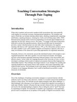
is shown in Figure 1. Wells of a high-binding microtiter
plate (TPP, Trasadingen, Switzerland) were coated with
250 ng/200 μL of activated MMP-2 or MMP-9. The wells
were washed three times with phosphate-buffered saline
(PBS) containing 0.1% Tween 20 (TPBS), and the wells
were blocked with 4% milk powder without fat for 2 h
at 4 °C. For subtractive screening, 200 µL of the Ph.D.12 library (containing 4 × 10¹⁰ phages) was first added to
wells coated with active MMP-9 figand incubated for 1 h
at room temperature. Unbound phages were recovered
and transferred to wells coated with active-MMP-2.
After 1 h of incubation, unbound phages were discarded
by intensive washing (30 times TPBS and then 30 times
with PBS). Phages bound to active-MMP2 were eluted
with 200 µL elution buffer (0.2 M glycine-HCl, pH = 2.2)
and amplified in E. coli TG1. The amplified phages were
subjected to another three rounds of selective screening
as aforementioned, to enrich for clones that are specific to
active MMP-2. After a total of four rounds of biopanning,
the MMP-2-specific clones were plated, and single pure
plaques were isolated through phage amplification (Smith
and Scott, 1993).
2.5. Phage amplification
Overnight E. coli TG1 cultures were refreshed (1/100
volume) in 20 mL nutrient broth, and phage solutions
were added to the cultures. The cultures were incubated
for 4 h at 37 °C with shaking at 220 rpm. The culture was
then centrifuged at 10.000 rpm for 10 min at 4 °C. The
supernatant was then centrifuged again under the same
conditions. Then, 80% of the supernatant was collected,
and polyethylene glycol solution (1/6 of total volume;
PEG8000 (Merck KGaA, Darmstadt, Germany)/ 2.5 M
NaCl) was added. The solution was incubated on ice for
1 h. The solution was centrifuged again at 10,000 rpm
for 15 min at 4 °C. The pellet was dissolved in 1 mL of
Tris-buffered saline (TBS), 1/6 volume of PEG8000/ 2.5
M NaCl solution was added, and the resuspensions were
incubated on ice for half an hour. Then, the mixture was
675
ÖZDEMİR BAHADIR et al. / Turk J Biol
centrifuged at 10,000 rpm for 15 min at 4 °C. The pellet
containing the phages was dissolved in a TBS. The number
of phages was determined as plaque-forming units per mL
(pfu/mL) through the phage titration method (Smith and
Scott, 1993; Bahadir et al., 2011).
2.6. ssDNA isolation from phages
After four rounds of biopanning, the final enriched MMP2 specific clones were plated, and single pure plaques
were isolated. Single-strand phage DNA was isolated
from 30 peptide clones. Overnight E. coli TG1 cultures
were refreshed (1/100 volume) in 5 mL nutrient broth.
Phage plaques were collected from the plates using a
pipet tip, added to E. coli TG1 cultures, and incubated for
4 h at 37 °C on a shaker. Infected cells were collected by
centrifugation at 4000 rpm for 10 min. Supernatants (1
mL) were collected, and PEG8000/ 2.5 M NaCl solution
were added. After incubation at room temperature for
10 min, the mixtures were centrifuged at 10,000 rpm for
10 min. The supernatants were discarded, and pellets
were dissolved in 160 μL NaI and 400 μL ethanol. After
incubation for 10 min, the mixtures were centrifuged
at 10,000 rpm for 10 min. The pellets were washed with
70% ethanol. The samples were centrifuged again under
the same conditions. Supernatants were discarded, and
the sediments were dissolved in 30 μL sterile distilled
water. The collected ssDNA were visualized through
electrophoresis on 1% agarose gel (Tomley, 1993).
2.7. Sequence analyses
To determine the peptide-encoding nucleotide sequence
contained in each phage clone, sequence analysis was
performed using the ssDNA of the phage as template. E. coli
TG1 infected with phages were plated on medium with X-gal
(5-bromo-4-chloro-3-indolyl-β-D-galactopyranoside)isopropyl β-D-1-thiogalactopyranoside (IPTG) to obtain
phage plaques. Thirty plaques were randomly selected.
Sequencing reactions were performed as described in
the Beckman Coulter Dye Terminator Cycle Sequencing
(DTCS) protocol. The purified sequencing reactions were
run on a CEQ 8000 DNA Sequencer (Beckman, Fullerton,
CA, USA).
2.8. Zymogram
MMP-2 (15 ng) was incubated overnight with 2 ×10¹¹ pfu/
mL phage clones that were enriched for binding against
active MMP-2. After 18 h, each mixture was run on 7.5%
SDS-PAGE with 20 mg/mL gelatin. After electrophoresis,
the gel was washed twice at room temperature with 2.5%
Triton-X 100 for 30 min. The gel was then kept overnight
in zymogram retention solution (6.06 g Tris-HCl, 1.47
g CaCl2, and 2.92 g NaCl per liter). The next day, the gel
was stained with Coomassie Blue R. The density of the
bands were analyzed using the Bio-Rad Multi-Analyst
program (Tajhya et al., 2017). The gel was scanned using
676
a digital scanner, and MMP-2 activity was determined by
measuring the peak area of each band. The peak percentage
was normalized relative to the activity of MMP-2 (positive
control) and wild-type M13 phage (negative control).
2.9. Peptide synthesis
Each MMP-2-specific peptide was synthesized (5
mg) and purified through high-performance liquid
chromatography (HPLC) at 95% purity by Peptron Inc.
(Daejeon, South Korea) for use in cell culture assays.
2.10. Cell culture assays
For cell culture study, human umbilical vein endothelial
cells (HUVECs; C2519A) were purchased from Lonza,
Basel, Switzerland. The cells were cultured in endothelial
complete media, which is composed of endothelial basal
media EBM-2 (CC-3156, Lonza) supplemented with
1% penicillin (10,000 units/mL)- streptomycin (10,000
units/mL) (Gibco, Cat No 15140122) and endothelial
cell growth medium 2 (EGM-2) SingleQuots (CC-4176,
Lonza, Switzerland) containing growth factors (insulinlike growth factor, human fibroblast growth factor,
human epidermal growth factor, and vascular endothelial
growth factor), and supplements (ascorbic acid, heparin,
hydrocortisone, and fetal bovine serum). Cell passage
was carried out three times per week, considering the cell
doubling time (Erdag et al., 2011).
2.11. Cytotoxicity assays
A cytotoxicity assay based on impedance was performed
using the xCELLigence real-time cell analyzer multiple
plates (RTCA MP; Agilent Technologies, CA, USA).
HUVECs were suspended in EBM-2 medium (CC-3156)
and seeded at a density of 5 × 10³ cells per well into a
disposable sterile 16-well E-plate of the xCELLigence
RTCA MP. Various concentrations of MMPs (50, 100, 500,
and 1000 μM) in EGM-2 complete medium were added
to the wells. The cells were maintained in a humidified
incubator at 37 °C with 5% CO2. The experiment was
terminated at the end of the time period (72h), and the
data were evaluated using the RTCA Software Pro software.
2.12. Cell migration assays
Cell migration was investigated using a wound-healing
assay. HUVEC cells were detached from the cell culture
flask using 0.25% trypsin-EDTA and harvested when cell
confluence was approximately 70%–80%. After confirming
that cell viability was at least 90%, the cells were suspended
in complete medium to a density of 1 × 10⁵ cells/mL. The
cells were seeded (1 × 10⁴ cells/well) into 96-well plates
(Greiner CellStar, USA), and incubated at 37 °C in a 5%
CO2 humidified incubator for 24 h. After incubation,
cell monolayers were gently scratched using AutoScratch
(BioTek, USA) equipment to create repeatable wound
areas. Cells were washed with medium to remove cell
debris and treated with MMP-2 peptides (100 μM) in
ÖZDEMİR BAHADIR et al. / Turk J Biol
complete medium. EDTA (2 mM) was used as the positive
control for MMP-2 inhibition. Cellular migration towards
the wound area was captured and investigated every hour
over a 24-h period using the 4× objective and the brightfield filter of the Cytation 5 (BioTek) cell imager. Wound
areas were measured using the Gen5 software, and the
relative wound area per time-point was calculated based
on the wound at 0 h.
3. Results
3.1. Biopanning and phage amplification
In order to select 12-mer peptides that selectively bind
active MMP-2, a 12-mer phage peptide library, which
includes 4 × 10¹⁰ phages, was screened against APMAactivated MMP-9 to first exclude MMP-9-specific
phages from further screening. Then, the remaining 12mer peptides were screened against active MMP-2, and
the peptides that bound active MMP-2 were recovered
through elution (Figure 1).
The biopanning cycle was repeated four times to
determine MMP-2-specific peptides. After each cycle
(except for the last cycle), the recovered and amplified
phages were titrated (Bahadir et al., 2011, Smith and Scott,
1993) to ensure that the same number of phages was used
for each biopanning cycle. Even with equal amounts of
phage (1 × 10¹² pfu/mL) used at the start of the selection
cycles against MMP-2, we detected approximately 103-fold
increase in the number of phages recovered from the 3rd
cycle relative to number recovered in the 1st cycle (Figure
2). Biopanning was stopped as the number of phages
obtained after the fourth round of biopanning did not
increase (Figure 2). The quantities of phages bound to
MMP-9 were also determined during the selection cycles.
After the fourth round of biopanning, the number of
phages bound to MMP-9 also increased.
3.2. Sequence and zymogram assay of phage plaques
Zymogram results were normalized relative to the activity
of the wild M13 phage, which displayed approximately
25% inhibition of gelatinase activity on an SDS-PAGE gel
with gelatin (Figure 3) (Lorenzl et al., 2003; Atkinson et
al., 2004).
Thirty plaques were randomly selected for sequencing.
The ssDNA from 30 phages were sequenced, and seventeen
unique peptide sequences were identified. The sequences
and copy numbers the peptides are shown in Figure 4.
Based on the 17 unique peptide-encoding sequences,
homologue sequence motifs, including WHW, HW, WH,
or HWW, were observed in all but three peptides (AOMP3,
AOMP4, and AOMP23).
The gelatinase inhibitory activities of all 17 phage
clones were tested. Based on the gelatin degradation value
obtained from the control sample (active-MMP-2), the
percentage of gelatin degradation value was calculated for
each phage clone. The peptides AOMP23, AOMP26, and
AOMP27 showed lower MMP-2 inhibitory, with MMP-2
activity remaining over 60% (Figure 4). The best MMP-2
inhibitory peptides were AOMP5, AOMP 12, AOMP 28,
AOMP 29, and AOMP 13, which reduced MMP-2 activity
Depletion of anti-MMP-9
binding phages
Phage display peptide
library
�
After 3 rounds of
biopanning phage plaques
were formed
Active MMP-9
Active MMP-2
Amplification
Bound phages
Figure 1. Schematic representation of the subtractive selection process on a phage display peptide library. The phage library was exposed
to active matrix metalloproteinase (MMP)-9 to deplete MMP-9-binding phages. Phages that did not bind MMP-9 were exposed to
active MMP-2. The bound pages were then eluted and amplified and exposed again to active MMP-9. This cycle was repeated four times.
After the last cycle, the recovered phage clones were amplified, titrated, and sequenced.
677
ÖZDEMİR BAHADIR et al. / Turk J Biol
4500
4000
Number of phage recovered
(x106 pu/ml)
3500
3000
2500
2000
1500
1000
500
0
1st
2nd
3rd
Round of biopanning
Phages bound to active MMP-9
4th
Phages bound to active MMP-2
Figure 2. Recovery of matrix metalloproteinase (MMP)-9- and MMP-2-binding
phages after each round of biopanning.
100
90
Relative activity of MMP-2 (%)
80
70
60
50
40
30
20
10
0
MMP-2
MMP-2 + 2mM
EDTA
MMP-2 + 1x10¹¹ MMP-2 + 2x10¹¹
pfu/ml phage
pfu/ml phage
Figure 3. Effects of the M13 bacteriophage on matrix metalloproteinase
(MMP)-2 activity.
to under 40%. These preliminary findings indicate that our
phage display selection of MMP-2 inhibitory peptides is a
suitable screening strategy.
3.3. Cell culture assays
The top 4 peptides that inhibited MMP-2 activity (AOMP5,
AOMP 12, AOMP 28, and AOMP 29) were synthesized
for use in wound healing assays. First, the toxicity of the
peptides on HUVECs were checked, and no toxicity was
observed (data not shown). To determine the inhibitory
effects of the peptides on HUVEC proliferation, a wound
healing experiment was performed. Without peptides
group was labeled as w/o. HUVEC monolayers were
scratched, and the cells were treated with 100 μM of each
peptide. After 24 h of incubation with 2 mM EDTA (as
678
a control), no significant wound healing was observed,
and 82% of the wound area was still uncovered (Figure
5a). Peptide AOMP28 did not inhibit wound healing.
However, peptides AOMP29 and AOMP12 left a wound
opening area of 42% and 40%, respectively.
4. Discussion
Gelatinases (MMP-2 and MMP-9) play essential roles in
cancer progression and metastasis, as they can break down
basal membranes. Therefore, the detection and inhibition
of gelatinases is essential for cancer treatment.
Several studies have been conducted on the selection
of MMP-2 and MMP-9 binding peptides from a peptide
library. Koivunen et al. selected several peptides from a
ÖZDEMİR BAHADIR et al. / Turk J Biol
Figure 4. The different phage clones and their effects on matrix metalloproteinase
(MMP)-2 activity. Amino acid sequences and copy numbers of the peptides are shown.
The zymogram assay was used to measure inhibitory effects of the peptides on the
gelatinase activity of MMP-2. EDTA (2 mM) was used as a control inhibitor of MMP-2
activity.
cyclic peptide library that bound both MMP-2 and MMP9. They found that HWGF-containing cyclic peptides act
as specific inhibitors of MMP-9 and MMP-2 (Koivunen et
al., 1999). Trexler et al. screened a peptide library against
the fibronexin 2 region of MMP-2, which interacts with
the substrate (Trexler et al., 2003; Jani et al., 2005). Peptides
obtained from these studies bound both gelatinases and
displayed no selectivity for either MMP-2 or MMP-9.
In this study, we used a substractive biopanning
strategy using a 12-mer peptide library for the selection of
MMP-2-specific peptides. During the biopanning process,
103-fold increase in phage production was observed
679
ÖZDEMİR BAHADIR et al. / Turk J Biol
b
100
90
80
WOUND AREA %
70
60
50
40
30
20
10
0
AOMP5
0
AOMP12
AOMP28
5
AOMP29
w/o
10
TIME (HOURS)
EDTA (2 mM)
15
24
Figure 5. Effects of specific matrix metalloproteinase (MMP)-2 inhibitory peptides on endothelial wound healing. Human umbilical
vein endothelial cells (HUVEC) monolayers were scratched using AutoScratch (BioTek) equipment to obtain repeatable and comparable
wounds. Cells were treated with 100 μM MMP peptides. (a) HUVEC migration was monitored per hour under the 4× objective of the
bright-field filter of Cytation 5 (BioTek) cell imager. (b) Relative wound areas at different time points were measured and calculated
based on the wound at 0 h. Results shown are the means and SD from three independent experiments.
680
ÖZDEMİR BAHADIR et al. / Turk J Biol
from the first to the third biopanning steps, showing an
enrichment against MMP-2-binding phages (Figure 2).
Since phages which bind strongly to MMP-2 can also
have a weakly affinity to MMP-9, these phages can also be
amplified at each biopanning cycle. After the last round of
panning, 30 phage plaques were randomly selected, and
their peptide-encoding DNA sequences were determined.
Sequencing identified 17 unique sequences and 13
duplicates of the sequences. The increase in the number
of identical clones is proof of the enrichment of the library
toward MMP-2. After sequencing, 14 peptide structures
showed similar a.a. motifs (WHW, HW, WH, or HWW).
The motifs we identified are similar to those that have been
previously reported, indicating that peptides with high
aromatic residue content are highly likely to bind MMP2 (Koivunen et al., 1999; Trexler et al., 2003; Lu et al.,
2012). A radiolabeled cyclic HWGF peptide, with similar
motif to our peptides, showed promising results about the
determination of gelatinase activity (Kuhnast et al., 2004).
We also performed zymogram analyses using the
peptides present on the phage surface. to test for their
activity against MMP-2. The M13 bacteriophage without
a 12-mer peptide on its surface did not inhibit MMP-2,
so that the phages that displayed the 12-mer peptides
could be used for the MMP-2 inhibition assay. After the
zymogram tests, four of the 17 phage clones inhibited
MPP-2 activity by 65%–70%. These four peptide structures
were synthesized for further in vitro analyses. Peptide
structures, all known to be non-toxic, were analyzed for
wound healing, and we found that peptides AOMP29
and AOMP12 showed approximately 40% wound-healing
inhibition (Figure 5b). Although EDTA, a chemical
inhibitor, has twice the inhibition effect, the inhibition rate
of the peptide structures is promising.
In conclusion, we have screened a 12-mer peptide
library to identify peptides that block gelatinase A and
isolated two new nanotechnological molecular tools
specific for MMP-2.
Cross-reactivity characterization
with other MMP’s and subsequently the in vivo effects of
anti MMP-2 peptides will be object of future work.
Acknowledgments
The authors wish to thank Aydin Bahar for his excellent
technical assistance and Dr. Filiz Kaya, Dr. Hasan Ümit
Öztürk, and MSc Melis Denizci for their comments on
this paper.
References
Atkinson SJ, English JL, Holway N, Murphy G (2004). Cellular
cholesterol regulates MT1 MMP dependent activation of MMP
2 via MEK-1 in HT1080 fibrosarcoma cells. FEBS Letters 566
(1–3): 65–70. doi: 10.1016/j.febslet.2004.04.040
Bahadir AO, Balcioglu BK, Uzyol KS, Hatipoglu I, Sogut I et al.
(2011). Phage displayed HBV core antigen with immunogenic
activity. Applied Biochemistry and Biotechnology 165 (7–8):
1437–1447. doi: 10.1007/s12010-011-9365-1
Cao Y (2001). Endogenous angiogenesis inhibitors and their
therapeutic implications. International Journal of Biochemistry
and Cell Biology 33 (4): 357–369. doi: 10.1016/S13572725(01)00023-1
Chakraborti S, Mandal M, Das S, Mandal A, Chakraborti T (2003).
Regulation of matrix metalloproteinases. An overview.
Molecular and Cellular Biochemistry 253 (1–2): 269–285. doi:
10.1023/A:1026028303196
Cieplak P, Strongin AY (2017). Matrix metalloproteinases – From the
cleavage data to the prediction tools and beyond. In Biochimica
et Biophysica Acta - Molecular Cell Research 1864 (11): 1952–
1963. doi: 10.1016/j.bbamcr.2017.03.010
Deryugina EI, Quigley JP (2006). Matrix metalloproteinases and
tumor metastasis. Cancer and Metastasis Reviews 25 (1): 9–34.
doi: 10.1007/s10555-006-78869
Dufour A, Overall CM (2013). Missing the target: Matrix
metalloproteinase antitargets in inflammation and cancer.
In Trends in Pharmacological Sciences 34 (4): 233–242. doi:
10.1016/j.tips.2013.02.004
Erdag B, Balcioglu BK, Bahadir AO, Serhatli M, Kacar O et al.
(2011). Identification of novel neutralizing single-chain
antibodies against vascular endothelial growth factor receptor
2. Biotechnology and Applied Biochemistry 58 (6): 412–422.
doi: 10.1002/bab.61
Giannopoulos G, Pavlakis K, Parasi A, Kavatzas N, Tiniakos
D (2008). The expression of matrix metalloproteinases-2
and -9 and their tissue inhibitor 2 in pancreatic ductal and
ampullary carcinoma and their relation to angiogenesis and
clinicopathological parameters. Anticancer Research 28 (3B):
1875–1882.
Guruvayoorappan C, Kuttan G (2008). Amentoflavone
inhibits
experimental
tumor
metastasisthrough
a
regulatory mechanism involving MMP-2, MMP-9, prolyl
hydroxylase, lysyl oxidase,VEGF, ERK-1, ERK-2, STAT1, nm23 and cytokines in lung tissues of C57BL/6 mice.
Immunopharmacology and Immunotoxicology 30 (4): 711–
727. doi: 10.1080/08923970802278276
Huang H (2018). Matrix metalloproteinase-9 (MMP-9) as a cancer
biomarker and MMP-9 biosensors: Recent advances. In
Sensors (Switzerland) 18 (10): 3249. doi: 10.3390/s18103249
Jani M, Tordai H, Trexler M, Bányai L, Patthy L (2005). Hydroxamatebased peptide inhibitors of matrix metalloprotease 2. Biochimie,
87 (3–4): 385–392. doi: 10.1016/j.biochi.2004.09.008
Koivunen E, Arap W, Valtanen H, Rainisalo A, Medina OP et al.
(1999). Tumor targeting with a selective gelatinase inhibitor.
Nature Biotechnology 17 (8): 768–774. doi: 10.1038/11703
681
ÖZDEMİR BAHADIR et al. / Turk J Biol
Kuhnast B, Bodenstein C, Haubner R, Wester HJ, Senekowitsch
Schmidtke R et al. (2004). Targeting of gelatinase activity with
a radiolabeled cyclic HWGF peptide. Nuclear Medicine and
Biology 31 (3): 337–344. doi: 10.1016/j.nucmedbio.2003.10.011
Kumar B, Koul S, Petersen J, Khandrika L, Hwa JS et al. (2010).
p38 Mitogen-activated protein kinase-driven MAPKAPK2
regulates invasion of bladder cancer by modulation of MMP2 and MMP-9 activity. Cancer Research 70 (2): 832–841. doi:
10.1158/0008-5472.CAN-09-2918
Levin M, Udi Y, Solomonov I, Sagi I (2017). Next generation
matrix metalloproteinase inhibitors — Novel strategies
bring new prospects. In Biochimica et Biophysica Acta Molecular Cell Research 1864 (11): 1927–1939. doi: 10.1016/j.
bbamcr.2017.06.009
Lorenzl S, Albers DS, LeWitt PA, Chirichigno JW, Hilgenberg SL et
al. (2003). Tissue inhibitors of matrix metalloproteinases are
elevated in cerebrospinal fluid of neurodegenerative diseases.
Journal of the Neurological Sciences 207 (1–2): 71–76. doi:
10.1016/S0022-510X(02)00398-2
Lu G, Zheng M, Zhu Y, Sha M, Wu Y, Han X (2012). Selection of
Peptide Inhibitor to Matrix Metalloproteinase-2 Using Phage
Display and Its Effects on Pancreatic Cancer Cell lines PANC-1
and CFPAC-1. International Journal of Biological Sciences 8
(5): 650–662. doi: 10.7150/ijbs.3897
Marqus S, Pirogova E, Piva TJ (2017). Evaluation of the use
of therapeutic peptides for cancer treatment. Journal of
Biomedical Science 24 (1): 1–15. doi: 10.1186/s12929-0170328-x
Nagaset H, Woessner JF (1999). Matrix metalloproteinases. In
Journal of Biological Chemistry 274 (31): 21491–21494. doi:
10.1074/jbc.274.31.21491
Ndinguri MW, Manishabrata Bhowmick M, Tokmina-Roszyk D,
Robichaud TK, Fields GB (2012). Peptide-Based Selective
Inhibitors of Matrix Metalloproteinase-Mediated Activities.
Molecules 17: 14230-14248. doi:10.3390/molecules171214230
Overall CM, López-Otín C (2002). Strategies for MMP inhibition
in cancer: Innovations for the post-trial era. Nature Reviews
Cancer 2 (9): 657–672. doi: 10.1038/nrc884
682
Pilcher BK, Wang M, Qin XJ, Parks WC, Senior RM et al. (1999). Role
of Matrix Metalloproteinases and Their Inhibition in Cutaneous
Wound Healing and Allergic Contact Hypersensitivity. Annals
of the New York Academy of Sciences 878 (1): 12–24. doi:
10.1111/j.1749-6632.1999.tb07671.x
Raeeszadeh-Sarmazdeh M, Do LD, Hritz BG (2020).
Metalloproteinases and Their Inhibitors: Potential for the
Development of New Therapeutics Cells 9: 1313. doi:10.3390/
cells9051313
Roy R, Yang J, Moses MA (2009). Matrix metalloproteinases as novel
biomarkers and potential therapeutic targets in human cancer.
In Journal of Clinical Oncology 27 (31): 5287–5297. doi:
10.1200/JCO.2009.23.5556
Sambrook J, Aniatis T, Fritsch EF (1989). Molecular cloning : a
laboratory manual Cold Spring Harbor Laboratory Press.
Smith GP, Scott JK (1993). Libraries of Peptides and Proteins
Displayed on Filamentous Phage. Methods in Enzymology 217
(C): 228–257. doi: 10.1016/0076-6879(93)17065-D
Tajhya RB, Patel RS, Beeton C (2017). Detection of Matrix
Metalloproteinases by Zymography. In: Charles A. Galea
(Editor). Methods in Molecular Biology Humana Press, New
York: NY pp. 231-244. doi: 10.1007/978-1-4939-6863-3_12
Tomley FM (1993). M13 phage growth and single-strand DNA
preparation. In: Griffin H.G., Griffin A.M. (Editor). Methods in
Molecular Biology. Humana Press, pp. 37–40. doi: 10.1385/089603-248-5:37
Trexler M, Briknarová K, Gehrmann M, Llinás M, Patthy L (2003).
Peptide ligands for the fibronectin type II modules of matrix
metalloproteinase 2 (MMP-2). Journal of Biological Chemistry
278 (14): 12241–12246. doi: 10.1074/jbc.M210116200
Vihinen P, Kähäri VM (2002). Matrix metalloproteinases in cancer:
Prognostic markers and therapeutic targets. In International
Journal of Cancer 99 (2): 157–166. doi: 10.1002/ijc.10329