Lec7 các vi khuẩn thấp tim, nội tâm mạc, xét nghiệm vi sinh
Bạn đang xem bản rút gọn của tài liệu. Xem và tải ngay bản đầy đủ của tài liệu tại đây (30.85 MB, 58 trang )
CÁC VI KHUẨN THƯỜNG GẶP GÂY THẤP TIM,
VIÊM NỘI TÂM MẠC VÀ
CÁC XÉT NGHIỆM VI SINH CHẨN ĐOÁN
Phạm Hồng Nhung
Bộ môn Visinh,Đại học YHà Nội
Khoa Visinh,Bệnh viện Bạch Mai
MỤCTIÊU
1. Trình bày được đặc điểm sinh học, cơ chế gây thấp tim của S. pyogenes.
2. Trình bày được xét nghiệm vi sinh sử dụng cho chẩn đoán thấp tim.
3. Trình bày được các căn nguyên gây viêm nội tâm mạc.
4. Phân tích được giá trị của các phương pháp chẩn đoán viêm nội tâm mạc.
CĂN NGUYÊN GÂY THẤP TIM
CÁC XÉT NGHIỆM VI SINH CHẨN ĐOÁN
Thấp tim
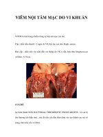
• Thấp tim tình trạng vantim bị thương tổn vĩnh viễn xảy ra sau một
haynhiều đợt viêm họng haysốt banđỏ donhiễm Streptococcus
pyogenes không được điều trị hoặc điều trị không đúng cách.
Phân loại học streptococci
Organism
Direct invasion
and inflammation
Local
spread
Distant
spread
Distant
toxin effects
Immune
mechanisms
Strep. pyogenes
Throat, wound and
burn infections
Puerperal sepsis
Erysipelas
Septicaemia
Scarlet fever
Rheumatic
fever
Glomerulonephritis
Strep. agalactiae
Neonatal
pneumonia
Puerperal sepsis
Abscess
Neonatal
meningitis
Enterococci
Urinary tract
infection
Abscess
Endocarditis
Septicaemia
Strep. pneumoniae
Bronchitis
Pneumonia
Septicaemia
Meningitis
Strep. viridans
Caries
resistant enterococci, are increasingly
common.
Phân loại học streptococci
• Dựa vào cấu trúc carbohydrateCở vách
• Dựa vào đặc điểm tanmáu khi nuôi cấy
Endocarditis
Bacteraemia
Streptococci
Control
A multivalent pneumococcal vaccine is
used to protect those particularly at risk,
Lancefield
groups
including splenectomised
and immunocompromised patients. Locating carriers
(nose, throat, skin or perineal carriage)
is important in controlling outbreaks,
A B orCclosed
D . com.. T
especially in hospitals
munities.
Others
U
α
Streptococcus bovis or
γ
Capsule
Hyaluronic acid
Cell wall
Protein antigens
Group-specific carbohydrate
Streptococci and enterococci
■
■
■
■
■
■
Peptidoglycan layer
Cytoplasmic membrane
Fig. 3 Structure of Strep. pyogenes.
■
Streptococcus agalactiae
β
Streptococci are Gram-positive cocci, usually
growing in chains,
Streptococcus
pyogenes β
facultative anaerobes, nutritionally fastidious and catalase negative.
Enterococci are more resistant than streptococci to bile, salt and
antibiotics.
Identification depends on Gram stain, haemolysis,
biochemicalpneumoniae
tests
α
Streptococcus
and Lancefield grouping.
The complex cell wall and enzymes and toxins
have importantmutans α
Streptococcus
or
functions, including adhesion, virulence and spread.
γ
Strep. pyogenes and Strep. pneumoniae are aggressive pathogens,
invasive and virulent even in normal hosts.Key:
Other streptococci are opportunistic pathogens, i.e. normal flora that
α
α hosts.
= α-Hemolytic
cause disease in abnormal sites or abnormal
Disease is caused by invasion and spread,βtoxin
effects and immune or = α or γ hemolytic
= β-Hemolytic
γ
mechanisms.
γ = γ-Hemolytic
II
S
p
p
m
in
S
a
e
s
ro
c
b
a
A
capsulated (Fig. 1c)
C
Đặc điểm sinh học
• Cầu khuẩn,Gramdương
• Xếp thành chuỗi
• Catalaseâm tính
ification of Significant Isolates
• Hiếu kỵ khí tùy tiện
Pe
St
(F
st
by
■
(a)
Capsule
T
Teichoic acid
S.pyogenes
Cell membrane
Liên cầu tanmáu ßnhómA
(b)
S
(b
S
S
E
S
S
S
3. Extracellular products: Like Staphylococcus aureus (see p. 70),
108
S. pyogenes secretes a wide range of exotoxins that often vary
from one strain to another and that play roles in the pathogenesis
of disease caused by these organisms (Figure 9.4).
Clinical Bacteriology
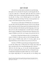
Yếu tố độc lực
108
Dissolves fibrin in
clots and thrombi
Apoptosis inhibits
phagocytosis
EGRATED REVIEW
OGY AND
BIOLOGY
Tissue
necrosis
Pyrogenic exotoxins
The only known reservoir for S. pyogenes in nature is the skin and
Inflammation
Immune
Activationdroplets or skin
mucous membranes
of theand
human
host. Respiratory
contact spreads group A streptococcal infection from person to person, especially in crowded environments such as classrooms and
children’s play areas.
Tissue
necrosis
Exotoxin B
Streptolysin O, S
Lysis of RBCs, WBCs, platelets
Lysis of RBCs, WBCs, platelets
Toxins and Hemolysins
Toxins and Hemolysins
Capsule,
ISBN: 978-0-323-07447-6
Damage mammalian cells, resulting in
cell lysis and release of lysosomal
enzymes.
Fibrin clot
Plasmin
Streptokinase
Catalyzes conversion of plasminogen
to plasmin, causing lysis of clots,
facilitating the rapid spread of organisms.
C5a
C5
C5a
C5a peptidase
Inactivates complement
component C5a.
Prevents phagocytosis,
Exotoxins,
M-protein
allows attachment : Pharyngitis is the most
1. Acute
pharyngitis or pharyngotonsilitis
to tissue
superantigens
common type of S. pyogenes infection. S. pyogenes pharyngitis
(exotoxin A)(“strep throat”) is associated with severe, purulent inflammation of
Exotoxins,
superantigens
the posterior oropharynx and tonsillar areas (see Figure 9.16).
(exotoxin
A) If a sunburnlike rash develops on the neck, trunk, and
[Note:
extremitiesactivator
in response to the release of pyrogenic exotoxin to
Mitogenic
which
the
patient
does not have antibodies, the syndrome is desof T cells
Mitogenic
ignated
scarletactivator
fever.] Many strep throats are mild, and many sore
of T cells
throats caused
by viruses are severe. Hence, laboratory confirmation is important for accurate diagnosis and treatment of streptococcal pharyngitis, particularly for the prevention of subsequent acute
rheumatic fever and rheumatic heart disease.
JeffreyFigure
K. Actor,
PhD
12-2. Pathogenic
formechanisms
group A streptococci
(Streptococcus
pyogenes).
RBCs,
red
cells;WBCs,
WBCs,
white
Figuremechanisms
12-2. Pathogenic
for group A streptococci
(Streptococcus
pyogenes).
RBCs,
redblood
blood cells;
white
Professor
ELSEVIER’S INTEGRATED
REVIEW IMMUNOLOGY
blood cells.
blood cells.
AND MICROBIOLOGY,
EDITION Medicine
partment
of Pathology SECOND
and Laboratory
Streptolysin O
Streptolysin S
phagocytosis,
peptidase Prevents
Inhibits
complement
D. Capsule,
ClinicalC5a
significance
anaphylatoxin
M-protein
allows attachment
S. pyogenes is a major cause
cellulitis. Other more specific syntooftissue
Streptococcus
dromes include:
Streptolysin O, S
Cause various effects, including the
rashes seen in scarlet fever and
streptococcal toxic shock disease.
C. Pathogenesis
Allows
spreading
in
S.
pyogenes
cells, perhaps
in an inhaled droplet, attach to the pharynsubcutaneous
tissue
Inflammation
and Immune
Activation
geal mucosa via
actions of protein
F, lipoteichoic
acid, and M protein.
The bacteria may simply replicate sufficiently to maintain themselves
without causing injury in which case the patient is then considered colStreptokinase
Dissolves fibrin in
onized. Alternatively, bacteria may grow and secrete toxins, causing
Erythrogenic toxins
(fibrinolysin)
clots and thrombi
damage to surrounding cells, invading the mucosa, and eliciting an
Apoptosis
inhibits
(pyrogenic
toxins)
Hyaluronidase
phagocytosis
inflammatory
with attendant influx of white cells, fluid leakAllows response
spreading in
subcutaneous
tissueThe patient then has streptococcal pharyngiage, and
pus formation.
tis. Occasionally, there is sufficient spread that the bloodstream is
Streptokinase
Streptodornase
Depolymerizes
significantly invaded, possibly
resulting in septicemia and/or seeding
Erythrogenic toxins
(fibrinolysin)
(DNAase)
DNA(acute
in necrotic
of
distant
sites,
where
cellulitis
inflammation of subcutaneous
(pyrogenic toxins)
Hyaluronidase
tissue
tissue), fasciitis (inflammation of the
tissue under the skin that covers a
surface of underlying tissue), or myonecrosis (death of muscle cells)
Toxemia,
Exotoxin
B
C5a peptidase
Inhibits complement
may develop rapidly or insidiously. However, direct inoculation of skin
skin rash
Streptodornase
Depolymerizes
anaphylatoxin
from another
person's infection
probably more common as the
(DNAase)
DNA is
in necrotic
Streptococcus
pathogenesis of streptococcal skintissue
and soft tissue infection.
SECOND EDITION
1600 John F. Kennedy Blvd. Ste 1800
Philadelphia, PA 19103-2899
Cytokines
B. Epidemiology
Clinical Bacteriology
Toxemia,
skin rash
Streptococcus pyogenes
2. Impetigo: Although S. aureus is recovered from most contemporary
cases of impetigo (see p. 72), S. pyogenes is the classic cause of
DNA
Streptodornases
DNAses that degrade the viscous
DNA in necrotizing tissue or exudates,
aiding the spread of infection.
Hyaluronic
acid
Hyaluronidase
Disrupts the organization of ground
substance, facilitating the spread of
infection.
(group A, β-hemolytic)
4All isolates remain sensitive to
penicillin G and ampicillin.
Acute pharyngitis or
5In life-threatening
infections, an aminopharyngotonsillitis
glycoside can be added to the regimen.
(group B, β-hemolytic)
•
• Acute rheumatic fever
• Erysipelas
sepsis
• Puerperal
group A streptococcal
• Invasive
disease
6Penicillin G has been the drug of choice,
but resistant
are regularly
seen.
Meningitis
andstrains
septicemia
in neonates
7Most resistant strains remain sensitive
Endometritis
to vancomycin.
•
• Septicemia or pneumonia
• in individuals with impaired
Khả năng gây bệnh
•
immune systems
Diabetic foot infections
• Acute bacterial pneumonia
• Otitis media
• Meningitis
1 Penicillin G6
1 Cefotaxime
1 Penicillin
1 Penicillin G1,2
(α-hemolytic)
G4
1 Ceftriaxone
2 Clarithromycin
• Viêm
họng
2 Azithromycin
3
2 An
aminoglycoside5
2 Vancomycin7
3
pyogenes has not acquired resistance to
•1S.penicillin
Nhiễm
khuẩn damủ
G.
2Clindamycin may be added to pencillin G for
soft tissue infection such as necrotizing fasciitis.
3For penicillin-allergic patient.
• Hội chứng shockđộc tố
Impetigo
• Các biến chứng sau nhiễm
4All isolates remain sensitive to
penicillin G and ampicillin.
5In life-threatening infections, an aminoglycoside can be added to the regimen.
6Penicillin G has been the drug of choice,
but resistant strains are regularly seen.
7Most resistant strains remain sensitive
to vancomycin.
Streptococcal pharyngitis
S.pyogenes
dicates first-line drugs; 2 indicates alternative drugs.
Facial erysipelas
Impetigo
Streptococcal pharyngitis
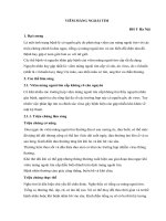
Cơ chế gây bệnh
• Tổnthươngvantim
• <3%sốngườibịviêmhọngGAS,vàituần
saukhibịviêmhọng
• Viêmkhớpdịchchuyển;tổnthươngvan
tim(50%),mộtsốtửvong
• Táiphátthườngxun,điềutrịbằng
penicillinsuốtđờikháng
• CấutrúckhángproteinM,myosintim
giốngnhau
• TấncơngcủatếbàoT,khángthểgâyviêm
vàtổnthươngvantim
Figure 2. Pathogenesis of carditis in acute rheumatic fever (VCAM1: Vascular cell adhesion molecule 1).
Figure reproduced with permission from (Carapetis, et al., 2016).
■
pneumococcal capsule gives resistance
to phagocytosis
every individual toxin with particular
clinical infections.
Chẩn đoán
Table 1 Classification and normal habitat
Species
(biochemical)
Serologic
Lancefield group
Haemolysis on
sheep blood agar
Normal flora (nf) or
asymptomatic carriage (ac)
Beta
Throat, nose (ac)
• Chẩn đốn trựcAB tiếp:Nuôi cấy,phân
lập
Beta (alpha, gamma)
Vagina, gut (nf)
Strep. pyogenes
Strep. agalactiae
E. faecalis
Strep. bovis, equinus
Strep. pneumoniae
Strep. viridans group (Strep. sanguis,
salivarius, mitis, ‘milleri’, mutans)
(a)
D
D
Ungroupable
Ungroupable
Gamma (alpha)
Gamma (alpha)
Alpha
Alpha (gamma)
(b)
Gut, perineum (nf)
Gut, perineum (nf)
Nasopharynx (ac)
Mouth (nf)
Chẩn đốn
Chẩn đốn gián tiếp:
• antistreptolysin O(ASO),antiDnase B
ASO Test
+
SO-Coated
Latex
Specimen
Containing ASO
Giá trị của ASO
• ASObắt đầu tăng sau 1tuần nhiễm trùng,đạt đỉnh sau 3-6tuần
• Hiệu giá ASOsau nhiễm trùng hơ hấp trên thường tăng cao cịn sau
viêm mủ dakhơng tăng cao
• S.dysagalactiae subsp.equisimilis cũng tạo ra SOnên tăng ASOkhơng
đặc hiệu cho nhiễm S.pyogenes.
Ngưỡng trên bình thường của hiệu giá ASO
Người lớn/người già:≤200IU/ml
6-15tuổi:240– 320IU/mL
AntiDnase B
• Có thể xuất hiện sớm hơn ASO
• Nhạy hơn trong hỗ trợ viêm cầu thận cấp sau nhiễm S.pyogenes
ngoài da.
Figure 2. Common antigenic proteins of S. pyogenes used for diagnostic and typing purposes.
Typing of Streptococcus pyogenes
• Có nhiều kỹ thuật khác nhau như ELISA,kỹ thuật trung hoà để phát
hiện antiDnase B.
In most clinical cases of acute infections, subtyping of group A streptococcal strains has
no immediate diagnostic or therapeutic consequences. Such typing is typically performed
by reference laboratories for epidemiologic surveys or in outbreak situations, and may
provide important information about the evolutionary relatedness of various strains.
Although classical antibody-dependent typing systems of surface proteins have been used
for many years, molecular methods have become more and more prevalent, since they do
not require the maintenance of rarely used large antibody panels or the establishment of
specialized techniques. As an additional advantage, the determination of DNA sequences
is independent from culture conditions and gene expression.
Conventional typing of S. pyogenes is based upon the antigenic specificity of the surfaceexpressed T and M proteins. (Johnson & Kaplan, 1993). The trypsin-resistant T protein is
part of the pilus structures (Mora, et al., 2005). T type identification can be achieved by
Đặc điểm đề kháng kháng sinh
• S.pyogene vẫn được coi là hồn tồn cịn nhạy cảm với β-lactamtừ 1940.
• Khó tiếp nhận các geneđề kháng ngoại lai,khơng có khả năng đề kháng tự
nhiên.
• Một số báo cáo về thất bại điều trị bằng penicilin do;
• S.pyogenes tồn tại dai dẳng nội bào
• Các vikhuẩn khác tiết β-lactamasebảo vệ cho S.pyogenes
• Có sự phối hợp M.catarrhalis và S.pyogenes giúp S.pyogenes dễ dàng bám vào tế
bào biểu mô
tissue infections, streptococcal toxic shock) and
nonsuppurative
diseases
(rheumatic fever,
Summary:
Streptococcus
pyogenes
(Group
A)
Summary: Streptococcus pyogenes (Group A)
glomerulonephritis)
Diagnosis
Biology, Virulence, and Epidemiology
Disease
Diagnosis
Biology, Virulence, and Disease
Microscopy is useful in soft-tis
Rapidly growing gram-positive cocci arranged in chains;
Microscopy
is
useful
in
soft-tissue
infections
but
not
Rapidly growing gram-positive cocci arranged
in
chains;
Transient
colonization
in
upper
respiratory
tract
skin
pharyngitis
or and
nonsuppurativ
group-specific carbohydrate (A antigen) and type-specific
pharyngitis
nonsuppurative
complications
group-specific carbohydrate (A antigen) proteins
and type-specific
caused by
recently
acquired
strains
(M protein) in surface
cell
wall withordisease
Direct tests
for the group
A an
proteins (M protein) in cell wall
Direct
tests
forphagocytosis
the group
A antigen
useful for
the
(before
protective
antibodies
areare
produced)
diagnosis
of streptococcal
ph
Virulence determined by ability
to
avoid
Diagnosis determined by ability to avoid phagocytosis
diagnosis
of
streptococcal
pharyngitis,
but
negative
Virulence
mustcaused
be confirmed
(mediated primarily by
capsule, M and
and M-like
proteins,
Pharyngitis
soft-tissue
infectionsresults
typically
by by
musthost
be confirmed
by culture
or molecular
(mediatedisprimarily
capsule, infections
M and M-like
proteins,adhere toresults
C5a
and invade
cells (M
Microscopy
useful inby
soft-tissue
butpeptidase),
not
Isolates
identified assays
by catalase (
strains
with
different
M
proteins
C5a
peptidase),
adhere
to
and
invade
host
cells
(M
protein,
lipoteichoic
acid,
F
protein),
and
produce
toxins
pharyngitis or nonsuppurative complications
Isolates identified by catalase (negative),
positive PYRarylamidase)
(L-pyrrolidonyl
Person-to-person
spread
by
respiratory
droplets
protein,
lipoteichoic
acid,
F
protein),
and
produce
toxins
(streptococcal
pyrogenic
exotoxins,
streptolysin
S,
(L-pyrrolidonyl arylamidase) reaction,
susceptibility
to
bacitracin,
and presence
of g
Direct tests for the group A antigen are useful for the
streptolysin
DNases)and
(streptococcal
pyrogenic exotoxins,
streptolysin
S, O, streptokinase,
(pharyngitis)
orpresence
throughofbreaks
in Askin
after
antigen)
bacitracin,
group-specific
antigendirect
(group
diagnosis of streptococcal
pharyngitis,
but
negative
streptolysin
DNases)or
Responsible
suppurative
diseases
(pharyngitis,
softcontact
with
infected
person, fomite,
or
A
antigen)
Antistreptolysin
O test is usefu
results must O,
be streptokinase,
confirmed by culture
molecularfor
assays
tissue
infections,
streptococcal
toxic
shock)
and
Responsible
for suppurative
(pharyngitis,
softfever or glomerulonephritis
a
arthropod vector
Antistreptolysin
O test is useful for confirming
rheumatic
Isolates identified
by catalasediseases
(negative),
positive
PYR
nonsuppurative
diseases (rheumatic fever,
anti-DNase B te
tissue
infections,arylamidase)
streptococcal
toxic shock)
and
fever or at
glomerulonephritis
with streptococcal
(L-pyrrolidonyl
reaction,
susceptibility
to
Individuals
higher risk for associated
disease pharyngitis;
include
children
5 to
glomerulonephritis)
glomerulonephritis
associated
nonsuppurative
diseases
(rheumatic
fever,
B test
should be
performed
for with
bacitracin, and presence of group-specific antigen (group
15pharyngitis;
years old anti-DNase
(pharyngitis);
children
2 to
5 years
old
soft-tissue
infections
Epidemiology
glomerulonephritis)
glomerulonephritis associated with pharyngitis or
A antigen)
poor
personal tract
hygiene
(pyoderma);
patients
with and Co
Transient colonization in upper
respiratory
and skin
Treatment,
Prevention,
soft-tissue
infections
Epidemiology
Antistreptolysin O test is useful for confirming
rheumatic
soft-tissue
infection
toxic Vshock
surface
with disease caused
by recently
acquired(streptococcal
strains
Penicillin
or amoxicillin used
Transient
colonization
in
upper
respiratory
tract
and
skin
Treatment,
Prevention,
and
Control
fever or glomerulonephritis associated with
streptococcal
(before protective antibodies
are produced)
syndrome);
patients with prior streptococcal
cephalosporinpharyngitis
or macrolide f
surface
withanti-DNase
disease caused
by should
recently
acquired
strains
pharyngitis;
B test
be
performed
for
Penicillin
V
or
amoxicillin
used
to
treat
pharyngitis;
oral plus cl
Pharyngitis and soft-tissue(rheumatic
infections typically
caused
by
intravenous
penicillin
fever,
glomerulonephritis)
or
soft-tissue
(before
protective antibodies
glomerulonephritis
associated are
withproduced)
pharyngitis
cephalosporin or macrolide for penicillin-allergic
patients;
strains or
with different Minfection
proteins
infections
(glomerulonephritis)
soft-tissue infections
BOX 19-1
BOX 19-1
Pharyngitis and soft-tissue infections typically
caused by spread by respiratory
intravenousdroplets
penicillin plus clindamycin
used for systemic
Person-to-person
Oropharyngeal
carriage occurri
strains
with
different
M
proteins
infections
Treatment, Prevention, and Control
(pharyngitis) or through breaks in skin after direct
re-treated; treatment is not i
Person-to-person
spread byused
respiratory
Oropharyngeal
treatment cancarriage
be
contact with
fomite, orcarriage occurring after asymptomatic
becau
Penicillin V or amoxicillin
to treat droplets
pharyngitis;
oralinfected person,
arthropod
vector
flora
(pharyngitis)
breaks
in skin after
direct
re-treated; treatment is not indicatedprotective
for prolonged
cephalosporinororthrough
macrolide
for penicillin-allergic
patients;
contact
withpenicillin
infected plus
person,
fomite, Individuals
orused for systemic
antibiotics
disrupt normal
at higher risk forasymptomatic
disease includecarriage
childrenbecause
5 to
Starting antibiotic
therapy with
intravenous
clindamycin
CĂN NGUYÊN GÂY VIÊM NỘI TÂM MẠC
CÁC XÉT NGHIỆM VI SINH CHẨN ĐOÁN
Định nghĩa
• Viêm nội tâm mạc nhiễm trùng là viêm bề mặt nội tâm mạc
• Phân loại chính:
• Viêm nội tâm mạc vantim tự nhiên
• Viêm nội tâm mạc vantim nhân tạo
• Đặc điểm:Sốt,vikhuẩn trong máu thường xuyên
Yếu tố nguy cơ VNTMdovikhuẩn
• Thủ thuật răng miệng
• Bệnh lý răng miệng (sâu răng,abscess)
• Nhiễm trùng ngồi tim (phổi,tiết niệu,da,xương)
• Dụng cụ canthiệp (đường tiết niệu,tiêu hố,tĩnh mạch)
• Phẫu thuật tim
• Sử dụng thuốc đường tiêm
• Khơng rõ
Các căn nguyên từ vãng khuẩn huyết
Đường vào
Vikhuẩn
Đánh răng
Streptococcitanmáu alphatừ vihệ họng
Ăn/Nhai
miệng
Thủ thuật răng miệng
Đường truyền tĩnh mạch
Staphylococcusaureus từ da/mũi
Sử dụng thuốc đường tĩnh mạch
Nhiễm trùng tại chỗ/abscess
Staphylococcusaureus, Streptococcus
pneumoniae
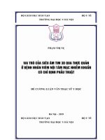
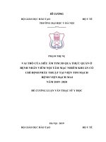
Panel 1: Proportion of cases of infective endocarditis
caused by different microorganisms from a French
population-based cohort of 497 patients2
Căn nguyên gây bệnh
Staphylococci
Staphylococcus aureus: 26·6%
Coagulase-negative staphylococci: 9·7%
• Căn nguyên thường gặp
Streptococci and enterococci
• Streptococciviridans – VNTMbán cấp
Oral streptococci: 18·7%
• Streptococcinhóm D(S.galllolyticus,S.bovis):khối uđại tràng tiềm ẩn Non-oral streptococci: 17·5%
Enterococci: 10·5%
(đường vào)
Other: 1·6%
• Enterococci- VNTMbán cấp (PVE,NVEBNcó bệnh lý mạn tính,người già) HACEK (haemophilus, aggregatibacter, cardiobacterium,
Eikenella corrodens, kingella) microorganisms
• CoN Staphylococci- VNTMbán cấp (PVEsớm,nhiễm trùng bệnh viện
1·2%
NEV,tạo biofilm)
Seminar
• Căn ngun ít gặp
Candida species
1·2%
• S.aureus – VNTMcấp (BNlọc máu,sử dụng thuốc đường tĩnh mạch)
• Streptococcitanmáu 𝛽
• S.pneumoniae
• Căn nguyên hiếm gặp
• Nấm
• Pseudomonas/Coliform
• HACEK
Other*
6·0%
Polymicrobial (≥2 microorganisms)
1·8%
Seminar
No microorganism identified
5·2%
Infec
*Includes small numbers of Enterobacteriaceae, Propionibacterium acnes, Pseudomonas
aeruginosa, Lactobacillus spp, Corynebacterium spp, Coxiella burnetii, Bartonella quintana,
Tropheryma whipplei, Gordonia bronchialis, Bacillus spp, Erysipelothrix rhusiopathiae, Neisseria
elongata, Moraxella catarrhalis, Veillonella spp, Listeria monocytogenes, Acinetobacter ursingii,
Campylobacter fetus, Francisella tularensis, and Catabacter hongkongensi.
Thomas J C
Infective endocarditis
Infective
Infective endocarditis
occurs worldwide,
defined by infectio
Published
Onlineand issurface,
Lancet
2016;
882–93
Thomas J Cahill,
Bernard D387:
Prendergast
infective endocarditis in high-income countries and
Lancet 2016; 387: 882–93
1,2
Căn nguyên gây bệnh
• Căn nguyên gây bệnh cấy máu âm tính
• Coxiella burnetti
• Bartonella spp.
• Tropheryma whipplei
VNTMvantim nhân tạo
(PVE- ProstheticValveEndocardititis)
• PVEsớm:trong vịng 60ngày
• Nhiễm trùng bệnh viện (S.epidermidis)
• PVEmuộn:sau 60ngày
• Nhiễm trùng cộng đồng (giống với NVE)
Cell structure and function
Streptococci have a complex cell wall
(Fig. 3). The biological principle that
structure relates to function is illustrated
by the components:
■
pneumococcal capsule gives resistance
to phagocytosis
Virulence factors (Table 3) are found in
the aggressive pathogens Strep. pyogenes
and Strep. pneumoniae and are related to
surface antigens and extracellular products. Some appear to help the spread of
disease, but it is not yet possible to link
every individual toxin with particular
clinical infections.
)
)
)
Table 1 Classification and normal habitat
Species
(biochemical)
Serologic
Lancefield group
Haemolysis on
sheep blood agar
Normal flora (nf) or
asymptomatic carriage (ac)
Strep. pyogenes
Strep. agalactiae
E. faecalis
Strep. bovis, equinus
Strep. pneumoniae
Strep. viridans group (Strep. sanguis,
salivarius, mitis, ‘milleri’, mutans)
A
B
D
D
Ungroupable
Ungroupable
Beta
Beta (alpha, gamma)
Gamma (alpha)
Gamma (alpha)
Alpha
Alpha (gamma)
Throat, nose (ac)
Vagina, gut (nf)
Gut, perineum (nf)
Gut, perineum (nf)
Nasopharynx (ac)
Mouth (nf)