A method for phenomenological and chemical kinetics study of autocatalytic reactive dissolution by optical microscopy. The case of uranium dioxide dissolution in nitric acid media
Bạn đang xem bản rút gọn của tài liệu. Xem và tải ngay bản đầy đủ của tài liệu tại đây (2.23 MB, 13 trang )
EPJ Nuclear Sci. Technol. 4, 2 (2018)
© P. Marc et al., published by EDP Sciences, 2018
/>
Nuclear
Sciences
& Technologies
Available online at:
REGULAR ARTICLE
A method for phenomenological and chemical kinetics study of
autocatalytic reactive dissolution by optical microscopy. The case
of uranium dioxide dissolution in nitric acid media
Philippe Marc1, Alastair Magnaldo1,*, Jérémy Godard1, and Éric Schaer2
1
2
CEA, Nuclear Energy Division, Research Department on Mining and Fuel Recycling Processes, Research Service for
Dissolution and Separation Processes, Laboratory of Dissolution Studies, 30207 Bagnols-sur-Cèze, France
Laboratoire Réactions et Génie des Procédés, UMR CNRS 7274, University of Lorraine, 54001 Nancy, France
Received: 14 December 2016 / Received in final form: 4 October 2017 / Accepted: 10 October 2017
Abstract. Dissolution is a milestone of the head-end of hydrometallurgical processes, as the stabilization rates
of the chemical elements determine the process performance and hold-up. This study aims at better
understanding the chemical and physico-chemical phenomena of uranium dioxide dissolution reactions in nitric
acid media in the Purex process, which separates the reusable materials and the final wastes of the spent nuclear
fuels. It has been documented that the attack of sintering-manufactured uranium dioxide solids occurs through
preferential attack sites, which leads to the development of cracks in the solids. Optical microscopy observations
show that in some cases, the development of these cracks leads to the solid cleavage. It is shown here that the
dissolution of the detached fragments is much slower than the process of the complete cleavage of the solid, and
occurs with no disturbing phenomena, like gas bubbling. This fact has motivated the measurement of dissolution
kinetics using optical microscopy and image processing. By further discriminating between external resistance
and chemical reaction, the “true” chemical kinetics of the reaction have been measured, and the highly
autocatalytic nature of the reaction confirmed. Based on these results, the constants of the chemical reactions
kinetic laws have also been evaluated.
1 Introduction
Dissolution is a key phenomenon encountered in various
processes, for example for drug delivery, quality control in
pharmacology [1] or in the food-processing industry [2,3].
Dissolution also takes part in many chemical processes in the
mining industry [4–7], batteries [8,9], fertilizer production
[10], or the recycling industry [11]. Among these chemical
processes, the Purex process is a hydrometallurgical process
involving the dissolution of spent nuclear fuels in nitric acid
in the head-end steps, before carrying out solvent extraction
steps allowing the recovery of uranium and plutonium [12]. In
an optimization approach of this dissolution step, its
modeling has recently become a source of interest. Given
that currently recycled spent nuclear fuels are made of about
95% of uranium dioxide [13], the modeling of the dissolution
of this chemical specie in nitric acid media represents a step
which cannot be overlooked.
An analysis of the state of knowledge of the dissolution
reaction of uranium dioxide in nitric acid media [14] shows
that despite the importance of this reaction in the
* e-mail:
hydrometallurgical reprocessing of spent nuclear fuels, its
chemical and physico-chemical mechanisms remain poorly
understood. The relationship between the fraction of
dissolved solid, which can be linked more or less simply
with the bulk concentration of the chemical elements
composing it, and the chemical reaction kinetics requires
the accurate knowledge of the surface of the dissolving solid
and the reactivity of each element of this surface over time.
As a result of the physico-chemical phenomena occurring
during the dissolution of uranium dioxide macroscopic solids
in nitric acid media, like the complex reactions and species
produced in nitric acid, the chemical reaction kinetics are
today impossible to relate to the evolution of the concentration of dissolved materials in the bulk.
However, a recent trend in dissolution mechanisms and
kinetics study is the use of optical microscopy. This technic
has already been used in several dissolution studies. Steiger
et al. used it for general observation of the growth and
dissolution of lithium mosses and needles in 1 mol lÀ1 LiPF6
[8] and during lithium electrodeposition on tungsten and
copper substrates [9]. Boetker et al. [15] studied the
concentration gradients and diffusion layer thickness around
amlodipine besylate dissolving in water, as well as
This is an Open Access article distributed under the terms of the Creative Commons Attribution License ( />which permits unrestricted use, distribution, and reproduction in any medium, provided the original work is properly cited.
2
P. Marc et al.: EPJ Nuclear Sci. Technol. 4, 2 (2018)
Østergaard et al. [16] for lidocaine dissolution in water and
Delwaulle et al. [17,18] for copper and uranium dioxide
dissolving in nitric acid. Mgaidi et al. [4] and Singh et al. [19]
used it for monitoring the evolution of the morphology of
sand and succinic acid crystals during dissolution. The
temporal studies vary from the measurement of total
dissolution time of sucrose crystals in melted sorbitol by
Bhandari et al. [2] to more complex studies which used
optical microscopy for measuring the dissolution rates of
several solids, such as those from Marabi et al. [3] for the
dissolution rates of pure sucrose spherical particles in
water, ethylene glycol, and polyethylene glycol, Forny
et al. [20] for those of milk powder particles in water, and
Dorozhkin [10,21] for single crystals of the natural Khibin
(Kola) fluorapatite. Prasad et al. [22] and Raghavan et al.
[23,24] have even measured the dependency of dissolution
rates of paracetamol and a lactose monohydrate crystals
in water depending on the crystal faces considered.
More recently, Svanbäck et al. [25–27] have addressed
papers summarizing the advantages of optical microscopy
as a method for dissolution kinetics measurements over the
macroscopic methods, and presenting interesting designs
for the cells and methods for the monitoring of such
reactions. Part of these advantages are the reduction of the
amounts of reagents required, the simpler experimental
preparation (no compound-specific method development,
calibration or evaluation is required for image analysis),
which reduced the time required for analysis and the interoperator variability error sources, and the low cost of the
optical microscopy equipment compared to other technics
such as HPLC-MS or GC-MS. However, the application of
the presented cells in the dissolution conditions used for
uranium dioxide (i.e. warm and concentrated nitric acid,
implying strong acidic and oxidizing conditions) has not
been possible as such, and dissolution cells fitting these
conditions have been developed and will be presented in
this paper.
It will also be shown that, during the dissolution of a
uranium dioxide pellet, fragments can detach from it. Even
if these fragments dissolved in a much simpler way than the
pellet itself, two issues make them remain unsuitable for
macroscopic chemical reaction rates studies. The first one
is that even at this scale, non-uniform attack occurs, as
documented by Briggs [28,29], Shabbir and Robbins [30]
and Zhao and Chen [31–33], and thus that the surface and
associated reactivity remain practically impossible to know
precisely over time. The second issue is that the
measurement of dissolving elements released in solution
would require the use of several fragments, and of a larger
volume of dissolution solution, thus rising the question of
the accumulation of dissolution products, and their
autocatalytic effect.
On the other hand, these fragments offer a good
opportunity to measure the dissolution rates in situ by
using optical microscopy and image processing. The
determination of the rate determining step during these
measurements allows to discriminate diffusion controlled
from chemically controlled dissolutions. The study of the
rates corresponding to the chemical reaction has shown that,
without doubt, it occurs through a strongly autocatalyzed
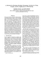
mechanism. Optical microscopy has also allowed measuring
Fig. 1. Microscopy installation in the glove box.
specifically the chemical reaction rates for the non-catalyzed
reaction, leading to the proposal of reactivity ratios between
the non- and the autocatalyzed reactions.
2 Experimental section
2.1 Microscope
The microscope used for this study is a reversed optical
microscope Zeiss .Z1m equipped with three lenses offering
magnification ratios of 5, 20 and 40. The reverse position
of the lenses is required by the production of nitrogen
oxides bubbles during the attack of uranium dioxide by
nitric acid: when these bubbles rise to the top of the liquid,
they hide the solid and make any observation by the top
impossible.
The microscope has been installed in a depressurized
glove box, in order to confine radioactive materials (Fig. 1).
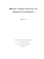
2.2 Dissolution cells
A first continuous dissolution cell is presented in Figure 2.
It is composed of a central well where the solid and the
solution are introduced. It is closed bottom-side by a
quartz pothole in order to ensure observation. The upper
part can be closed by rings system, which can be changed
depending on the kind of experiments. The dissolution
volume is 15 ml. This central well is surrounded by a
jacket in which water can flow to maintain a stationary
temperature in the central well. A coil, guaranteeing an
optional continuous feed of the well with dissolution
solution, circulates in this jacket so as to heat the solution
inflow at the working temperature. Another pipe crosses
the jacket in a straight line, allowing outflow and also
placing a temperature sensor in the well. This device is well
P. Marc et al.: EPJ Nuclear Sci. Technol. 4, 2 (2018)
3
Fig. 2. Pictures of the continuous dissolution cell.
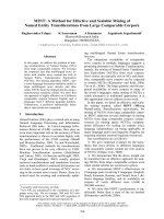
Fig. 3. Thermoelectric device for the observation of the dissolution of microscopic solids.
adapted for dissolution of macroscopic solids, dissolution
under continuous flow, or batch dissolution of microscopic
solids requiring important liquid/solid ratios.
The solution feed is controlled by a KD Scientific Legato
270 Push/Pull Syringe Pump coupled with a Gemini 88
Valve Box for long time ranging experiments.
A second device is presented in Figure 3. It consists in a
quartz disc at the center of which a well has been
manufactured. Around the well, a groove receives an Oring seal, and a quartz disk placed over the system closes it.
This device is placed on a thermoelectric heating stage
Linkam PE100 adapted for the microscope. The control of
the temperature is realized by a Linkam T95 system
controller. In order to insulate the system, a polydimethylsiloxane cover designed to fit the heating stage has been
manufactured by moulding.
Temperature stabilizing is more difficult with this noncirculating device, due to the configuration of the thermoelectric system: the time require for stabilizing the
temperature is long (several hours), and there are important
differences between the temperature set and the effectively
reached temperature once the system is stabilized.
2.3 Reagents
Uranium dioxide powder was provided by CEA Cadarache.
The uranium dioxide purity of the powder is 99.6%, and
detailed analysis of the powder is given in Table 4 in the
Supplementary Material.
This powder is also used for the manufacturing of the
uranium dioxide pellets. The pellets have been pressed at a
pressure of 518 MPa before being sintered at 1100 °C during
4 h under Ar-H2 (4%) atmosphere. Resulting sintered
pellets have an average diameter of 4.66 mm, height of
4 mm and mass of 0.5 g.
Nitric acid solutions have been prepared by dilution of
68% HNO3 provided by VWR (ref. 20422.297). Each
diluted solution have been titrated three times by mean of a
848 Titrino Plus, fed with 1 mol lÀ1 sodium hydroxide
Titrinorm provided by Prolabo (ref. 180.031627.60).
2.4 Dissolutions in solutions containing reaction
products
The autocatalytic component of the dissolution reaction of
uranium dioxide in nitric acid media has been widely
documented in the literature [14]. The ratio of the volume
of dissolution solution over dissolved amount of solid in the
dissolution cells is an advantageous condition for studying
this component under well known dissolution products
concentrations and temperature conditions.
The solutions for the measurement of dissolution
kinetics in presence of various amounts of reaction products
have been prepared by pre-dissolving uranium dioxide
powder in fresh nitric acid (Fig. 4). The dissolution is
realized in a bottle containing a known volume of fresh
nitric acid initially at room temperature, with a known
mass of uranium dioxide powder introduced in the bottle,
4
P. Marc et al.: EPJ Nuclear Sci. Technol. 4, 2 (2018)
Fig. 4. Diagram of the experimental protocol for the study of the
autocatalyzed reaction kinetics.
whose opening is immediately covered with a cork after the
introduction of the powder, in order to limit the evacuation
of gaseous reaction products. The bottle is not hermetically
closed to avoid overpressure troubles during the reaction.
Four solutions with a pre-dissolved amount of uranium
dioxide of 0.1, 10, 50, and 100 g lÀ1 have been prepared in
fresh 4.73 mol lÀ1 nitric acid.
The time required (about 10 min) for the transfer of the
solution to the microscope glove box insures that
potentially remaining undissolved uranium dioxide gets
completely dissolved. The solution is then continuously
pumped at a 5 ml hÀ1 flow rate into the dissolution cell, and
the dissolution of the uranium dioxide fragments under the
microscope starts. Even if this continuous flow contributes
to guarantee the stability of the concentrations of the
reagents and products in the cell at the values of the predissolved uranium diocide solutions, it is primarily used, in
the absence of a consolidated knowledge on the autocatalytic species and their stabilities, to counter as much as
possible a potential degradation of the autocatalytic
species.
2.5 Measurement of dissolution kinetics by optical
microscopy observation and image processing
The methodologies used in previous dissolution kinetics
measurement studies for calculating the dissolution rates
from a set of images are usually not detailed [3,10,21].
These methods consist in measuring the distance between
the profiles of one dissolving solid at different times. This
distance corresponds to Dl on Figure 5, without stating if
only one or several measurements are done along the
profile.
A different method, based on the measurement of the
projected area and the associated perimeter of a dissolving
particle on each image, is developed here and detailed in the
following paragraphs. The geometric evolution of the
projected area of a uniformly dissolving solid is represented
in Figure 5.
Fig. 5. Evolution of the projected area and associated perimeter
of a uniformly dissolving particle.
In the particular case of a weak dissolution of the
particle, and in the absence of neo-formed phases at the
solid/liquid interface, a mathematical link can be drawn
between the variation of its projected area (A) between
times t and t ỵ Dt, the perimeter (P) of its projected area at
t, and the progression of the dissolution front (Dl), which
corresponds to the apparent dissolution rate (r) over Dt,
considered as constant over Dt (Eq. (1)) :
At ỵ Dtị Atị À P ðtÞDl À P ðtÞrDt:
ð1Þ
Thus, one of the advantages of this method is to focus
on the measurement of the external perimeter of the solid,
and to be able to make dissolution rates measurement
without the issue of the internal porosity disturbance.
equation (1) leads to the expression of the variation of the
area at a time t (Eq. (2)) :
DA
ðtÞ ≈ À P ðtÞr:
Dt
ð2Þ
Therefore, it is possible to extract the dissolution rate of
a dissolving solid by measuring its area and perimeter on
each image of a time sequence set of images. In practice, the
integrated form of equation (2) (Eq. (3)) will be used on the
sets of images, since this form allows smoothing the
variations which can appear in the case of images with a
poor quality, for example when the images are acquired
under reflected light conditions.
AðtÞ A0ị
tX
Dt
P tịrDt:
3ị
tẳ0
Considering the dissolution of the solid as uniform, and
taking place under stationary conditions, it comes that the
dissolution rate is constant over the time, and can be
extracted from the sum sign, as well as the time interval Dt
between two images, since this value is fixed by the
experimenter, and thus is also constant over the acquisition. This leads to express the projected area of the particle
at a time t as a linear function of the sum of the perimeters
P. Marc et al.: EPJ Nuclear Sci. Technol. 4, 2 (2018)
5
Fig. 6. Example of image thresholding and holes filling: original image (a), binarised image (b), and binarised image with holes filled (c).
of the projected area from t = 0 to t Dt (Eq. (4)).
tX
Dt
P tị:
Atị A0ị rDt
4ị
tẳ0
It is important to insist on the fact that these equations
are practicable in the case of a uniform attack of the
fragments. Nevertheless, it is not impossible that, even if no
porosity development was detected at the scale of the
grains we have been working with, microporosity development occurs at a smaller scale than the resolution of the
microscope. It should be noted that in this case, if microporosity were created, it would also disappear at the same
rate during dissolution: the dissolution would fatally
appear non-uniform in the other case. Thus, the dissolution
front moves globally uniformly at the resolution of the
microscope.
In this case, the dissolution kinetics is given as a speed,
in distance per time units. Assuming the density is known,
the relationship between the reactive surface and the
measured surface is linear, and equation (5) enables to
convert these kinetics into more common units system for
dissolution kinetics.
r ½msÀ1 ẳ
1
Mi
r ẵkg m2 s1 ẳ
r ẵmol m2 s1 :
ri
ri
5ị
The measurement of area and perimeter used in this
method raises the issue of the relevance of the dissolution
kinetics measured in the case of a non-uniform attack of the
solid. Once more, the problematic of the evolution of the
rugosity and porosity of the surface is one of the main
problem which has to be dealt with when measuring the
chemical dissolution reaction kinetics, whatever the
method applied [34–37], since there is no method for insitu measurement of the surface evolution on such a short
period of time.
A first fact to take into consideration is that in any case,
porosity appears, but also disappears. This results in a
stabilization of surface roughness after a given period of time.
In the case of microscopic observations and image processing,
two different cases must be considered depending on the scale
they occurred at, and regarding the resolution of the images.
The first case applies when the surface roughness evolves
at a smaller scale than the resolution of the microscope. In
this case, the effect of the development of surface roughness
on the measured area and perimeter of a given particle is null,
or at least weak. What is more important is the case where the
evolution of the surface roughness of the solid is detectable
with the microscopic observations. In this case, the initial
dissolution rate measured by this method will be greater than
the average of the different reaction kinetics. Nevertheless,
while the surface roughness will stabilize, the measured
dissolution rates will get closer to the expected average of the
dissolution rates.
Thus, concerning the method presented in this paper,
one can draw the conclusions that the dissolution rates
measured with this method are at least as good as those
measured by classical macroscopic method, and in many
cases even better since they only take under consideration
the external surface, and not the complex and disrupting
contribution of internal porosity.
2.6 Image processing for the extraction of the area
and perimeter of the particles
The analysis of the images is realized through a three-step
process which consists of image binarisation, extraction of
the area and perimeter of the particle, compilation of the
data, and linear regression to calculate the dissolution rate.
The processing of a series of images is realized by the
mean of a program developed in-house1 for the automation
of this process.
2.6.1 Image binarisation
After turning the images from colored to 8-bits grayscale
images, the luminosity of each pixel of the image varies from 0
(black) to 255 (white). The histogram representing the
number of pixels composing the image as a function of their
luminosity is a bimodal curve. One of the two peaks
corresponds to the pixels of the background (black on Fig. 6),
and the other to the pixels of the object (white on Fig. 6).
In order to measure the area and perimeter of a particle,
it is first required to clearly separate the pixels of the image
in two categories: object and background. This issue is
1
This code was written in Scilab 5.5.0, free open source software
distributed under CeCILL license (GPL compatible), developed
by Scilab Enterprises. Available on gr17.
6
P. Marc et al.: EPJ Nuclear Sci. Technol. 4, 2 (2018)
Fig. 8. Possible configurations of the neighborhood of a pixel
belonging to the perimeter of the object.
Fig. 7. Example of threshold establishing.
widely documented in image treatment literature [38–43]
and several methods have been proposed to define the
threshold value.
The method selected defines the threshold as the
luminosity for which the pixels population reaches a
minimum between the two peaks. For this purpose the
histogram is first smoothed by a moving average with a
subset of nine values (Fig. 7).
This treatment results in a binary image, where pixels
value is 0 if they belong to the background or 1 if they
belong to the object. It can led pixels belonging to the solid
to be categorized as background pixels, which would falsify
the calculation of the solid area. This calculation relies on
the counting of the pixels belonging to the solid, and thus
requires to fill these holes before going further in the image
treatment. Figure 6 presents the result of the complete
treatment applied to a reflected light image.
2.6.2 Extraction of the area and perimeter
The calculation of the area of the object on the segmented
image consists in counting the number of pixels which
belong to the object, and multiplying this number by the
area of a pixel.
For the perimeter, it requires the determination of
border pixels. It is assumed in the analysis of the images
that if a pixel of the object has one of its neighboring pixels
belonging to the background, then it belongs to the border.
Once the border pixels have been identified, their
contribution to the total perimeter is refined depending
on their environment, as shown in Figure 8.
2.6.3 Calculation of the dissolution rate and identification
of the rate-determining step
The measured areas are plotted as a function of the sum of
the perimeters, according to equation (4). A linear
regression, given the time lapse between the images, gives
the corresponding dissolution rate. An example of the
result of this treatment is presented in Figure 9 for a set of
images of a dissolving uranium dioxide fragment in
4.93 mol lÀ1 nitric acid at ∼343.15 K.
Fig. 9. Result of the processing of a set of images of a dissolving
uranium dioxide fragment in 4.93 mol lÀ1 nitric acid at ∼343.15 K.
Once the dissolution rate has been measured, it is
important to ascertain if this rate corresponds to the
chemical reaction rate or to a diffusion rate.
For this purpose, the stoichiometric equation (9),
identified in a former paper as the most likely taking place
[14], has been retained for the balance of the reaction:
8
2
4
UO2 þ HNO3 ! UO2 ðNO3 Þ2 þ NO þ H2 O :
3
3
3
ð6Þ
Evaluating the rate determining step can be achieved
by evaluating the concentrations ratio at the surface of the
solid to the bulk, by means of the external resistance ratio
fe, also known as Mear’s criterion (Eq. (6), where i stands
for the reacting specie diffusing through the diffusion layer)
[44–46].
fe ¼ 1 À
C i;s
:
C i;b
ð7Þ
Considering stationary conditions in the diffusion layer,
a mass balance gives:
nHNO3
r ¼ jHNO3 ¼ kd; HNO3 ðC HNO3 ; b À C HNO3 ; s Þ:
nUO2
ð8Þ
P. Marc et al.: EPJ Nuclear Sci. Technol. 4, 2 (2018)
7
Namely:
nHNO3
r
:
nUO2 kd; HNO3 C HNO3 ; b
fe ẳ
9ị
The mass transfer conductivity can be estimated
through Ranz and Levenspiel formulas [47,48]:
Sh ¼
kd; HNO3 2 Rp
ẳ 2:0 ỵ 1:8 Re1=2 Sc1=3 :
DHNO3
10ị
Given that the acquisitions are made in a lowly agitated
medium, equation (9) comes down to:
kd; HNO3 ẳ
DHNO3
:
Rp
11ị
Leading to the expression of fe presented in equation
(12):
nHNO3
r Rp
:
12ị
fe ẳ
nUO2 DHNO3 C HNO3 ; b
In this study, the chemical reaction is considered to be
the rate determining step if the value of fe is smaller than
0.05 [45]. In practice, the measured rates will be drawn with
the rate rf e ¼0:05 , which is the rate for which fe = 0.05. If the
measured rates are smaller than this rate, this means they
correspond to the chemical reaction rates. The calculations
of rf e ¼0:05 have been realized for nitric acid taking the
values below. The retained radius of the particle is a high
value, in order to be conservative when affirming that a
dissolution rate corresponds to the chemical reaction rate:
– DHNO3 ¼ 1 Â 10À9 m2 sÀ1 ; [49,50],
– Rp ¼ 25 mm;
–
nHNO3
nUO2
¼ 83 :(Eq. (6))
2.7 Error in the measure of the dissolution rates
The results obtained with this method contain a certain
amount of measurement errors. These measurement errors
have not been calculated in this work, due to the
complication of the identification of the sources of the
errors, and of the evaluation and quantification of their
contribution to the total measurement errors.
Nevertheless, it is possible to suggest some elements
which need to be taken into account for such an assessment.
These elements stem from the two steps of the experimental procedure:
– when acquiring the images:
• the calibration of the microscope, which enables the
calculation of the size of a pixel,
• the optical quality of the glass and quartz used in the
microscope lenses and dissolution cells, which can
impact the final quality of the images,
• the acquisition of the images, which are dot matrices
filled with the grayscale of the considered pixel. Figure
10 represents a schematization of the disparities which
can occurs when representing a real object under the
form of dot matrix,
Fig. 10. Comparison between the projected area of a particle and
its representation in the form of a dot matrix.
• it is also possible that the object moves during the
acquisition, which would distort the measurements of
the perimeter and the area.
– when treating the images:
• the choice of the threshold will necessarily lead to the
omission of some pixel belonging to the solid, and vice
versa,
• the calculation of the contribution of a border pixel to
the total perimeter of the object, which is based on an
approximation depending on the neighbouring environment of the pixel.
Thus, the determination of the measurement error of
the method presented in this paper constitutes an
interesting and key subject for future developments.
3 Results and discussion
3.1 Mechanism of the attack of the solid by nitric acid
The first experiment realized consists in observing the
attack of a UO2 pellet by optical microscopy. The pellet has
been placed on a microscope glass including wells, and a few
drops of a 4.93 mol lÀ1 nitric acid solution at glove box
temperature (i.e. 298.15 K) have been introduced in the
well.
The uranium dioxide pellet before the addition of the
nitric acid solution is presented in Figure 11a. About 1 or
2 s after the addition of the nitric acid solution, the first
NOx bubbles appear at the solid-liquid interface, indicating
that the reaction has started (Fig. 11b). The reaction keeps
running, and the first detachment of macro-bubbles can be
observed. These macro-bubbles are formed from coalescence of smaller ones (Fig. 11c). Finally, bubbling comes to
an intense stationary regime, and maintaining the focus
becomes very complicated. It is possible to see uranium
dioxide fragments detaching from the pellet, and falling at
the bottom of the vessel (Fig. 11d).
These fragments have been sampled and introduced in
another microscope glass well with the same fresh solution
as used for the pellet attack. Figure 12 shows the
dissolution of the fragments: after more than 22 h of
contact with the nitric acid solution, there are still some
fragments which are not completely dissolved.
8
P. Marc et al.: EPJ Nuclear Sci. Technol. 4, 2 (2018)
Fig. 11. Microscopic observations of the dissolution of a uranium dioxide pellet in nitric acid (corresponding times are indicated on top
right of the images).
Fig. 12. Microscopic observations of the dissolution of the uranium dioxide detached fragments in nitric acid (corresponding times are
indicated on top right of the images).
These two series of observations highlight at least a
two-steps mechanism for the dissolution of uranium
dioxide sintered solids by nitric acid solutions. Based on
the observations documented in former articles [30–
33,51,52], it is likely that the first step of the attack
consists in the formation and development of preferential
attack sites. As the result of the development of the biggest
sites, as observed in particular in Uriarte and Rainey
technical report [52], fragments detached from the solid,
which disintegrates. These fragments dissolve much more
slowly in the solution, and through a much simpler
dissolution mechanism than the pellet one. Indeed, the
fragments dissolve without the production of bubbles,
likely because of the absence of compatible nucleation sites,
and seemingly through a uniform attack. Nevertheless, it is
likely that preferential attack sites are formed at the
surface of the fragments, and would be closer from the
etching pits already reported in previous articles [28,30–
33,51]. Thus, these sites cannot be observed by optical
microscopy, and do not interfere with the dissolution
kinetics measurements.
This last point is of primary importance: one of the
main defaults which can be noticed concerning the
measurements of dissolution kinetics of uranium dioxide
in nitric acid media found in the literature is that they are
made at a macroscopic scale, using pellets. At this scale, the
P. Marc et al.: EPJ Nuclear Sci. Technol. 4, 2 (2018)
9
evolution of the concentration of dissolving materials in the
bulk is practically impossible to relate to the chemical
reaction kinetic. Indeed, it results from the complex
coupled phenomena of the chemical reaction and mass
transport, complicated by others elements such as the
reactive surface area evolution during dissolution and
bubbling at the surface of the solid [34–37,53–56].
3.2 Chemical kinetics measurement
The knowledge of the chemical kinetic laws of the
dissolution reaction of uranium dioxide in nitric acid
media is necessary for determining dissolution residence
times at industrial scales. Regarding the complication of
the dissolution mechanism at the microscopic scale, the
previously reported data, measured at a macroscopic scale
[14], seem to be questionable.
The uniform attack and absence of bubbling during the
dissolution of the fragments, as well as the possibility of
ascertaining the rate determining step during the dissolution rates measurements, encourage their use for chemical
kinetics measurements.
3.2.1 Autocatalysis
As presented earlier [14], several experimental observations
seem to indicate that the mechanism of the chemical
reaction between uranium dioxide and nitric acid is
autocatalytic. Nevertheless, the scale at which these
observations were made implies that the conclusions
drawn could result from the disturbance of other important
phenomena like transport phenomena or bubbling [4]. As
fragments dissolve in the absence of these potentially
disturbing phenomena, the measurements of dissolution
rates in solutions containing various amounts of dissolution
products allow to conclude on the existence, or not, of an
autocatalyzed mechanism. Moreover, the important liquid/solid ratios during these experiments limit catalyzer
accumulation, enabling the measurement of the effect of
the concentration of dissolution products on the dissolution
rates.
Figure 13 shows the dissolution rates of uranium
dioxide fragments as a function of the pre-dissolved mass of
uranium dioxide, for a 4.73 mol lÀ1 fresh nitric acid solution
at 343.15 K. The nitric acid concentration drawn on this
figure corresponds to the initial nitric acid concentration in
the solution containing the pre-dissolved mass of uranium
dioxide. Thus, its variation is due to the consumption of
this reagent by the pre-dissolution of uranium dioxide. Due
to the large excess of solution relative to the mass of solid
used, the concentration of nitric acid and reaction products
can be considered as constant over the time.
It can be seen on this figure that the dissolution rates
strongly increase with the increase of the amount of predissolved uranium dioxide, and rapidly reach the limit
imposed by mass-transport. Thus, it can be concluded
without doubt that the reaction is strongly autocatalyzed.
Indeed, considering the global balance equation presented
in equation (9), it can be concluded that the total amount
of uranium dioxide which can be dissolved in a 4.73 mol lÀ1
Fig. 13. Dissolution rates as a function of the pre-dissolved mass
of uranium dioxide.
nitric acid solution is about 479 g lÀ1. This concentration
makes any saturation issue hypothetic, since the solubility
of uranyl nitrate in water is about 1.27 kg lÀ1 at 25 °C. The
observations of nitric acid gradients around dissolving
copper and uranium dioxide particles in this media made
by Delwaulle et al. [17,18] also consolidate the conclusion
that the slowdown of the increase of the dissolution rates is
related to a mass-transport limitation of the nitric acid.
These experiments show that the dissolution rate
increases from 2.87 nm sÀ1 to 70.43 nm sÀ1, representing
about a 25 times increase, while only 10 g lÀ1 out of the
possible 479 g lÀ1 of uranium dioxide have been predissolved in one liter of a 4.73 mol lÀ1 nitric acid solution.
The evidence of the existence of an autocatalyzed
mechanism also reinforces the interest in measuring
dissolution kinetics using microscopic fragments and
optical microscopy: the possibility of working with a large
excess of solution allows considering that the concentrations of the species, including the products, remain
constant over the experiment. Additionally, when the
chemical reaction is the rate determining step, the
concentrations at the solid/liquid interface can be considered as equal to the concentrations in the bulk. This implies
that this method allows, for the first time, measuring the
rates of the non-catalyzed reaction and the rates of the
catalyzed one separately for various reaction products
amounts.
3.2.2 Chemical kinetics of the non-catalyzed reaction
Dissolution rates measurements have been realized in
condition of large excess of fresh nitric acid solution at
several temperatures and nitric acid concentrations
(Fig. 14). The large excess of nitric acid solution is
guaranteed by the volume of nitric acid in the well of the
dissolution cell, which is about 5, and the fact that the
uranium dioxide fragments dissolved for each measurement represent few micrograms of uranium dioxide. The
volumetric liquid/solid ratio of these experiments is
calculated as presented in equation (12).
mUO2 1
S
ẳ
:
L
rUO2 V l
13ị
10
P. Marc et al.: EPJ Nuclear Sci. Technol. 4, 2 (2018)
Fig. 15. Linear regression of ln(r) as a function of lnðC HNO3 Þ.
Fig. 14. Non-catalyzed dissolution rates as a function of nitric
acid concentration and temperature.
Considering a quantity of 10 mg of uranium dioxide
dissolved for each run, it results in a final concentration of
dissolved uranium of about 7:4 Â 10À6 mol lÀ1 , and a
volumetric liquid/solid ratio of 5.5 Â 106. These experimental conditions assure that no accumulation of reaction
products, responsible for the autocatalysis, occurs in the
bulk.
The comparison of the measured dissolution rates with
the rate at which the rate determining step switch between
chemical reaction and mass-transport (rf e ¼0:05 ) shows that
these rates have been measured under chemical reaction
control. Thus, they correspond to the chemical reaction
rates.
The rate determining step being the chemical reaction,
this means that the transportation of the reagents and
products through the external diffusion layer is much faster
than the chemical reaction. Thus, this confirms that there
is neither depletion of the reagents nor accumulation of the
products in the external diffusion layer. The absence of
accumulation of the reaction products in the external layer
is of importance since it justifies the absence of autocatalysis contribution to the measured dissolution rates.
Aberrations appear for some results, as well as
important disparities in the measured rates for given
conditions. Two facts could explain these defaults:
– The difficulties for the management of the temperature
encountered when using the thermoelectric device
probably explain the differences between the dissolution
rates measured at the same given nitric acid concentration and temperature with the thermoelectric device and
the continuous flow cell.
– The acquisitions which have been realized under reflected
light conditions, which gives poor quality images, due to
the little amount of light reflected, and to troubles for
Table 1. Non-catalyzed reaction order of nitric acid.
Device
Temperature n
(°C)
30
Thermoelectric device 40
50
50
Continuous cell
70
4.21
4.45
4.17
3.43
3.10
knc
2.85 Â 10À22
4.79 Â 10À23
1.44 Â 10À21
3.94 Â 10À19
1.16 Â 10À16
maintaining a constant contrast on the images over the
experiment. This point definitely encourages to work
under transmitted light conditions, which has given
much better quality images.
Despite these negative aspects, these measurements
give an order of magnitude of the chemical kinetics in
presence. They also enable a first estimation of the key
parameters of the rate law.
3.2.3 Partial order of nitric acid in the non-catalyzed
reaction
Considering the rate law presented in equation (13) for the
non-catalyzed reaction:
rnc ¼ knc C nHNO3 :
ð14Þ
A linear regression of ln(r) as a function of lnðC HNO3 Þ
gives the value of the order of nitric acid in the rate law n
and the rate constant of the reaction (Fig. 15 and Tab. 1).
Based on the data collected in this work, the value of n
varies between 3.10 and 4.45, which is in good agreement
with previously reported values [14], while the disparities of
P. Marc et al.: EPJ Nuclear Sci. Technol. 4, 2 (2018)
Fig. 16. Arrhenius plot of the dissolution rates.
Table 2. Activation energies (kJ molÀ1) of the dissolution
reaction.
Device
Temperature (°C)
C HNO3
(mol lÀ1) 30–40 40–50 50–70 70–90
4.93
Thermoelectric 5.92
6.97
device
7.91
4.93
Continuous cell 7.91
29.2
16.0
45.5
29.3
–
À
108.2
63.3
15.2
121.1
À
À
À
À
À
À
134.7
127.7
À
À
À
À
À
12.6
the measured dissolution rates are likely explaining the
variations of the calculated n values.
3.2.4 Activation energy of the non-catalyzed reaction
The Arrhenius plot of the dissolution rates (Fig. 16 and
Tab. 2) shows the same dependence of activation energy
upon temperature as reported in literature, and, not taking
into consideration abnormal values, the magnitude of the
calculated activation energies are also in good agreement
with the literature [14,57–59]. This confirms that this
reaction does not follow the Arrhenius law, which could be
due to a change in the chemical step limiting the overall
dissolution rates.
4 Conclusions
This study of uranium dioxide dissolution in nitric acid
media has been realized by means of in situ optical
microscopy. This technique has required the development
of devices allowing the observation of the dissolution and
control of the temperature which are presented in this
paper.
11
It first enables progress in the understanding of the
dissolution mechanism of a macroscopic sintered solid of
uranium dioxide. Microscopic observations show that the
development of the cracks at the surface of the solid results
in its cleavage. There is a large difference in the dissolution
of the whole pellet, which disintegrates in smaller
fragments with non-addressed complex phenomena and
under a short time and the further dissolution of these
fragments, which occurs in the absence of bubbling and
seems to be uniform.
The simple way the fragments dissolve through has
motivated their use for a kinetic study by the means of
optical microscopy. Thus, a complete methodology for the
treatment of the images of the dissolving fragments has
been developed.
The comparison of optical microscopy over the classical
macroscopic techniques has shown that it is particularly
efficient in other domains, and offers even more advantages
in the case of uranium dioxide dissolution in nitric acid
media. One of these advantages relates to environmental
and safety issues, since this method requires smaller
amount of reagent in a comparison with macroscopic ones:
this is particularly important in the nuclear chemistry field,
where the cost of waste treatment is expensive.
The other points are of primary interest since they
concern the specificity of the measurement. The dissolution
mechanism of sintered macroscopic uranium dioxide solids
is comprised of phenomena which make highly complex,
even impossible, any link between measured dissolution
kinetics at a macroscopic scale and chemical reaction
kinetics. The microscopic experiments presented in this
paper show that the dissolution of small fragments occurs
in absence of potentially disturbing phenomena, such as
bubbling, or non-uniform surface evolution. They also give
possibility to easily work with a large excess of liquid,
ensuring, when chemical reaction is the rate determining
step, that the concentrations of the reagents and products
are unchanged over time both in the bulk and at the solid/
liquid interface. This means that the concentrations and
temperature corresponding to the measured dissolution
rates can be accurately known. These last points make
microscopy a reliable method for the measurement of the
dissolution chemical reaction kinetics.
The series of measurements realized in this work
demonstrate that the chemical mechanism is strongly
autocatalyzed. The microscopy method enables the
measurement of chemical rates of the non-catalyzed
reaction, giving a first approximation of the parameters
of the rate law. Problems of disparity and some aberrant
results imply that further studies will be required in order
to measure dissolution rates and determine the parameters
of the rate law more accurately, including the catalysed
reaction rates.
The measurements presented in this work constitute
encouraging preliminary results, which have to be clarified,
but remain interesting for approximating some key
parameters for modeling the dissolution of uranium dioxide
in nitric acid media.
P. Marc et al.: EPJ Nuclear Sci. Technol. 4, 2 (2018)
12
Table 3. List of symbols.
Symbol
Description
S.I. Units
A
Ci,b
Ci,s
Di
Mi
P
R
Re
Rp
Sc
Sh
T
Vl
fe
ji
kd,i
knc
mi
n
Projected surface of the particle
Concentration of the specie i in the bulk solution
Concentration of the specie i at the external surface of the solid
Molecular diffusivity of the specie i
Molar mass of the specie i
Perimeter of the projected surface of the particle
Universal gas constant
Reynolds number
Particle radius
Scriven number
Sherwood number
Temperature
Volume of liquid
External resistance to mass transfer ratio
Diffusion flow of the specie i in the external diffusion layer
Mass transfer conductance of the specie i in the external diffusion layer
Non-catalyzed chemical reaction constant
Mass of the specie i
Partial order relating to nitric acid in the non-catalyzed chemical reaction
r
Dissolution rate
rnc
rf e ¼0:05
t
Dl
ni
ri
Non-catalyzed chemical reaction rate
Dissolution rate of swinging between chemical and diffusional control
Time
Displacement of the solid/liquid interface
Stoichiometric coefficient of the specie i
Density of the specie i
m2
mol mÀ3
mol mÀ3
m2 sÀ1
g molÀ1
m
J molÀ1 KÀ1
À
m
À
À
K
m3
À
mol mÀ2sÀ1
m sÀ1
m sÀ1
kg
À
m sÀ1
mol mÀ2 sÀ1
kg mÀ2 sÀ1
m sÀ1
m sÀ1
s
m
À
kg mÀ3
Supplementary Material
Table S1 Uranium dioxide powder analysis.
Table S2 Uranium dioxide pellets manufacturing conditions.
The Supplementary Material is available at https://www.
epj-n.org/10.1051/epjn/2017026/olm.
This work was financed by the French Alternative Energies and
Atomic Energy Commission and AREVA NC. The authors are
thankful to Thibaud Delahaye from the CEA, Nuclear Energy
Division, Research Department on Mining and Fuel Recycling
Processes, Research Service for Actinide-based Materials
Manufacturing Processes, Laboratory of Actinide Conversion
Processes Studies, for providing the uranium dioxide pellets and
powder.
References
1. V. Pillay, R. Fassihi, J. Pharm. Sci. 88, 843 (1999)
2. B.R. Bhandari, Y.H. Roos, Carbohydr. Res. 338, 361
(2003)
3. A. Marabi et al., Chem. Eng. J. (Amsterdam, Neth.) 139, 118
(2008).
4. A. Mgaidi et al., Hydrometallurgy 71, 435 (2004)
5. R. Gilligan, A.N. Nikoloski, Miner. Eng. 71, 34 (2015)
6. H.R. Watling, Hydrometallurgy 140, 163 (2014)
7. H.R. Watling, Hydrometallurgy 146, 96 (2014)
8. J. Steiger, D. Kramer, R. Mönig, J. Power Sour. 261, 112
(2014)
9. J. Steiger, D. Kramer, R. Mönig, Electrochim. Acta 136, 529
(2014)
10. S.V. Dorozhkin, Ind. Eng. Chem. Res. 35, 4328 (1996)
11. M. Joulié, R. Laucournet, E. Billy, J. Power Sour. 247, 551
(2014)
12. B. Boullis, Treatment and recycling of spent nuclear fuels.
DEN Monographs, (Paris, Éditions du Moniteur, 2008), pp.
7–10
13. C. Poinssot et al., Procedia Chem. 7, 349 (2012)
14. P. Marc et al., Dissolution of uranium dioxide in nitric acid
media: what do we know? EPJ Nuclear Sci. Technol. 3, 13 (2017)
15. J.P. Boetker et al., Mol. Pharm. 8, 1372 (2011)
16. J. Østergaard et al., J. Pharm. Sci. 100, 3405 (2011)
17. C. Delwaulle et al., Chem. Eng. J. (Amsterdam, Neth.) 174,
383 (2011)
P. Marc et al.: EPJ Nuclear Sci. Technol. 4, 2 (2018)
18. C. Delwaulle, Étude de la dissolution du dioxyded’uranium
en milieu nitrique: unenouvelle approchevisant à la compréhension des mécanismes interfaciaux. PhD thesis. Institut
National Polytechnique de Lorraine, 2011
19. M.R. Singh et al., Cryst. Growth Des. 14, 5647 (2014)
20. L. Forny, A. Marabi, S. Palzer, Powder Technol. 206, 72 (2011)
21. S.V. Dorozhkin, J. Cryst. Growth 182, 125 (1997)
22. K.V.R. Prasad et al., Int. J. Pharm. (Amsterdam, Neth.)
238, 29 (2002)
23. S.L. Raghavan et al., J. Pharm. Sci. 91, 2166 (2002)
24. S.L. Raghavan et al., J. Pharm. Sci. 92, 439 (2003)
25. S. Svanbäck, H. Ehlers, J. Yliruusi, Int. J. Pharm.
(Amsterdam, Neth.) 469, 10 (2014)
26. S. Svanbäck et al., Anal. Chem. (Washington, DC, US) 87,
5041 (2015)
27. S. Svanbäck et al., Anal. Chem. (Washington, DC, US) 87,
11058 (2015)
28. A. Briggs, Dislocation etching and chemical polishing studies
on UO2 single crystals. Technical document Ref. AERE-M
859. (United Kingdom Atomic Energy Authority, 1961)
29. A. Briggs, Brit. Ceram. Trans. J. 60, 505 (1961)
30. M. Shabbir, R.G. Robins, J. Nucl. Mater. 25, 236 (1968)
31. Y. Zhao, J. Chen, J. Nucl. Mater. 373, 53 (2008)
32. Y. Zhao, J. Chen, Radiochim. Acta 96, 467 (2008)
33. Y. Zhao, J. Chen, Sci. China. Ser. B. 51, 700 (2008)
34. J.Y. Park, O. Levenspiel, Chem. Eng. Sci. 30, 1207 (1975)
35. H. Grénman et al., Chem. Eng. Sci. 66, 4459 (2011)
36. H. Grénman, T. Salmi, D.Y. Murzin, Rev. Chem. Eng. 27, 53
(2011)
37. T. Salmi et al., Chem. Eng. Process. 50, 1076 (2011)
38. J.S. Weszka, Comput. Vision Graph. 7, 259 (1978)
39. K.S. Fu, J.K. Mui, Pattern Recogn. 13, 3 (1988)
40. R.M. Haralick, L.G. Shapiro, Comput. Vision Graph. 29, 100
(1985)
41. P.K. Sahoo et al., Comput. Vision Graph. 41, 233 (1988)
42. N.R. Pal, S.K. Pal, Pattern Recogn. 26, 1277 (1993)
43. N.R. Pal, D. Bhandari, Signal Process. 33, 139 (1993)
13
44. D.E. Mears, Ind. Eng. Chem. Process Des. Dev. 10, 541
(1971)
45. J.-L. Houzelot, Réacteurs chimiques polyphasés-Couplage
réaction/diffusion, in Opérations unitaires. Génie de la
réaction chimique (Paris, Techniques de l’ingénieur, 2000)
46. M. Mohagheghi, G. Bakeri, M. Saeedizad, Chem. Eng.
Technol. 30, 1721 (2007)
47. J. Villermaux, Génie de la réactionchimique: conception
etfonctionnement des réacteurs. Techniques et documentation À Lavoisier, 1985.
48. O. Levenspiel, Chemical Reaction Engineering (John Wiley
& Sons, New York, 1999)
49. R.S. Ondrejcin, Physical properties of uranium process
solutions. Technical document Ref. DP-653.E. I. du Pont de
Nemour& Co., 1961.
50. H.-S. Yeh, G.B. Wills, J. Chem. Eng. Data 16, 76 (1971)
51. A. Portnoff, H. Frisby, Comptes-rendus hebdomadaires des
séances de l’académie des sciences 250, 1486 (1960)
52. A.L. Uriarte, R.H. Rainey, Dissolution of high-density UO2,
PuO2 and UO2-PuO2 pellets in inorganic acids. Technical
document Ref. ORNL-3695 (Oak Ridge National Laboratory, USA, 1965)
53. L.D. Datsevich, Catal. Today 294, 22 (2005)
54. B. Blümich et al., Chem. Eng. J. (Amsterdam, Neth.) 134, 35
(2007)
55. T. Oehmichen, L. Datsevich, A. Jess, Chem. Eng. Technol.
33, 911 (2010)
56. T. Oehmichen, L. Datsevich, A. Jess, Chem. Eng. Technol.
33, 921 (2010)
57. B. Hermann. Dissolution of unirradiated UO2-pellets in nitric
acid. Technical document Ref. 3673. (Karlsruher Institut für
Technologie, Germany, 1984)
58. R.F. Taylor et al., Processing in limited geometry. Part III.
The dissolution of uranium dioxide sintered pellets in nitric
acid. Technical document Ref. AERE-R 3678. (United
Kingdom Atomic Energy Authority, UK, 1962)
59. R.F. Taylor et al., J. Appl. Chem. 13, 32 (1963)
Cite this article as: Philippe Marc, Alastair Magnaldo, Jérémy Godard, Éric Schaer, A method for phenomenological and
chemical kinetics study of autocatalytic reactive dissolution by optical microscopy. The case of uranium dioxide dissolution in nitric
acid media, EPJ Nuclear Sci. Technol. 4, 2 (2018)