Bài tập tổng hợp: B1-13, B2-13, B3-13
Bạn đang xem bản rút gọn của tài liệu. Xem và tải ngay bản đầy đủ của tài liệu tại đây (96 KB, 6 trang )
<span class='text_page_counter'>(1)</span><div class='page_container' data-page=1>
<b>Introduction</b>
Chicken infectious anemia (CIA) caused by chicken
infectious anemia virus (CIAV) is a disease of young
chickens (1,2). The causative agent was first isolated by
Yuasa et al. (2) in 1978. Since then, the disease has been
demonstrated by serological, virus isolation, and PCR
methods in various countries (3-12). CIA is characterized
by anemia, marked atrophy of bone marrow, thymus,
and bursa of Fabricius and severe immunosuppression
(13,14). Additionally, specific symptoms are observed
such as haemorrhages in leg and chest muscles, focal
necrosis in liver, ulcerative erosions in gizzard, and
necrosis of wing skin (15).
<b>Investigation of Chicken Infectious Anemia Virus Infection by PCR</b>
<b>and ELISA in Chicken Flocks*</b>
H. Hüseyin HAD‹ML‹1,<sub>**, Osman ERGAN‹fi</sub>1<sub>, Leyla GÜLER</sub>2<sub>, U. Sait UầAN</sub>1
1<sub>Department of Microbiology, Faculty of Veterinary Medicine, Selỗuk University, 42075 Kampüs, Konya - TURKEY </sub>
2<sub>Konya Veterinary Control and Research Institute, Konya - TURKEY</sub>
Received: 04.10.2006
<b>Abstract:</b>Presence of the chicken infectious anemia virus (CIAV) infection in chicken flocks in some provinces of Turkey was
investigated by ELISA and PCR in this study. From 38 flocks (16 commercial layer, 10 commercial broilers, 4 breeder layers and 8
breeder broilers), 922 serum samples were tested for CIAV-specific antibodies using a commercially available competitive ELISA.
CIAV antibodies were positive in 609 (66.0%) sera from 34 flocks (89.5%). A total of 95 thymus samples from 25 flocks, 57
samples from 15 commercial layers, and 38 samples from 10 commercial broiler flocks were tested by PCR. In 53 (55.8%) thymus
samples from 20 flocks (80.0%) CIAV-specific DNA was detected. In commercial layers antibodies were detected in 15 (93.7%) of
16 flocks and viral DNA was detected in 13 (86.6%) of 15 flocks. In commercial broilers both CIAV antibodies and DNA were
detected in 7 (70%) of 10 flocks tested. While seropositivity was detected in 12 (100%) of breeding broilers (8) and layer (4) flocks
tested, no PCR was performed in these flocks. The study showed high prevalence of subclinical CIAV infection in the investigated
chicken flocks.
Key Words:Chicken infectious anemia virus, ELISA, PCR, chicken
<b>Tavukỗuluk ‹flletmelerinde Chicken Infectious Anemia Virus Enfeksiyonunun</b>
<b>PCR ve ELISA ile Araflt›r›lmas›</b>
<b>Özet:</b>Bu çal›flmada, Türkiye’nin baz› illerinde tavukçuluk iflletmelerinde chicken infectious anemia virus (CIAV) enfeksiyonu ELISA ve
PCR ile araflt›r›ld›. Toplam 38 kümesten (16 ticari yumurtac›, 10 ticari broyler, 4 dam›zl›k yumurtac› ve 8 damzlk broyler) 922
serum ửrneÔi, ticari bir kompetetiv ELISA kiti kullanarak CIAV antikorlar› yönünden test edildi. Otuz dört iflletmeden 609 (% 66,0)
serum ửrneÔinde CIAV antikorlar pozitif bulundu. Yirmi be iletmeden 95 timus ửrneÔi (15 ticari yumurtac iletmesinden 57 ửrnek
ve 10 ticari broyler iflletmesinden 38 örnek) PCR ile test edildi. Yirmi iflletmeden (% 80,0) 53 (% 55,8) timus ửrneÔinde
CIAV-spesifik DNA tespit edildi. Ticari yumurtac›larda, 16 iflletmeden 15’inde (% 93,7) antikorlar ve 15 iflletmeden 13’ünde (% 86,6)
DNA belirlendi. Ticari broylerlerde, test edilen 10 iflletmeden 7’sinde (% 70,0) hem CIAV antikorlar› hemde DNA tespit edildi. On iki
broyler (8) ve yumurtac› (4) dam›zl›k kümeslerin hepsinde (% 100) sero-pozitiflik belirlenirken, bu kỹmeslerde PCR yaplmad. Bu
ỗalma, aratrlan tavuk iletmelerinde CIAV enfeksiyonunun subklinik olarak yỹksek prevalansta olduÔunu gösterdi
Anahtar Sözcükler:Chicken infectious anemia virus, ELISA, PCR, tavuk
</div>
<span class='text_page_counter'>(2)</span><div class='page_container' data-page=2>
CIAV in young chicken causes aplastic anemia,
generalised lenfoid depletion and immunosuppression
(16). The immunosuppression is responsible for
increased mortality, reduced performance and decreased
resistance to viral and bacterial diseases in breeding
period (17,18). CIA appears mostly in subclinic form (14)
and complicated secondarily with viral, bacterial, fungal
and
parasitic
diseases
(19).
The
effect
of
immunosuppression was subclinically altered with CIAV
and Infectious Bursal Disease virus (IBDV), Marek’s
Disease virus (MDV), Adenovirus (AV), and Reovirus
(REV) (13,14,17,20) to increase pathogenicity (19,21).
Diagnosis by virus isolation is time-consuming and
requires a well-equipped laboratory and experienced
personnel
(3,7).
However,
serology
using
immunofluorescent antibody (IFA), ELISA, and
neutralisation tests can detect antibodies to CIAV (6,8,9).
Indirect diagnosis, using serological tests solely, is not
reliable because of some disadvantages of these tests.
Thus, serological data needs to be evaluated in the light
of other diagnostic tests (9,11). Because PCR is rapid,
sensitive, specific, and gives an opportunity to diagnose
diseases using tiny amount of tissue or biological liquid
samples, it could be used to monitor flock diseases
(4,5,7,12,22,23).
The aim of this study was to detect CIAV infection,
together with other infections under field conditions, by
ELISA and PCR in Turkey.
<b>Materials and Methods</b>
Serum Samples
From 38 flocks with different age of chickens located
in several provinces of Turkey, 922 blood samples were
collected (Table 1). These flocks were 16 commercial
layer (4-36 weeks of age), 10 commercial broiler (11-39
days of age), 4 breeder layer (20-42 weeks of age), and
8 breeder broiler breeding (30-63 weeks of age). From
each flock, 20 to 25 serum samples were taken. None of
the flocks has had a history of vaccination against CIAV.
Table 1. History of the tested flocks.
Layers Broilers
Flock Location Age Other Flock Location Age Other
number (week) Infections number (day) Infections*
1 Afyonkarahisar 8 - 1 Afyonkarahisar 15 Salmonellosis
2 Afyonkarahisar 12 - 2 Afyonkarahisar 36
-3 Afyonkarahisar 18 Salmonellosis 3 Konya 22
-4 Afyonkarahisar 16 IBD 4 Konya 11 E. coli
5 Afyonkarahisar 36 Leucosis 5 Karaman 21
-6 Konya 4 IBD 6 Ankara 24
-7 Konya 12 IBD 7 Konya 39 IBD
8 Konya 16 IBD 8 Ankara 31
-9 ElazÔ 23 - 9 Konya 34 IBD
10 Konya 10 MD 10 Karaman 25
-11 Konya 9 MD
12 Konya 10 MD, E. coli
13 Çorum 21 MD
14 Çorum 10 MD, E. coli
15 Konya 9 IBD
16 Bursa One day.
</div>
<span class='text_page_counter'>(3)</span><div class='page_container' data-page=3>
Tissue Samples
A total of 95 thymus samples for amplification of
CIAV DNA were collected from 15 commercial layer (57
samples) and 10 broiler flocks (38 samples). However,
sampling for thymus could not be performed from a flock
(number 16 of flock).
ELISA
Sera were tested for CIAV-specific antibodies using a
commercial competitive ELISA Kit (Chicken Infectious
Anemia Test Kit, IDEXX Laboratories, Inc., Westbrook,
Maine 04092, USA). Sera were diluted 1/10 and tested
according to the manufacturer’s instructions.
PCR Assay
DNA Extraction:
CIAV DNA was extracted from
thymus samples of 25 mg, using a commercial kit
(DNeasy Blood&Tissue Kits, Qiagen Ltd., Qiagen House,
Fleming Way, Crawley, West Sussex, RH 10 9NQ). DNA
extraction processes were made according to
manufacturer’s instructions.
Primers:
PCR was performed using primers specific
for ORF3, which coded VP3 and produced a band of 298
bp (12). They were purchased from Genosys
Biotecnologies (Cambridge, UK).
5’ ACG CTC TCC AAG AAG ATA CTC CAC CC-3’
5’ TTT AGC TCG CTT ACC CTG TAC TCG GAG G-3’
DNA Amplification:
The PCR assay was carried out in
a final volume of 50 ml mixture consisted of PCR buffer
(10 mM Tris-HCl (pH 8.3)), 50 mM KCl, 0.001% gelatin
and 1.5 mM MgCl
2(Sigma), 200 µM each of the
deoxynucleoside triphosphates, 1 mM each of the
primers, 1.25 U Taq DNA polymerase (Sigma), and 2 µL
template.
The amplification was performed under the following
conditions in a thermal cycler (MJ Research, Inc.,
Watertown, MA, USA): a denaturation step of 94 ºC for
3 min followed by 35 cycles of 94 ºC for 1 min, 59 ºC for
1 min, 72 ºC for 2 min, with a final extension at 72 ºC
for 5 min. The PCR product was then analysed by
electrophoresis in 1.5% agarose gel and visualised under
ultraviolet light after staining with ethidium bromide
(12).
<b>Results</b>
ELISA Results.
Out of 922 samples from a total of
38 flocks, 609 (66.0%) were found positive while 313
(33.9%) were observed negative by ELISA (Table 2). In
16 commercial layer flocks, 278 (70.9%) were positive
out of 392 serum samples. When evaluating flocks, 15 of
16 flocks (93.7%) were positive. In commercial broiler
flocks (n=10), out of 240 sera 50 (20.8%) were positive
and 190 (79.2%) were negative. In addition, 7 broiler
flocks (70.0%) were positive. Analysis of 90 samples
taken from 4 layer breeding flocks showed that all had
100% positive results. Out of 200 sera, obtained from 8
broiler breeding flocks, 191 (95.5%) were positive and 9
(4.5%) negative. On the other hand, all broiler-breeding
flocks (100%) were determined positive.
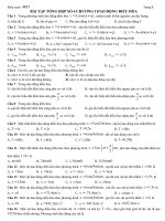
PCR Results.
Bands with the weight of 298 bp were
evaluated as positive (Figure). PCR analysis revealed that
Table 2. ELISA results of chicken sera.
Number of tested Number of flocks Number of sera
Flock type Flocks Sera Positive Negative Positive Negative
n % n % n % n %
Commercial layer 16 392 15 93.7 1 6.3 278 70.9 114 29.1
Commercial broiler 10 240 7 70.0 3 30 50 20.8 190 79.2
Broiler breeding 8 200 8 100 - 0 191 95.5 9 4.5
Layer breeding 4 90 4 100 - 0 90 100 0 0
</div>
<span class='text_page_counter'>(4)</span><div class='page_container' data-page=4>
53 (55.8%) of 95 samples from a total of 25 flocks (15
commercial laying and 10 commercial broiler flocks) were
positive (Table 3). While 38 (66.7%) of 57 thymus
samples were positive taken from 15 commercial layer
flocks and 15 (39.5%) of 38 thymus samples taken from
10 commercial broiler flocks were positive (Table 3).
<b>Discussion</b>
CIA is a very common infection taking a part in the
aetiology of some multifactorial diseases in layer and
broiler flocks of both commercial and breeding types
throughout the world (18,24). One of the most
outstanding features of the CIAV is to cause
immunosuppression by itself directly or by participating
indirectly
with
other
viruses
in
chickens
(13,19,21,24,25).
Some serological (Neutralisation, ELISA, and IFAT)
(6,8,9,26) and molecular studies (PCR) (7,12,23,27) on
determining either presence of antibodies to the agent or
amplification of causatives’ DNA have been reported.
Ergün et al. (8) examined 4 different broiler flocks by
ELISA and observed that 68.2% of the flocks were
positive. Positive results were also reported in 4
(27.7100%) of 10 parent stocks by ELISA. KuyucuoÔlu
et al. (26) reported that a high degree of positivity
(85.7%) was also found by ELISA from a total of 21
commercial layer flocks. It was also stated that CIAV
antibodies were detected in 66.0% of commercial broiler
and layer, broiler-breeding of flocks by ELISA in provinces
‹zmir and Bandırma (28). By virus neutralization test,
more than 89% of the flocks (either commercial broiler
or layer commercial or layer parent) were found to be
positive (6). Finally, CIA is one of the common infections
according to serologic results in commercial flocks in
Turkey. This study is in line with previous studies.
Some researchers (9,29) reported positive levels of
antibodies to CIAV by IFA in more than 89.5% of broilers
or layers (both type of breeding) in the USA. These and
other studies from all over the world showed that the
infection is common in all chicken types
(8,10,11,26,28-30).
PCR, used widely due to some difficulties in the
isolation of virus (5,7,12), gives the flexibility to
researchers for both in vivo and in vitro studies (30) and
an opportunity to select right strain of virus for vaccine
production (4). Additionally, CIAV contaminations in cell
cultures or various vaccines can be demonstrated by PCR
(5,23). Thymus is the most suitable organ for
determining CIAV’s DNA by PCR (4).
M 1 2 3 4 5 6 7
Table 3. PCR results in thymus samples.
Number of tested Number of flocks Number of thymus samples
Flock type Flocks Thymus Positive Negative Positive Negative
n % n % n % n %
Commercial layer 15 57 13 86.6 2 13.4 38 66.7 19 33.3
Commercial broiler 10 38 7 70.0 3 30.0 15 39.5 23 60.5
Total 25 95 20 80.0 5 20.0 53 55.8 42 44.2
</div>
<span class='text_page_counter'>(5)</span><div class='page_container' data-page=5>
Yılmaz et al. (12) investigated the presence of virus
DNA in limited numbers of thymus samples from broilers
in 8 different flocks and reported that the lowest
occurrence (8.5%) was in Marmara Region in Turkey. On
the other hand, this study is more comprehensive because
CIA positive chickens were detected using PCR and ELISA.
In the current study, the number of anti-CIAV antibody
positive chickens from any type of breeding is actually
much higher than those reported by Yılmaz et al. (12).
CIAV is known to act synergically with IBDV and
Marek (17,19,25). When the history of flocks evaluated,
we found out that some of the CIAV positive flocks had
also showed other infections (IBD, MD, and Leucosis).
Thus, which infection within the flock triggered by which
infection is not clear (17,19,25). Not only viral but also
some bacterial infections like Salmonella sp. and E. coli in
commercial chicken flocks were also reported. When the
ages of the animals were taken into account from
commercial layers and broilers, the occurrence of CIA
with IBD or some bacterial infections may be explained by
a possible immunosuppression caused by CIAV prior to
IBD or bacterial infection.
In conclusion, the results of our study showed that
CIAV infection in Turkey is more common than expected
in poultry. Therefore, CIAV infected positive chicken
flocks may have already been infected with other
causative agents and we strongly recommend using
vaccines to CIA in a vaccination schedule in parent stocks
countrywide. Vaccination may prevent further virus
spread and can also minimize vertical transmission of
virus in field conditions.
<b>Acknowledgment</b>
This work was granted by The Scientific and
Technological Research Council of Turkey (TÜB‹TAK)
(Project no. VHAG-1932)
<b>References</b>
1. Barbour, E.K., Farran, M.T., Hamadeh, S.K., Bouljihad, M.,
Faroon, O., Kreydiyyeh, S.: Comparative impact of live chicken
infectious anemia virus vaccine versus natural exposure in meat
chicken breeders on immunity to infectivity by CIA and inclusion
body hepatitis viruses in their offspring. Vet. Res. Commun.
2002; 26: 397-405.
2. Yuasa, N., Taniguchi, T., Yoshida, I.: Isolation and some
characteristics of an agent inducing anemia in chics. Avian Dis.,
1979; 23: 366-385.
3. Brentano, L., Mores, N., Wentz, I., Chandratilleke, D., Schat,
K.A.: Isolation and identification of chicken infectious anemia virus
in Brazil. Avian Dis., 1991; 35: 793-800.
4. Todd, D., Mawhinney, K.A., McNulty, M.S.: Detection and
differentiation of chicken anemia virus isolates by using the
polymerase chain reaction. J. Clin. Microbiol., 1992; 30:
1661-1666.
5. Soiné, C., Watson, S.K., Rybicki, E., Lucio, B., Nordgren, R.M.,
Parrish, C.R., Schat, K.A.: Determination of the detection limit of
the polymerase chain reaction for chicken infectious anemia virus.
Avian Dis., 1993; 37: 467-476.
6. Drén, C., Farkas, T., Németh, I.: Serological survey on the
prevalence of chicken anemia virus infection in Hungarian chicken
flocks. Vet. Microbiol., 1996; 50: 7-16.
7. Zhou, W., Shen, B., Yang, B., Han, S., Wei, L., Xiao, B., Zhou, V.:
Isolation and identification of chicken infectious anemia virus in
China. Avian Dis., 1997; 41: 361-364.
8. Ergün, A., Yurtman, M., Nalbantsoy, A.: Civciv anemisi hastalÔ
(CAV) ỹzerinde ELISA testi ile sero-sửrvey ỗalma. ầiftlik Derg.,
1998; 5: 44-46.
9. Cardona, C., Lucio, B., O’Connell, P., Jagne, J., Schat, K.A.:
Humoral immune responses to chicken infectious anemia virus in
three strains of chickens in a closed flock. Avian Dis., 2000; 44:
661-667.
10. De Herdt, P., Van den Bosch, G., Ducatelle, R., Uyttebroek, E.,
Schrier, C.: Epidemiology and significance of chicken infectious
anemia virus infections in broilers and broiler parents under
nonvaccinated European circumstances. Avian Dis., 2001; 45:
706-708.
11. Ledesma, N., Fehervari, T., Casaubon, M.T., Lucio, E., Ratz, F.:
Chicken infectious anemia in Mexico: virus identication and
serology survey. Avian Dis., 2001; 45: 788-796.
12. Yılmaz, H., Turan, N., Özgür, N. ., Helps, C.R., Akay, Ö.:
Detection of chicken anemia virus DNA in the thymus of naturally
infected chicks in Turkey. Avian Dis., 2001; 45: 529-533.
13. Cloud, S.S., Rosenberger, J.K., Lillehoj, H.S.: Immune dysfunction
following infection with chicken anemia agent and infectious
bursal disease virus. II. Alteriations of in vitro lymphoproliferation
and in vivo immune responses. Vet. Immunol Immunopathol.,
1992; 34: 353-366.
</div>
<span class='text_page_counter'>(6)</span><div class='page_container' data-page=6>
15. Smyth, J.A., Moffett, D.A., McNulty, M.S., Todd, D., Mackie,
D.P.: A sequantial histopathologic and immunocytochemical study
of chicken anemia virus infection at one day of age. Avian Dis.,
1993; 37: 324-338.
16. Toro, H., Ramirez, A.M., Larenas, J.: Pathogenicity of chicken
anemia virus (isolate 10343) for young and older chickens. Avian
Pathol., 1997; 26: 485-499.
17. Rosenberger, J.K., Cloud, S.S.: The effects of age, route of
exposure, and coinfection with infectious bursal disease virus on
the pathogenicity and transmissibility of chicken anemia agent
(CAA). Avian Dis., 1989; 33: 753-759.
18. McIlroy, S.G., McNulty, M.S., Bruce, D.W., Smyth, J.A., Goodall,
E.A., Alcorn, M.J.: Economic effects of clinical chicken anemia
agent infection on profitable broiler production. Avian Dis., 1992;
36: 566-574.
19. Otaki, Y., Nunoya, T., Tajima, M., Saito, K., Nomura, Y.:
Enhanced pathogenicity of chicken anemia agent by infectious
bursal disease virus relative to the occurence of Marek’s disease
vaccination breaks. Jpn. J. Vet. Sci., 1989; 51: 849-852.
20. Jørgensen, P.H., Otte, L., Nielsen, O.L., Bisgaard, M.: Influence of
subclinical virus infections and other factors on broiler flock
performance. Br. Poult. Sci., 1995; 36: 455-463.
21. Toro, H., Gonzalez, C., Cerda, L., Hess, M., Reyes, E., Geissea, C.:
Chicken anemia virus and fowl adenoviruses: association to induce
the inclusion body hepatitis/hydropericardium syndrome. Avian
Dis., 2000; 44: 51-58.
22. Tham, K.M., Stanislawek, W.L.: Polymerase chain reaction
amplification for direct detection of chicken anemia virus-DNA in
tissues and sera. Avian Dis., 1992; 36: 1000-1006.
23. Markowski-Grimsrud, C.J., Miller, M.M. , Schat, K.A.:
Development of strain-spesific real-time PCR and RT-PCR assays
for quantitation of chicken anemia virus. J. Virol. Methods, 2002;
101: 135-147.
24. Hagood, L.T., Kelly, T.F., Wright, J.C., Hoerr, F.J.: Evaluation of
chicken infectious anemia virus and associated risk factors with
disease and production losses in broilers. Avian Dis., 2000; 44:
803-808.
25. Miles, A.M., Reddy, S.M., Morgan, R.W.: Coinfection of
spesific-pathogen-free chickens with Marek’s disease virus (MDV) and
chicken infectious anemia virus: effect of MDV pathotype. Avian
Dis., 2001; 45: 9-18.
26. KuyucuoÔlu, Y., Hadimli, H.H., Kenar, B., Uỗan, U.S.: Detection of
chicken infectious anemia virus antibody in layer operations by
using ELISA in Afyon region. Vet. Hek. Mikrobiyol. Derg., 2003;
3: 21-26.
27. Noteborn, M.H.M., Koch, G.: Chicken anemia virus infection:
molecular basis of patogenicity. Avian Pathol., 1995; 24: 11-31.
28. Tỹrkylmaz, S., MsrloÔlu, O.Z.: ‹zmir ve Bandırma yörelerinde
chicken infectious anemia virus antikorlarının ELISA ile
saptanması. Pendik Vet. Mikrobiyol. Derg., 2004; 35: 13-19.
29. Lucio, B., Schat, K.A., Shivaprasad, H.L.: Identification of the
chicken anemia agent, reproduction of the disease, and serological
survey in the United States. Avian Dis., 1990; 34: 146-153.
30. Yamaguchi, S., Kaji, N., Munang’andu, H.M., Kojima, C., Mase,
</div>
<!--links-->