Bài tập tổng hợp - C12
Bạn đang xem bản rút gọn của tài liệu. Xem và tải ngay bản đầy đủ của tài liệu tại đây (534.54 KB, 11 trang )
<span class='text_page_counter'>(1)</span><div class='page_container' data-page=1>
Molecular Responses of Macrophages to Porcine Reproductive
and Respiratory Syndrome Virus Infection
Xuexian Zhang,* Jinho Shin,† Thomas W. Molitor,† Lawrence B. Schook,* and Mark S. Rutherford*,1
*Department of Veterinary Pathobiology and†Department of Clinical and Population Sciences,
<i>College of Veterinary Medicine, University of Minnesota, St. Paul, Minnesota 55108</i>
<i>Received April 1, 1999; returned to author for revision June 29, 1999; accepted July 20, 1999</i>
The detailed mechanism(s) by which porcine reproductive and respiratory syndrome virus (PRRSV) impairs alveolar Mø
homeostasis and function remains to be elucidated. We used differential display reverse-transcription PCR (DDRT-PCR) to
identify molecular genetic changes within PRRSV-infected Mø over a 24 h post infection period. From over 4000 DDRT-PCR
amplicons examined, 19 porcine-derived DDRT-PCR products induced by PRRSV were identified and cloned. Northern blot
analysis confirmed that four gene transcripts were induced during PRRSV infection. PRRSV attachment and penetration alone
did not induce these gene transcripts. DNA sequence revealed that one PRRSV-induced expressed sequence tag (EST)
encoded porcine<i>Mx1, while the remaining 3 clones represented novel ESTs. A full-length cDNA clone for EST G3V16 was</i>
obtained from a porcine blood cDNA library. Sequence data suggests that it encodes an ubiquitin-specific protease (UBP) that
regulates protein trafficking and degradation. In pigs infected<i>in vivo, upregulated transcript levels were observed forMx1</i>
and<i>Ubp</i>in lung and tonsils, and for<i>Mx1 in tracheobronchial lymph node (TBLN). These tissues correspond to sites for</i>
PRRSV persistence, suggesting that the<i>Mx1 andUbp</i>genes may play important roles in clinical disease during PRRSV
infection. © 1999 Academic Press
INTRODUCTION
Porcine reproductive and respiratory syndrome (PRRS)
is prevalent in Europe, North America, and Asia, and
leads to significant economic losses in the swine
indus-try. PRRS virus (PRRSV), the causative agent, was
iden-tified in 1991 in the Netherlands (Wensvoort<i>et al.,1991)</i>
and in 1992 in the United States (Collins <i>et al.,1992).</i>
PRRSV infection presents as reproductive failures
through premature farrowing and/or interstitial
pneumo-nia characterized by alveolar wall thickening with
mac-rophage (Mø) and necrotic cell debris. PRRSV is a small
enveloped RNA virus of the family<i>Arteriviridae</i>
(Conzel-mann<i>et al.,</i>1993), order<i>Nidovirales</i>(Cavanaugh, 1997),
and contains an approximately 15 kb positive strand RNA
genome. PRRSV structural proteins encoded from open
reading frames (ORF) 2 to 7 were identified as
glycopro-tein (GP)2 (29–30 kDa), GP3 (45–50 kDa), GP4 (31–35
kDa), major envelop protein (E; 24–26 kDa), a viral
mem-brane protein (M; 18–19 kDa), and a nucleocapsid (N; 15
kDa) (Meulenberg<i>et al.,</i>1996; Meulenberg <i>et al.,</i>1995;
van Nieuwstadt <i>et al.,</i> 1996). The E protein is strongly
cytotoxic via induction of apoptosis<i>in vitro</i>(Sua´rez<i>et al.,</i>
1996). As yet, the functions for GP2, GP3, and GP4 have
not been elucidated.
<i>Arteriviridae</i>replicate primarily within Mø, and porcine
alveolar Mø are a primary target cell for PRRSV
replica-tion<i>in vivo</i>(Molitor<i>et al.,</i>1996; Wensvoort <i>et al.,</i>1991).
PRRSV replication in alveolar Mø is associated with
cytopathic effects (CPE; Rossow, 1998). PRRSV infection
decreases alveolar Mø release of superoxide anion
(Thanawongnuwech <i>et al.,</i>1997) and the number of
al-veolar Mø in the lung (Plana<i>et al.,</i>1992). It is presumed
that altered alveolar Mø function is linked to the apparent
increased incidence of pulmonary bacterial co-infections
in PRRSV-infected herds (Kobayashi<i>et al.,</i>1999;
Thana-wongnuwech <i>et al.,</i>1997). The high incidence of
respi-ratory microbial co-infection in chronically infected herds
suggests that PRRSV interferes with host Mø activities
used to clear respiratory pathogens. However, the
mo-lecular pathways by which PRRSV infection disrupts
nor-mal Mø homeostasis have not been elucidated.
Once an intracellular pathogen such as a virus
in-vades a host cell, the interactions become physiological
and biochemical as well as immunological. Pathogens
that replicate within host cells usurp host biological
processes for their own benefit. In response, the host
cell manipulates gene expression to inhibit those
path-ways or processes required or induced by the pathogen.
Viruses in particular subvert host cell metabolism in such
a way that viral components can be synthesized via host
cell pathways to initiate viral replication. Viral particles,
viral components, and virus-induced cellular factors all
have the potential to alter host cell gene expression.
1<sub>To whom reprint requests should be addressed at 1988 Fitch</sub>
Avenue, Room 295. Fax: (612) 625-0204. E-mail:
Article ID viro.1999.9914, available online at on
0042-6822/99 $30.00
Copyright © 1999 by Academic Press
All rights of reproduction in any form reserved.
</div>
<span class='text_page_counter'>(2)</span><div class='page_container' data-page=2>
Molecular genetics and cell biology can be implemented
to define the specific host cell molecules and cellular
components with which virus-encoded molecules
inter-act.
Differential display reverse transcription polymerase
chain reaction (DDRT-PCR) is a powerful approach used
to directly compare gene expression between cells or
tissues at specific physiological states (Bhattacharjee<i>et</i>
<i>al.,</i> 1998; Liang <i>et al.,</i> 1992). DDRT-PCR provides an
unbiased mRNA fingerprint for direct observation of
cDNAs PCR amplified to different levels reflective of
relative transcript levels within a total RNA sample. By
using a reverse transcription/PCR primer that anchors on
the nucleotide just 5<sub>9</sub>of the poly(A) tail, DDRT-PCR
spe-cifically reverse transcribes only a subset of the mRNAs.
PCR is then performed using the 3<sub>9</sub>-anchor primer and
an arbitrary 5<sub>9</sub>primer to amplify 100–400 fragments of the
cDNAs generated by reverse transcription. Recently,
DDRT-PCR has been used to describe host cell genetic
responses to infection by pseudorabies virus (Hsiang<i>et</i>
<i>al.,</i>1996), cytomagolovirus (Zhu<i>et al.,</i>1997), HIV (Sorbara
<i>et al.,</i>1996), herpes simplex virus (Tal-Singer<i>et al.,</i>1998)
and rhabdovirus (Boudinot<i>et al.,</i>1999). We hypothesized
that PRRSV infection alters alveolar Mø homeostatic
gene expression, leading to compromised host defenses
in the lungs of infected animals. The effect of PRRSV
infection on alveolar Mø gene expression was therefore
observed by monitoring changes in gene expression
using DDRT-PCR. We identified 19 porcine Mø-derived
DDRT-PCR amplicons induced during a 24 h <i>in vitro</i>
infection period. Many of these transcripts appear to
encode previously unknown gene products. Further, we
confirmed that four of these genes are induced during
PRRSV infection, and are induced <i>in vivo</i> in tissues
where PRRSV persistently resides, suggesting that they
may provide insight for understanding host cell
molecu-lar responses during PRRSV pathogenesis.
RESULTS
PRRSV replication and altered gene expression
in alveolar Mø
Alveolar Mø were infected using PRRSV strain VR2332
(m.o.i.<sub>5</sub> 0.1)<i>in vitro. CPE was not observed until 16 h</i>
post infection and was less than 10% at 24 h post
infec-tion. At 72 h post infection, CPE was more than 70% (data
not shown). To identify differentially expressed mRNAs
during PRRSV infection, we collected total cellular RNA
from mock- and PRRSV-infected porcine alveolar Mø at 4,
12, 16, and 24 h post infection. PRRSV genome
replica-tion was confirmed via RT-PCR detecreplica-tion of accumulareplica-tion
for ORF 7 sequences of PRRSV genomic RNA (Fig. 1).
PRRSV ORF 7 transcript levels increased with time,
dem-onstrating active viral genomic replication in alveolar Mø.
Mø mRNAs differentially expressed during PRRSV
in-fection were detected by DDRT-PCR comparison against
mock-infected alveolar Mø at various times post
infec-tion. For each DDRT-PCR primer pair, duplicate RNA
samples from each time point were reverse-transcribed,
PCR amplified, and fractionated on adjacent lanes to
ensure DDRT-PCR accuracy and reproducibility.
Repre-sentative DDRT-PCR reactions are shown in Fig. 2.
PRRSV infection induced (Fig. 2A) or suppressed (data
not shown) several alveolar Mø transcripts. Using 16 of
the possible upstream various septamer H-AP primers
(GenHunter), over 4000 DDRT-PCR products were
visu-ally compared for band intensity. Twenty DDRT-PCR
products that were reproducibly induced (<sub>.</sub>twofold
dif-ference compared to mock-infected cultures) in both
DDRT-PCR reactions for a given RNA sample during a
FIG. 1.PRRSV replication in PRRSV-infected alveolar Mø. RT-PCR was performed with 59primer/39primer (PRRSV ORF 7-specific primers). Blank is
a negative PCR control without RT mixture. M is 100 bp DNA ladder. Arrow indicates the PCR product (508 bp).
</div>
<span class='text_page_counter'>(3)</span><div class='page_container' data-page=3>
24 h PRRSV infection<i>in vitro</i> (Table 1). All differentially
expressed DDRT-PCR products were extracted from
acrylamide gels, reamplified, and cloned into pBluescript
SKII (Stratagene). DDRT-PCR amplicons that showed
al-tered levels in only one of two identical samples were
not considered further.
Amplicon sequence analysis
To determine whether DDRT-PCR clones were derived
from porcine cellular genes or from the PRRSV genome,
all 20 PRRSV-induced DDRT-PCR amplicons were
se-quenced. One DDRT-PCR clone encoded a portion of the
PRRSV ORF2 (data not shown) and was removed from
further study. Only 5 of the remaining 19 PRRSV-induced
transcripts matched previous GenBank submissions
(Ta-ble 1). Our particular DDRT-PCR application utilizes a
reverse transcription primer anchored at the last
nucle-otide 5<sub>9</sub>to the poly(A) tail, and PCR conditions that favor
amplification of 200–400 bp amplicons. As expected for
short cDNAs derived from the 3<sub>9</sub> end of mRNAs, no
significant open reading frames or known conserved
protein functional domains could be detected in the
PRRSV-induced, novel cDNAs. Clone G12V24
repre-sented the porcine <i>Mx1 cDNA as determined by 99%</i>
nucleotide sequence similarity. <i>Mx1 is a previously </i>
de-scribed interferon-inducible protein with allelic
associa-tion to viral resistance/susceptibility phenotypes in mice
(Horisberger, 1995).
Confirmation of DDRT-PCR results
DDRT-PCR is a powerful approach to identify and
iso-late uniquely expressed genes (Liang and Pardee, 1992),
but it is a semi-quantitative technique with a high false
positive rate and artifacts. Template abundance, primer
sequence specificity, primer availability, PCR cycle
num-ber, and amplification efficiency can each affect
DDRT-PCR amplicon accumulation. Therefore, in addition to
simultaneously comparing duplicate RNA samples and
DDRT-PCR reactions, it was necessary to confirm the
expression patterns observed in the DDRT-PCR profiles.
All 19 PRRSV-induced porcine Mø DDRT-PCR clones
were screened by Northern blot analysis against total
cellular RNA from mock- and PRRSV-infected porcine
alveolar Mø. Three clones gave no signal on Northern
TABLE 1
PRRSV-Induced DDRT-PCR Amplicons
Clone
DDRT-PCR
levels (24 H)<i>a</i>
Insert
size (bp) Identity or similarity (%)
Mock PRRSV
A2V16-22<i>b</i> <sub>1</sub> <sub>11</sub> <sub>255</sub> <sub>Novel EST</sub><i>c</i>
A2V16-23 1 11 188 Novel EST
A4V12-22 1 11 187 BovineaS1-casein (84%)
A5V12-11 2 111 214 Novel EST
A12V24-11 1 111 307 Novel EST
A12V24-21 1 11 307 Novel EST
C3V16-11 1 11 188 Novel EST
C3V16-32 1 11 252 Novel EST
C7V16-11 1 11 238 Novel EST
C7V16-31 1 11 259 Human thioredoxin (87%)
C7V16-41 1 11 341 Bovine NADH-ubiquinone
oxidoreductase (93%)
C7V16-52 1 111 436 Human galactin-3 (90%)
C12V16-21 1 11 352 Novel EST
G2V12-11 1 11 208 Novel EST
G3V16-11 1 111 240 Novel EST<i>d</i>
G4V12-11 1 11 290 Novel EST
G12V24-11 1 11 316 Novel EST
G12V24-12 2 111 317 Porcine Mx1 (99%)
G13V16-21 1 11 264 Novel EST
<i>a</i><sub>The intensity of the DDRT-PCR products was graded visually from</sub>
DDRT-PCR gels exposed to film. Relative band intensity is denoted1
to111, and2denotes no DDRT-PCR product.
<i>b</i><sub>Clones in bold were subsequently confirmed by Northern blotting to</sub>
be induced by PRRSV infection of porcine alveolar Mø.
<i>c</i><sub>DDRT-PCR amplicons were considered novel ESTs if they had</sub>
,
70% identity over a continuous 100-bp sequence.
<i>d</i><sub>Clone G3V16 was subsequently determined to encode a ubiquitin</sub>
protease.
</div>
<span class='text_page_counter'>(4)</span><div class='page_container' data-page=4>
blot analysis (data not shown), suggesting that they were
either cloning artifacts or represented sequences
ex-pressed at levels too low to detect by this technique. Our
previous experience has shown that as many as 40% of
DDRT-PCR clones require more sensitive RT-PCR or
ri-bonuclease protection assays (RPA) for quantification
(Bhattacharjee <i>et al.,</i> 1998). An additional 12
PRRSV-induced DDRT-PCR clones showed less than twofold
induced expression on Northern blots (data not shown)
and their expression was not further investigated. The
four remaining DDRT-PCR clones were confirmed by
Northern blot analysis to be induced by PRRSV infection
(Fig. 3). All 4 transcripts were induced by PRRSV
infec-tion, exhibited distinct expression levels, and were of
different sizes (data not shown). These data support that
each clone was derived from a transcript representing a
unique amplicon.
Temporal accumulation of the molecular markers
iden-tified by DDRT-PCR during PRRSV infection was
deter-mined. Total cellular RNA was isolated from alveolar Mø
from 0 to 36 h after treatment with medium alone,
con-ditioned medium from CL2621 cells used to generate
infectious PRRSV preparations, UV-irradiated PRRSV, and
PRRSV. Transcripts detected by DDRT-PCR clones
A5V12, G3V16, G2V12, and G12V24 increased
concomi-tant with PRRSV replication (Fig. 3). Transcripts detected
by A5V12, G2V12, and G3V16 were not detected until 16 h
post infection, whereas G12V24 transcripts were induced
as early as 8 h post infection (Fig. 3). Gene transcripts
were not induced in medium control cultures or in Mø
treated with CL2621 cell-conditioned medium.
Impor-tantly, Mø infected with UV-irradiated PRRSV, which can
bind to and penetrate Mø but not replicate, did not
express detectable levels for any of the transcripts
ex-amined (Fig. 3). Together, these data indicate that active
PRRSV genomic replication within Mø is required for
induction of gene expression for these selected
ampli-cons.
Common molecular responses of porcine alveolar Mø
To determine whether the DDRT-PCR clones identified
and characterized from PRRSV-infected Mø were specific
to this pathogen or instead result from a general viral
response molecular program, transcript expression was
determined for porcine alveolar Mø infected with
pseu-dorabies virus (PRV)<i>in vitro. As was observed for PRRSV,</i>
all 4 transcripts were induced by PRV infection (Fig. 4).
However, the kinetics of expression were different.
Tran-scripts appeared sooner and peak expression levels
were higher compared to PRRSV-infected cultures,
per-haps reflecting the more severe disruption of Mø
ho-meostasis and CPE for PRV (data not shown). Further,
transcripts for G2V12 dissipated by 24 h in PRV-infected
cultures, suggesting that it is only transiently expressed
by virally-infected cells. Thus, induction of these
tran-scripts appears to reflect a generalized Mø molecular
response to intracellular viral replication.
Identification of a full-length cDNA clone
To further characterize PRRSV-induced porcine Mø
genes, a porcine peripheral blood cell cDNA library was
screened using clone G3V16 as a probe. A phage clone
was isolated via plaque lift hybridization and contained
an insert (1.7 kb) of approximately the same size as the
mRNA (data not shown), suggesting that it contained a
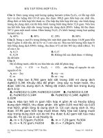
FIG. 3.Temporal transcript expression in PRRSV-infected Mø determined by Northern blot. Total cellular RNAs (10mg per lane) were collected from
mock- and PRRSV-infected Mø at 0 - 36 h post infection, and hybridized against DDRT-PCR cDNAs as shown. Medium indicates cultures treated with
RPMI 1640 with 10% fetal bovine serum. CL2621 denotes cultures receiving only supernatant from mock-infected CL2621 cells. UV-PRRSV denotes
cultures receiving UV-inactivated PRRSV infection. GAPDH transcripts were quantitated to normalize for RNA loading.
</div>
<span class='text_page_counter'>(5)</span><div class='page_container' data-page=5>
full-length cDNA. DNA sequence determination
(Acces-sion number AF134195) of the isolated cDNA clone
con-firmed that it contained a full-length coding sequence,
which included a 39 untranslated region (UTR, 572bp),
coding sequence (966 bp), and a 59 UTR (172bp).
De-duced amino acid sequence identified a conserved Cys
domain (block entry, BL00972A) and a His domain (block
entry, BL00972D) (Fig. 5A), which are thought to act as
active sites for ubiquitin-specific proteases (UBPs;
Wilkinson, 1997). Further, a GenBank data search and
analyses by Blast (NCBI, NIH) and FEX (Find exon
pro-gram, Sanger Center, UK) indicated that a putative
hu-man homolog is located at chromosome 22q11.2. The
putative human UBP has eight ORFs derived from
ap-proximately 15 kb of genomic DNA (Fig. 6). The amino
acid similarity and identity of porcine UBP with the
pu-tative human UBP were 81% and 75%, respectively (Fig.
5B). These results indicate that porcine UBP is a novel
member of an UBP superfamily, and suggest that
intra-cellular protein trafficking, turnover or degradation may
be altered during PRRSV infection of porcine alveolar
Mø. The identities or putative function for PRRSV-induced
ESTs A5V12 and G2V12 have not yet been established.
Tissue specific expression in<i>in vivo</i>PRRSV-infected
animals
To determine whether expression of DDRT-PCR
prod-ucts identified <i>in vitro</i> reflected events during PRRSV
infection<i>in vivo, we examined tissue-specific expression</i>
of DDRT-PCR amplicons in PRRSV-infected pigs. Whole
tissues were collected at 14 days post infection from 2
PRRSV-infected pigs and 2 mock-infected pigs.
Quanti-tative RT-PCR demonstrated the presence of PRRSV
genomic RNA in lungs, lymph nodes, and tonsils (data
not shown). Tissue RNAs from identically treated animals
were pooled to minimize animal-to-animal variation, and
RT-PCR was performed for 14 and 17 cycles, which we
have determined is in the linear range of amplification
(data not shown). Products were transferred onto
mem-branes for Southern blot analysis via hybridization to
cDNA probes for porcine <i>Mx1 and</i> <i>Ubp</i> (Fig. 7). PCR
amplicon levels for each tissue sample were normalized
to HPRT amplicon levels, and normalized values for each
tissue were compared between mock- and
PRRSV-in-fected animal (Fig. 7). Porcine <i>Ubp</i> transcripts were
greatly upregulated during PRRSV infection in the lungs
(4.5-fold) and tonsils (11.4-fold), but were reduced 30% in
TBLN from PRRSV-infected animals. In contrast, <i>Mx1</i>
transcripts were greatly induced in all three tissues (Fig.
7). Taken together, these data show (1) constitutive
ex-pression for these genes<i>in vivo; (2) tissue-specific </i>
reg-ulation of gene expression; and (3) PRRSV-induced
up-regulation of transcript levels in tissues where PRRSV is
persistent.
DISCUSSION
PRRSV infection causes significant losses in the swine
industry, in part due to poor growth associated with
interstitial pneumonia (Rossow, 1998). PRRSV infection<i>in</i>
<i>vivo</i>is thought to be a contributing factor that results in
increased secondary pulmonary bacterial infections
fol-lowing impairment of Mø function in the lungs. Toward
</div>
<span class='text_page_counter'>(6)</span><div class='page_container' data-page=6>
this end, recent evidence describes slightly impaired
killing of<i>Haemophilus parasuis</i>and<i>Staphylococcus </i>
<i>au-reus</i>by PRRSV-infected alveolar Mø (Solano<i>et al.,</i>1998;
Thanawongnuwech <i>et al.,</i> 1997). However, conflicting
data exists. While PRRSV-infected Mø demonstrate
re-duced reactive oxygen product formation (Done and
Pa-ton, 1995; Thanawongnuwech<i>et al.,</i>1997) and late-stage
inhibition of bacterial phagocytosis (Solano<i>et al.,</i>1998),
phagocytosis of opsonized <i>S. aureus</i>
(Thanawongnu-wech<i>et al.,</i>1997) or<i>H. parasuis</i>(Segale´s<i>et al.,</i>1998) is
not impaired. Pro-inflammatory cytokine gene
expres-sion, including TNF-<sub>a</sub>, IL-8, IFN-<sub>a</sub> and IL-1<sub>b</sub> does not
appear to be significantly altered (Buddaert<i>et al.,</i>1998;
Trebichavsky and Valicek, 1998; Zhang and Rutherford,
1997). Further, a systemic impairment of host immunity
during persistent PRRSV infection is not supported
(Al-bina <i>et al.,</i> 1998). Hence, we have used a DDRT-PCR
mRNA fingerprinting approach to identify molecular
re-sponses during PRRSV infection of porcine alveolar Mø.
We now report that PRRSV infection alters host Mø gene
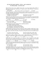
FIG. 5.Conserved domains and human homology of porcine UBP amino acids. (A) Positions of the conserved amino acid sequence domains that
contains Cys residue and His residue in ubiquitin-specific protease (UBP) superfamily are shown in standard single letter code. Bold letters indicate
identity with porcine UBP with other members in these two domains. The aligned gene accession numbers are: UBPH-human, Q93009; FAFX-human,
Q93008; UBP41-human, AF079564; UBPY-human, P40818; UBP41-mouse, AF079565; UBP41-chicken, AF016107; FAF-flies, A49132; UBPE-flies, Q24574;
UBP8-yeast, P50102; UBPF-yeast, P50101; UBPB-schpo, Q09738; UCH-putative, AL021889. Numbers in parentheses are the amino terminus position
of conserved domain in UBPs. (B) Alignment of porcine UBP amino acid and a putative human UBP homolog with Gap program (GCG). Blast search
(NCBI, NIH) was performed for the entire G3V16 cDNA sequence. Further, FEX (find exon, http://genomic. sanger.ac.uk/) was used to find the human
homolog. Human sequence was derived from genomic DNA sequence (AC005500). Letters in bold denote the conserved Cys and His domains.
Asterisks denote translational stop.
</div>
<span class='text_page_counter'>(7)</span><div class='page_container' data-page=7>
expression programs, including a ubiquitinated protein
degradation pathway and the induction of novel genes of
unknown function.
Molecular characterization of PRRSV infection will
per-mit us to identify and isolate important host cell
molec-ular responses associated with PRRSV infection.
How-ever, it is clear from our<i>in vivo</i>studies (Fig. 7) that factors
within individual tissues can impact the molecular
phe-notype of the tissues and the infected cells therein.
Further, temporal effects<i>in vivo</i>are difficult to gauge on
a per cell basis due to the continuous influx of immune
cells through secondary lymphoid organs and
inflamma-tory sites, leading to asynchronous infection times. The
<i>in vivo</i>molecular status of a tissue reflects a wide range
of effects, particularly for tissue Mø that display
signifi-cant functional and molecular heterogeneity between
tissues and stages of differentiation (Rutherford <i>et al.,</i>
1993). Consistent with this premise, PRRSV tropism for
Mø is greatly dependent on Mø origin, state of
differen-tiation, and level of activation (Duan<i>et al.,</i>1995; Molitor
<i>et al.,</i>1996). Thus, tissue-specific regulation of <i>Mx1 and</i>
<i>Ubp</i>gene expression is not unexpected, and may reflect
changes in the number of tissue macrophages. Further,
while these gene transcripts were identified in Mø
cul-tures, the cell(s) that express these transcripts <i>in vivo</i>
and their spatial distribution in relation to
virus-contain-ing cells await elucidation.
Initial experiments used both cell culture medium and
CL2621 cell-conditioned medium as control treatments
for confirming DDRT-PCR results. This was necessary in
that PRRSV viral stocks were propagated using the
CL2621 cell line. Our results showing that CL2621
con-ditioned medium or UV-inactivated PRRSV stocks did not
induce Mø expression of these genes suggests that
components within the CL2621 cell culture supernatant
itself do not account for our DDRT-PCR findings. On the
FIG. 6.Putative human<i>Ubp</i>gene structure. Eight exons were mapped in human genomic DNA from 1 to 8. Each solid box indicates an exon. ATG
is the start codon and TGA is the stop codon.
</div>
<span class='text_page_counter'>(8)</span><div class='page_container' data-page=8>
other hand, PRRSV attachment and penetration also
likely impacts host cell gene expression. Again, our data
suggest that PRRSV attachment and penetration alone
was not sufficient to induce expression of the four genes
examined here (Fig. 3). This is in contrast to Zhu<i>et al.,</i>
(1997) who reported that human cytomegalovirus
in-duces host cell mRNA accumulation via original viral
particles and not viral replication in host cells. Recently,
Boudinot<i>et al.,</i>(1999) reported that a glycoprotein from
hemorrhagic septicemia virus (VHSV) in fish directly
in-duced<i>vig-1 gene expression. Failure to observe </i>
attach-ment-induced transcripts may be due to the fact that,
compared to actively infected cultures, a lower
percent-age of host cells experience viral attachment and
pene-tration in cultures treated with UV-inactivated PRRSV
(m.o.i. is 0.1), with subsequently fewer
attachment-in-duced transcripts being present in these cultures. A
higher titer infection may help identify viral attachment
effects on host cell transcripts. We are also incorporating
a more sensitive RT-PCR screening for DDRT-PCR clones
that do not detect transcripts by Northern blot screening.
Finally, we have used only 16 of the possible 80
DDRT-PCR primer pairs, and further characterization of
addi-tional DDRT-PCR clones may also reveal transcripts
al-tered as a direct result of PRRSV attachment.
Interferons (IFN) play an important role in host defense
against viruses, in part via the induction of cellular genes
(Staeheli, 1990; Zhu<i>et al.,</i>1997) that include<i>Mx</i>genes.
The <i>Mx1 gene was originally isolated as a viral </i>
resis-tance gene from mice (Lindenmann, 1964) having two
alleles, Mx1 <sub>(resistant, dominant) and Mx</sub>- <sub>(susceptible,</sub>
recessive).<i>Mx1 gene homologues have been described</i>
in other mammalian species, including pigs (Horisberger
and Gunst, 1991). Mx1 inhibits primary transcription of
parental influenza viral genomes in mice (Krug <i>et al.,</i>
1985). In humans, the <i>Mx1-encoded MxA protein is </i>
in-duced by type I IFNs, double-stranded RNA, and several
viruses, including influenza virus and Newcastle disease
virus in human embryonic cells (Aebi<i>et al.,</i>1989) and HIV
in monocytes (Baca <i>et al.,</i> 1994). We now report that
PRRSV infection of Mø induces porcine<i>Mx1 expression,</i>
either directly or subsequent to PRRSV-induced IFN
pro-duction in infected cultures.
The kinetics of<i>Mx1 gene activation are very fast, and</i>
MxA accumulation is detectable within 4 h post infection
(Horisberger, 1995), consistent with our observations.
The functional significance for porcine<i>Mx1 gene </i>
expres-sion in PRRSV infection is unknown. Based on <i>Mx1</i>
activities in other species and PRV induction of <i>Mx1</i>
expression in porcine Mø, this gene product is likely to
be involved in host cell protection against viruses in
general, either following infection directly or via IFN
released by neighboring cells which harbor the virus.
However, <i>Mx1 mRNA accumulation did not prevent</i>
PRRSV or PRV replication in porcine alveolar Mø. Thus,
its importance during PRRSV infection, maintenance of
Mø homeostasis, and development of CPE is unclear. It
is interesting to note a recent report that describes pig
breed differences in tissue lesions to a high virulence
strain of PRRSV (Halbur <i>et al.,</i> 1998). It is presently
unknown whether<i>Mx1 alleles exist in pigs and whether</i>
they associate with viral resistance/susceptibility
pheno-types as observed in mice (Lindenmann, 1964).
UBP comprises a protein superfamily in which more
than 60 UBPs have been identified in different species
(Wilkinson, 1997). Identification of a PRRSV-induced UBP
is the first such protein described in pigs. UBPs
specif-ically hydrolyze ester, thiol ester, and amide bonds to the
carboxyl group of G76 of ubiquitin in which ubiquitin
conjugates with target proteins that will be degraded by
proteasome 26 (Hochstrasser, 1995; Goldberg, 1995;
Pickart, 1997). Ubiquitin modification and
deubiquitina-tion by UBPs is increasingly recognized as important
protein regulatory strategies that impact cell cycle
regu-lation (Pagano, 1997), cellular growth moduregu-lation (Zhu<i>et</i>
<i>al.,</i>1996), transcription activation (Trier <i>et al.,</i> 1994),
an-tigen presentation by MHC class I (Rock<i>et al.,</i>1994), and
DNA repair and differentiation (Hochstrasser, 1995).
Por-cine <i>Ubp</i> gene expression induced by PRRSV may be
involved in regulating protein metabolism via a
ubiquitin-conjugated pathway. This could benefit the host cell in
that removing ubiquitin from host proteins prevents them
from being moved to the proteasome, helping to maintain
Mø protein levels in the face of viral disruption of host
translation. Conversely, the virus may induce UBP to
prevent newly synthesized viral proteins from being
de-graded. Finally, <i>Ubp</i> gene induction may disrupt Mø
antigen presentation, thereby compromising host
im-mune responses to subsequent bacterial challenge.
By sequence data analysis, we determined that
por-cine<i>Ubp</i>is homologous to a putative human<i>Ubp</i>that is
located at the DiGeorge critical region (DGCR) on
chro-mosome 22q11 (Fig. 6). The recently identified<i>Ufd1 gene</i>
encodes a protein involved in degradation of
ubiquiti-nated proteins (Yamagishi <i>et al.,</i>1999), suggesting that
regulation of ubiquitinated protein degradation
contrib-utes to congenital heart and craniofacial defects in the
mouse embryo (Yamagishi <i>et al.,</i> 1999). The detailed
molecular mechanism by which PRRSV infection leads to
sow abortion is unknown and porcine <i>Ubp</i> may play a
role in fetal death. However, similar cardiac and
cranio-facial defects in PRRSV-aborted fetuses have not been
reported.
</div>
<span class='text_page_counter'>(9)</span><div class='page_container' data-page=9>
ruses and host cells can be delineated via DDRT-PCR
cloning of novel genes and subsequent characterization
of gene expression involved in the altered host cell
homeostasis.
MATERIALS AND METHODS
Cells, viruses, and pigs
Six- to eight-week old pigs were selected from healthy
and PRRSV-negative pig populations. Alveolar Mø were
collected by lung lavage (Lee<i>et al.,</i> 1996). Lungs were
washed 2–4 times with phosphate-buffered saline (PBS,
pH 7.2). Each wash was centrifuged at 1200 rpm at 4°C
for 10 min. Cell pellets were mixed, washed again in PBS,
and then resuspended in 20–50 ml of RPMI 1640. Mø
were incubated overnight at 37°C, 5% CO2in RPMI 1640
medium supplemented with 10% fetal bovine serum, 1
mM L-glutamine, 0.1 mM nonessential amino acids, 25
mM HEPES, and antibiotics before viral infection.
ATCC PRRSV strain VR2332 (passage 9, 53106<sub>PFU/</sub>
ml) and CL2621 cell culture supernatant were obtained
from Dr. K. S. Faaberg (University of Minnesota). PRRSV
suspension (m.o.i.50.1) or medium was inoculated after
washing Mø monolayers. For UV inactivation, PRRSV
stock placed in a 10 cm-diameter petri dish was
irradi-ated using an UV-Crosslinker (Stratagene Corp., La Jolla,
CA) with 120mJ/cm2<sub>for 15 min. Pseudorabies virus (PRV)</sub>
strain 086 was used to infect porcine alveolar Mø (m.o.i.
50.1)<i>in vitro.</i>
For<i>in vivo</i>infection, six-week-old pigs obtained from a
PRRSV seronegative farm were infected intranasally with
105 <sub>TCID50</sub><sub>of PRRSV strain VR2332 or PBS as a control.</sub>
Serum samples were collected at Day 0, 2, 5, 7, 10, and
14 post-infection, and stored at -80°C (data not shown).
Tissues were collected at 14 days post-infection and
immediately placed into TRIzol (Life Technologies, Grand
Island, NY) reagent and frozen in dry ice/ethanol. All
tissues were stored at280°C until used.
Total cellular RNA isolation and Northern blot
analysis
Total cellular RNA was extracted from alveolar Mø
cultures and tissues using TRIzol Reagent (Life
Technol-ogies) according to the manufacturer’s protocol. RNA
integrity was evaluated on 1% agarose gels with
formal-dehyde (0.4 M) after staining with ethidium bromide. For
DDRT-PCR analyses, trace genomic DNA contamination
was removed with MessageClean (GenHunter Corp.,
Nashville, TN) before performing reverse transcription.
For Northern blots, total cellular RNAs (10<sub>m</sub>g per lane)
were fractionated on 1% agarose-0.4 M formaldehyde
gels, transferred to nylon membranes (Schleicher &
Schuell, Keene, NH), and cross-linked using a
UV-Crosslinker (Stratagene). The cDNA probe was labeled
by random primer labeling (Life Technologies) following
the manufacturer’s protocol. Hybridization was carried
out at 42°C in 10 ml of solution containing 5 x SSPE, 50%
formamide, 0.5% SDS, 5 x Denhardt’s reagent, and 100
mg/ml sonicated salmon sperm DNA overnight. The
hy-bridized membrane was washed twice with 2 x SSC/0.1%
SDS for 15 min at room temperature, followed by 0.1 x
SSC/0.1% SDS at 55°C for 20 min. Blots were exposed to
film overnight at -80°C or quantitated by
phosphorimag-ery (Molecular Dynamics, Sunnyvale, CA).
Differential display assays
DDRT-PCR was performed as previously described
(Bhattacharjee <i>et al.,</i> 1998). First-strand cDNAs (20 <sub>m</sub>l)
were synthesized for each RNA sample separately using
one of three H-T11M anchor primers (where M is G, A, or
C, GenHunter Corp.), 0.2–0.4<sub>m</sub>g total cellular RNA, 4<sub>m</sub>l
5 x RT buffer, 20<sub>m</sub>M dNTPs, and 200 U of Superscript II
reverse transcriptase (Life Technologies) at 42°C for 1 h.
PCR reactions (10<sub>m</sub>l) were performed using the
RNAim-age Kit (GenHunter) and contained 1 x PCR buffer, 2<sub>m</sub>M
dNTPs, 0.2 <sub>m</sub>M 5<sub>9</sub> H-AP primer/3<sub>9</sub> H-T11M anchored
primer, 0.15<sub>m</sub>l [<sub>a</sub>-33<sub>P] dATP (2500 Ci/mM, Amersham), 1</sub>
ml reverse transcription product, and 1 U AmpliTaq DNA
polymerase (Perkin–Elmer). The PCR cycling profile was
94°C for 2 min, [94°C for 30 s, 40°C for 2 min, 72°C for
30 s] for 40 cycles, then 72°C for 5 min. Denatured
DDRT-PCR products were loaded onto a 6% denaturing
polyacrylamide DNA sequencing gel, and run for 3.5 h.
The gel was blotted onto filter paper, dried under vacuum
on a gel dryer at 80°C for 1 h, and then exposed to film
at room temperature for 16–24 h. Amplicon intensities
were compared visually for each infection time across
duplicate samples, and differentially expressed
ampli-cons were prepared as described (Bhattacharjee<i>et al.,</i>
1997). Briefly, bands were excised from acrylamide gels,
placed in 100<sub>m</sub>l of dH2O for 10 min, and then boiled for
15 min. DDRT-PCR products were collected by
centrifug-ing for 2 min and stored at<sub>2</sub>20°C. Reamplified cDNAs
were purified from the 2% agarose gel using the QIAEX II
kit (Qiagen Corp., Chatsworth, CA), and then stored at
220°C for cloning and hybridizing analysis.
DDRT-PCR Amplicon cloning and sequencing
</div>
<span class='text_page_counter'>(10)</span><div class='page_container' data-page=10>
Plasmid DNA from clones with insert was prepared by
miniprep (Qiagen). DNA sequencing was performed on
an Applied Biosystem 377 Automatic DNA sequencer
(Perkin–Elmer) in the Advanced Genetic Analysis Center,
College of Veterinary Medicine, University of Minnesota.
Sequences were analyzed by a BLAST search (NCBI,
NIH). The accession numbers are: AF102503 for clone
A5V12, AF102504 for clone G3V16, AF102505 for clone
G2V12, AF102506 for clone G12V24 and AF134195 for
porcine<i>Ubp.</i>
Reverse transcription PCR assay
Reverse transcription (20ml) was performed as above
using total cellular RNA (2mg). The reaction was stopped
by heating to 70°C for 10 min, and RT products were
treated with RNase H (Promega Corp., Madison, WI) for
20 min at 37°C. PCR reactions (25ml) were performed
with RT product (1ml), 10 x PCR buffer, 25mM dNTPs, 0.2
mM each of 5<sub>9</sub>primer and 3<sub>9</sub>primers, and 1 U of
Ampli-Taq DNA polymerase (Perkin–Elmer). The primer pairs
used were:<i>Mx1: 5</i><sub>9</sub>primer GCTTGAGTGCTGTGGTTG/3<sub>9</sub>
primer GGACTTGGCAGTTCTGTGGAG; <i>Ubp: 5</i><sub>9</sub> primer
AGGGGCCAAGCTCATGTGAC/3<sub>9</sub> primer
GTGGCCAG-CATACCATCTCC. Primer sequences and PCR profile for
porcine hypoxanthine phosphoribosyltransferase (HPRT)
have been described (Foss<i>et al.,</i>1998). Each cDNA was
amplified for 14 cycles and 17 cycles (linear range, data
not shown). Amplicons were analyzed by Southern blot
hybridization against DDRT-PCR probes. Signals were
quantified by phosphorimagery (Molecular Dynamics).
Isolation of cDNA clones
A pig cDNA library (kindly provided by Dr. C. W.
Beat-tie, University of Minnesota) prepared from peripheral
blood cells and cloned in Uni-ZAP XR Vector (Stratagene)
was screened with DDRT-PCR clone G3V16. The probe
was labeled with [a232<sub>P] dATP by the random priming</sub>
(Life Technologies), and hybridization was performed as
described for Northern blots. A total of 1 <sub>3</sub> 106 <sub>phage</sub>
plaques were screened using G3V16 cDNA, and a single
positive clone was identified. By sequence analysis, the
clone identified by G3V16 probe was found to contain a
full-length coding sequence.
ACKNOWLEDGMENTS
This research was supported by the National Pork and Producers
Council (M.S.R), the Minnesota Pork Producers Association (T.W.M), the
U.S.D.A. grant 95–3205-3846 (L.B.S.), and the University of Minnesota
Agricultural Experiment Station (M.S.R.). The authors thank Drs. M. P.
Murtaugh and K. S. Faaberg for supplying the PRRSV VR2332 strain,
ORF7 PCR primers, and CL2621 cell culture, and Dr. C. W. Beattie for
porcine cDNA library. The authors further acknowledge Dr. J. E. Collins
for assistance with experimental design, and Drs. A. Bhattacharjee and
A. Rink for technical advice on the DDRT-PCR assays and cDNA library
screening.
REFERENCES
Aebi, M., Fah, J., Hurt, N., Samuel, C. E., Thomis, D., Bazzigher, L.,
Pavlovic, J., Haller, O., and Staeheli, P. (1989). cDNA structures and
regulation of two interferon-induced human Mx proteins.<i>Mol. Cell.</i>
<i>Biol.</i>9, 5062–5072.
Albina, E., Piriou, L., Hutet, E., Cariolet, R., and L’Hospitalier, R. (1998).
Immune responses in pigs infected with porcine reproductive and
respiratory syndrome virus (PRRSV).<i>Vet. Immunol. Immunopathol.</i>61,
49–66.
Baca, L. M., Genis, P., Kalvakolanu, D., Sen, G., Meltzer, M. S., Zhou, A.,
Silvermann, R., and Gendelman, H. E. (1994). Regulation of
interferon-a-inducible cellular genes in human immunodeficiency
virus-in-fected monocytes.<i>J. Leukoc. Biol.</i>55,299–309.
Bhattacharjee, A., Lappi, V. R., Rutherford, M. S., and Schook, L. B.
(1998). Molecular dissection of dimethylnitrosamine (DMN)-induced
hepatotoxicity by mRNA differential display.<i>Toxicol. Appl. Pharmacol.</i>
150,186–195.
Bhattacharjee, A., Rutherford, M. S., Abrahamsen, M. S., Lappi, V. R.,
and Schook, L. B. (1997). Refinements in re-amplification and cloning
of DDRT-PCR products.<i>BioTechniques</i>22,1048–1051.
Boudinot, P., Massin, P., Blanco, M., Riffault, S., and Benmansour, A.
(1999).<i>vig-1, a new fish gene induced by the Rhabdovirus </i>
glycopro-tein, has a virus-induced homologue in humans and shares
con-served motifs with the MoaA family.<i>J. Virol.</i>73,1846–1852.
Buddaert, W., van Reeth, K., and Pensaert, M. (1998).<i>In vivo</i>and<i>in vitro</i>
interferon (IFN) studies with the porcine reproductive and respiratory
syndrome virus (PRRSV).<i>Adv. Exp. Med. Biol.</i>440,461–467.
Cavanagh, D. (1997).<i>Nidovirales: a new order comprising</i>
<i>coronaviri-dae</i>and<i>arteriviridae. Arch. Virol.</i>142,629–633.
Collins, J. E., Benfield, D. A., Christianson, W. T., Harris, L.,
Christopher-Hennings, J., Shaw, D. P., Goyal, S. M., McCullough, S., Morrison,
R. B., Joo, H. S., Gorcyca, D., and Chladek, D. (1992). Isolation of
swine infertility and respiratory syndrome virus (isolate ATCC
VR-2332) in North America and experimental reproduction of disease in
gnotobiotic pigs.<i>J. Vet. Diagn. Invest.</i>4,117–126.
Conzelmann, K. K., Visser, N., Van Woensel, P., and Thiel, H. J. (1993).
Molecular characterization of porcine reproductive and respiratory
syndrome virus, a member of the arterivirus group.<i>Virology</i> 193,
329–339.
Done, S. H., and Paton, D. J. (1995). Porcine reproductive and
respira-tory syndrome: clinical disease, pathology and immunosuppression.
<i>Vet. Rec.</i>136,32–35.
Duan X., Nauwynck, H. J., and Pensaert, M. B. (1997). Effects of origin
and state of differentiation and activation of
monocytes/macro-phages on their susceptibility to porcine reproductive and respiratory
syndrome virus (PRRSV).<i>Arch. Virol.</i>142,2483–2497.
Foss, D. L., Baarsch, M. J., and Murtaugh, M. P. (1998). Regulation of
hypoxanthine phosphoribosyl transferase,
glyceraldehyde-3-phos-phate dehydrogenase and b actin mRNA expression in porcine
immune cells and tissues.<i>Anim. BioTechnol.</i>9,67–78.
Goldberg, A. L. (1995). Functions of the proteasome: the lysis at the end
of the tunnel.<i>Science</i>268,522–523.
Halbur, P. G., Rothschild, M. F., Thacker, B. J., Meng, X.-J., Paul, P. S. and
Bruna, J. D. (1998). Differences in susceptibility of Duroc, Hampshire,
and Meishan pigs to infection with a high virulence strain (VR2385)
of porcine reproductive and respiratory syndrome virus (PRRSV).<i>J.</i>
<i>Anim. Breed. Genet.</i>115,181–189.
Hochstrasser, M. (1995). Ubiquitin, proteasomes, and the regulation of
intracellular protein degradation.<i>Cur. Opinion Cell Biol.</i>7,215–223.
Horisberger, M. A. (1995). Interferons,<i>Mx</i>genes, and resistance to
influenza virus.<i>Am. J. Respir. Crit. Care Med.</i>152,S67–S71.
Horisberger, M. A., and Gunst, M. C. (1991). Interferon-induced proteins:
identification of Mx proteins in various mammalian species.<i>Virology</i>
180,185–190.
</div>
<span class='text_page_counter'>(11)</span><div class='page_container' data-page=11>
Analysis of upregulated cellular genes in pseudorabies virus
infec-tion: use of mRNA differential display.<i>J. Virol. Methods</i>62,11–19.
Kobayashi, H., Morozumi, T., Miyamoto, C., Shimizu, M., Yamada, S.,
Ohashi, S., Kubo, M., Kimura, K., Mitani, K., Ito, N., and Yamamoto, K.
(1996).<i>Mycoplasma hyorhinis</i>infection levels in lungs of piglets with
porcine reproductive and respiratory syndrome (PRRS).<i>J. Vet. Med.</i>
<i>Sci.</i>58,109–113.
Krug, R. M., Shaw, M., Broni, B., Shapiro, G., and Haller, O. (1985).
Inhibition of influenza viral mRNA synthesis in cells expressing the
interferon-induced<i>Mx</i>gene product.<i>J. Virol.</i>56,201–206.
Lee, J.-K., Schook, L. B. and Rutherford, M.S. (1996). Molecular cloning
and characterization of the porcine CD18 leukocyte adhesion
mole-cule.<i>Xenotransplantation</i>3,222–230.
Liang, P., and Pardee, A. B. (1992). Differential display of eukaryotic
messenger RNA by means of the polymerase chain reaction.<i></i>
<i>Sci-ence</i>257,967–971.
Lindenmann, J. (1964). Inheritance of resistance to influenza virus in
mice.<i>Proc. Soc. Exp. Biol. Med.</i>116,506–509.
Meulenberg, J. J. M., and Petersen-den Besten, A. (1996). Identification
and characterization of a sixth structural protein of Lelystad virus: the
glycoprotein GP2 encoded by ORF2 is incorporated in virus particles.
<i>Virology</i>225,44–51.
Meulenberg, J. J. M., Petersen-den Besten, A., de Kluyve, E. P.,
Moor-mann, R. J. M., Schaaper, W. M. M., and Wensvoort, G. (1995).
Characterization of proteins encoded by ORFs 2 to 7 of Lelystad
virus.<i>Virology</i>206,155–163.
Molitor, T. W., Xiao, J., and Choi, C. S. (1996). PRRS virus infection of
macrophages: regulation by matuation and activation state. <i>Proc.</i>
<i>Annu. Meet. Am. Assoc. Swine Pract.</i>27,563–569.
Pagano, M. (1997). Cell cycle regulation by the ubiquitin pathway.
<i>FASEB J.</i>11,1067–1075.
Pickart, C. M. (1997). Targeting of substrates to the 26 S proteasome.
<i>FASEB J.</i>11,1055–1066.
Plana, D. J., Vayreda, M., Vilarrasa, J., Bastons, M., Porquet, L., and
Urniza, A. (1992). PEARS (mystery swine disease)- summary of the
work conducted by our group. <i>Am. Assoc. Swine Pract. News</i> 4,
16–18.
Rock, K. L., Gramm, C., Rothstein, L., Clark, K., Stein, R., Dick, L., Hwang,
D., and Goldberg, A. L. (1994). Inhibitors of the proteasome block the
degradation of most cell proteins and the generation of peptides
presented on MHC class I molecules.<i>Cell</i>78,761–771.
Rossow, K. D. (1998). Porcine reproductive and respiratory syndrome.
<i>Vet. Pathol.</i>35,1–20.
Rutherford, M. S., Witsell, A. L., and Schook, L. B. (1993). Mechanisms
generating macrophage functionally heterogeneous macrophages:
chaos revisited.<i>J. Leukoc. Biol.</i>53,602–618.
Segale´s, J., Domingo, M., Balasch, M., Solano, G. I., and Pijoan, C.
(1998). Ultrastructural study of porcine alveolar macrophages
in-fected<i>in vitro</i>with porcine reproductive and respiratory syndrome
(PRRS) virus, with and without <i>Haemophilus parasuis. J. Comp.</i>
<i>Pathol.</i>118,231–243.
Solano, G. I., Bautista, E., Molitor, T. W., Segales, J., and Pijoan, C.
(1998). Effect of porcine reproductive and respiratory virus infection
on the clearance of<i>Haemophilus parasuis</i>by porcine alveolar
mac-rophages.<i>Can. J. Vet. Res.</i>62,251–256.
Sorbara, L. R., Maldarelli, F., Chamoun, G., Schilling, B.,
Chokekij-cahi, S., Staudt, L., Mitsuya, H., Simpson, I. A., and Zeichner, S. L.
(1996). Human immunodeficiency virus type I infection of H9 cells
induces increased glucose transporter expression. <i>J. Virol.</i> 70,
7275–7279.
Staeheli, P. (1990). Interferon-induced proteins and the antiviral state.
<i>Adv. Virus Res.</i>38,147–200.
Sua´rez, P., Dı´az-Guerra, M., Prieto, C., Esteban, M., Castro, J. M., Nieto,
A., and Ortin, J. (1996). Open reading frame 5 of porcine reproductive
and respiratory syndrome virus as a cause of virus-induced
apopto-sis.<i>J. Virol.</i>70,2876–2882.
Tal-Singer, R., Podrzucki, W., Lasner, T. M., Skokotas, A., Leary, J. J.,
Fraser, N. W., and Berger, S. L. (1998). Use of differential display
reverse transcription-PCR to reveal cellular changes during stimuli
that result in herpes simplex virus type I reactivation from latency:
upregulation of immediate-early cellular response genes TIS7,
inter-feron, and interferon regulatory factor-1.<i>J. Virol.</i>72,1252–1261.
Thanawongnuwech, R., Thacker, E. L., and Halbur, P. G. (1997). Effect of
porcine reproductive and respiratory syndrome virus (PRRSV)
(iso-late ATCC VR-2385) infection on bactericidal activity of porcine
pul-monary intravascular Mø (PIMs):<i>in vitro</i>comparisons with
pulmo-nary alveolar macrophages (PAMs).<i>Vet. Immunol. Immunopathol.</i>59,
323–335.
Trebichavsky, I., and Valicek, L. (1998). Immunoreactivity of interleukin-8
and absence of interferon-alpha in porcine bronchoalveolar lavage
cells infected with PRRS virus.<i>Vet. Medicina</i>43,7–10.
Treier, M., Staszewski, L. M., and Bohmann, D. (1994).
Ubiquitin-depen-dent c-Jun degradation<i>in vivo</i>is mediated by theddomain.<i>Cell</i>78,
787–798.
van Nieuwstadt, A. P., Meulenberg, J. J. M., van Essen-Zandbergen, A.,
Petersen-den Besten, A., Bende, R. J., Moormann, R. J. M., and
Wensvoort, G. (1996). Proteins encoded by open reading frames 3
and 4 of the genome of Lelystad virus (Arteriviridae) are structural
proteins of the virion.<i>J. Virol.</i>70,4767–4772.
Wensvoort, G., Terpstra, C., Pol, J. M. A., ter Lack, E. A., Bloemaraad, M.,
de Kluyver, E. P., Kragten, C., van Buiten, L., den Besten, A.,
Wa-genaar, F., Broăekhuijsen, J. M., Moonen, P. L. J. M., Zetstra, T., de Boer,
E. A., Tibben, H. J., de Jong, M. F., van’t Veld, P., Groenland, G. J. R., van
Gennep, J. A., Voets, M. T. H., Verheijden, J. H. M., and Braamskamp,
J. (1991). Mystery swine disease in the Netherlands: the isolation of
Lelystad virus.<i>Vet. Q.</i>13,121–130.
Wilkinson, K. D. (1997). Regulation of ubiquitin-dependent processes by
deubiquitinating enzymes.<i>FASEB J.</i>11,1245–1256.
Yamagishi, H., Garg, V., Matsuoka, R., Thomas, T., and Srivastava, D.
(1999). A molecular pathway revealing a genetic basis for human
cardiac and craniofacial defects.<i>Science</i>283,1158–1161.
Zhang, X., and Rutherford, M. S. (1997). Gene expression in
PRRSV-infected alveolar Mø,<i>In Proc. 78th Conference of Research Workers</i>
<i>Animal Disease 1997. Chicago, IL. pp. 94.</i>
Zhu, H., Cong, J.-P., and Shenk, T. (1997). Use of differential display
analysis to assess the effect of human cytomegalovirus infection on
the accumulation of cellular RNAs: induction of interferon-responsive
RNAs.<i>Proc. Natl. Acad. Sci. USA</i>94,13985–13990.
</div>
<!--links-->