Importance of complete pathology reporting for neuroendocrine carcinoma who guidelines are a good start but not enough neuroendocrinology 2020
Bạn đang xem bản rút gọn của tài liệu. Xem và tải ngay bản đầy đủ của tài liệu tại đây (430.53 KB, 7 trang )
..
Research Article
Neuroendocrinology
DOI: 10.1159/000505920
Received: January 21, 2019
Accepted after revision: January 11, 2020
Published online: January 28, 2020
Importance of Complete Pathology Reporting for
Neuroendocrine Carcinoma: WHO Guidelines Are
a Good Start but Not Enough
Wouter T. Zandee a Jan Maarten van der Zwan b Wouter W. de Herder a
Marie-Louise F. van Velthuysen c
a Department
of Internal Medicine, Sector Endocrinology, Rotterdam, The Netherlands; b Department of Research,
Netherlands Comprehensive Cancer Organisation (IKNL), Utrecht, The Netherlands; c Department of Pathology,
ENETS Centre of Excellence, Erasmus University Medical Center and Erasmus MC Cancer Institute, Rotterdam,
The Netherlands
Keywords
Neuroendocrine carcinoma · Pathology reporting · Quality
of care
Abstract
Background: Neuroendocrine carcinomas (NECs) are diagnosed through a combination of immunohistochemistry
(IHC) and morphology according to WHO guidelines. The
presence of these crucial components for classification in
the pathology report is critical for appropriate understanding of the report especially since terminology and definitions
of NEC have been changing a lot lately. Objectives: The aim
of this study is to assess the effect of WHO 2010 on the quality of pathology reporting for NEC and to assess the relevance of the criteria demanded. Methods: Patients registered with a NEC (gastrointestinal or unknown origin) in the
Netherlands Cancer Registry (NCR) between 2008 and 2012
were included. Local pathology reports were reviewed for
reporting of morphology and IHC comparing 2008–2010
(baseline) with 2011–2012. The diagnosis of NEC was confirmed according to WHO 2010, if synaptophysin or chromogranin were positive in a majority of cells and Ki-67 or mitotic count confirmed a grade 3 tumour. Results: 591 patients were registered with a NEC in the NCR. 436 pathology
reports were reviewed. 62.2% of reports described morphol-
© 2020 The Author(s)
Published by S. Karger AG, Basel
www.karger.com/nen
This article is licensed under the Creative Commons AttributionNonCommercial-NoDerivatives 4.0 International License (CC BYNC-ND) ( />Usage and distribution for commercial purposes as well as any distribution of modified material requires written permission.
ogy, IHC and grading in accordance with WHO 2010. Reporting of these parameters increased from 50.0% in 2008 to
69.2% in 2012. Large-cell NEC could be confirmed in 45.0%
of patients, increasing from 31.7% in 2008 to 56.7% in 2012
(p = 0.02). Other diagnoses included neuroendocrine tumour (NET) G1/2 13.3%, small-cell carcinoma 2.8%, no neuroendocrine neoplasm (NEN) 17.7%, NEN grade unknown
21.3%. Mean survival was 1.1 years in large cell NEC versus
2.2 years in NET G1/2 (p = 0.005). Conclusion: Implementation of the WHO 2010 guideline is associated with a significant increase in reporting parameters needed for classification. Stratification of patients is more reliable based on reports containing all parameters. Guidelines alone however
are not enough to warrant complete reporting; synoptic reports might be needed.
© 2020 The Author(s)
Published by S. Karger AG, Basel
Introduction
Histopathology is fundamental for the diagnosis of
neuroendocrine neoplasms (NENs). Biomarkers and imaging can certainly provide circumstantial evidence, but
a biopsy is needed to confirm the diagnosis and for prognostic stratification [1]. The histopathological diagnosis
is mainly based on neuroendocrine immunohistochemWouter T. Zandee, MD
Erasmus Medical Center and Erasmus MC Cancer Institute
Department of Internal Medicine, Sector Endocrinology, Dr. Molenwaterplein 40
NL–3015 GD Rotterdam (The Netherlands)
w.zandee @ erasmusmc.nl
istry (IHC) (chromogranin A and synaptophysin) followed by grading using the Ki-67 and mitotic index. The
correct diagnostic classification is of great importance for
several reasons: firstly, for the individual patient as diagnosis and grading is critical for the selection of correct
treatment. Secondly, a uniform diagnosis is needed to interpret clinical trials and cohort studies. Due to frequently changing definitions and nomenclature around this
topic, it is essential that all necessary parameters for the
diagnosis of a NEN are mentioned in the pathology report
to ensure a reliable and reproducible diagnosis. It is often
assumed that publication of a guideline ensures this much
needed correct and uniform diagnosing. In the last decades the classification of NENs has evolved from a classification based on embryological origin [2] via a classification based on morphology (well vs. poorly differentiated) and size [3] to a classification based on proliferative
activity [4]. In the most recent WHO classification of
2017 of neuroendocrine tumours (NETs) of the pancreas,
high-grade tumours are separated again based on morphology [5]. In 2010 the classification of NENs changed,
parting with a system defining a high-grade malignant
group based on metastases, invasion and differentiation.
Simultaneously, the pathology reports should have
evolved along with the evolution of NEN classification.
Therefore, we set out to investigate whether the implementation of the WHO 2010 guideline actually changed
pathology reporting for non-lung neuroendocrine carcinoma (NEC) in the Netherlands and whether the guideline resulted in uniform and complete pathology reporting. To evaluate the pertinence of complete reporting, the
relation with survival was investigated.
Methods
In the Netherlands, information on all patients with cancer is
recorded in the Netherlands Cancer Registry (NCR), which covers
95% of all cancers. Primary notification occurs through the histopathological diagnosis made by the local pathologist. Demographics, tumour characteristics and treatment are also registered. Morphology and topography of the tumour are recorded using the International Classification of Disease for Oncology, third edition
(ICD-O3). The data manager of the NCR is obligated to follow the
conclusion of the local pathologist. The data manager selects an
ICD-O3 code based on the histopathological conclusion by the
pathologist. For this study all patients with a large-cell extrapulmonary NEC (ICD-O3 code M8013) or a NEC not otherwise specified (ICD-O3 code M8246) were included if registered from 2008
to 2012. Only tumours from the gastrointestinal tract or unknown
primary tumour were included. From the NCR, vital status, extent
of disease, primary origin, age at diagnosis and gender were collected. Using an anonymized link via a third trusted party between
2
Neuroendocrinology
DOI: 10.1159/000505920
PALGA [6] (the nationwide network and registry of histo- and
cytopathology in the Netherlands) and the NCR, pathology reports
of these patients were obtained from the time of diagnosis. Microscopy text was reviewed for morphological appearance, IHC, Ki-67
index, mitotic figures, necrosis and differentiation.
As the aim of this study was to demonstrate a change in pathology reporting after the implementation of WHO 2010, we used
pathology reports from the period 2008–2010 as baseline to compare with reports after WHO 2010 in the period 2011–2012. This
was possible because the ICD-O3 codes did not change in 2010.
Percentages of cases with complete reporting of neuroendocrine
markers, mitotic index and Ki-67 index were calculated for the period 2008–2010 (baseline) and 2011–2012 to potentially demonstrate differences in diagnostic strategy [7]. Thereafter we aimed
to study whether the implementation of the WHO 2010 resulted
in a more uniform diagnostic pattern. Using the reports from patients registered in the NCR as well as the information from the
original pathology report we classified NECs (M8013 or M8246)
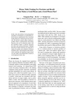
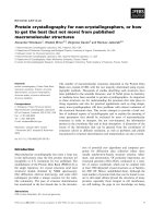
using the flow chart shown in Figure 1. We aimed to reproduce the
diagnosis of a NEC with use of the described morphology, IHC and
grading. First only patients concluded to have a NEC by the local
pathologist were included. Pathology conclusions diagnosing
small-cell carcinoma, low-grade NEN or carcinoma with neuroendocrine differentiation were regarded to be registered incorrectly.
They were excluded because the quality standard defined for NEC
did not apply for these tumours.
If no IHC was described at all, the patient was excluded, as IHC
might have been performed but could not be tracked with the
method used. Then, reports were classified as reporting a smallcell or large-cell carcinoma, either on the basis of the morphological description mentioning moulding of nuclei or lack of cytoplasm or based on the histopathological conclusion. As small-cell
NECs can be (focally/faintly) positive or negative for neuroendocrine IHC, these cases were not analysed for the presence of immunohistochemical parameters.
As a third step, large-cell carcinomas required a report of a majority of tumour cells positive with immunohistochemical staining
for either chromogranin or synaptophysin. Immunohistochemical
staining for CD56 was also registered but was not used for classification as NEN as it is an adhesion molecule and not a neuroendocrine protein [8]. If a neuroendocrine stain was not described,
while other (non-neuroendocrine) markers were described, the
neuroendocrine marker was assumed not to be performed. If all
neuroendocrine stains were negative or weakly positive, the diagnosis was revised to “Large cell carcinoma not otherwise specified”
(LCC NOS).
As a fourth step proliferative indexes, mitosis and Ki-67 index,
were evaluated. The Ki-67 index was required to be higher than
20% or the mitotic count needed to be higher than 20 per 10 highpower fields (HPF) in accordance with ENETS/WHO 2010 grading [8], for the tumours to be classified as NEC. When there was a
discrepancy between Ki-67 and mitotic count, the highest grade
was used for stratification. Patients with a Ki-67 index smaller than
20% and mitotic count below 20 per 10 HPF were recorded as a
low-grade NET. If no Ki-67 assessment was done and mitotic index not mentioned, NENs were classified as “NEN, unknown
grade.” In certain cases, the Ki-67 or mitotic count was described
subjectively (e.g. high/low or abundant). Cases with high or abundant proliferation parameters were classified as NEC and with low
levels as NET.
Zandee/van der Zwan/de Herder/
van Velthuysen
Exclusion criteria
591 cases registered in NCR
- NEN before 2008 (n = 7)
- Missing microscopy/IHC
(n = 55)
Pathology report conclusion:
- other than NEC (n = 93)
NEC conclusions (n = 436)
Small-cell carcinoma
(n = 12)1
Morphology
Large-cell carcinoma
(n = 424)
Fig. 1. Flow chart of neuroendocrine carci-
noma (NEC) diagnosis. 1 Small-cell carcinoma. 2 Large-cell carcinoma, not otherwise specified (NOS; negative IHC). 3 Lowgrade neuroendocrine tumour (NET;
positive immunohistochemistry, IHC, Ki67 <21% or mitotic count <21 per 10 HPF).
4
Large-cell neuroendocrine neoplasm
(NEN), not graded (positive IHC, missing
Ki-67 and mitotic count). 5 True NEC (positive IHC and Ki-67 >20% or mitotic count
>20 per 10 HPF).
Large-cell carcinoma NOS
(n = 77)2
Immunohistochemistry
Large-cell carcinomas with
pos. NET IHC (n = 347)
Large-cell NEN, no grade
(n = 93)4
Grading
To determine the adherence to the WHO 2010 guideline, percentages of cases with complete reporting of neuroendocrine
markers, mitotic index and Ki-67 index were calculated. A χ2 test
was performed to test whether the proportion of cases in which all
WHO 2010 parameters were reported increased for the years
2008–2012. To validate the model, survival of the different diagnostic groups was estimated with a Kaplan-Meier estimation, and
difference in survival was tested with a log-rank test. A univariate
analysis was performed to calculate hazard ratios (HRs).
Results
Low-grade NET
(n = 58)3
NEC (n = 196)5
port assigned different conclusions included small-cell
carcinoma (n = 15, 2.8%), carcinoma with neuroendocrine
differentiation (n = 29, 5.5%), low-grade NET (n = 42,
7.9%) or other carcinomas (n = 7, 1.3%). These tumours
were regarded to be misclassified. As the defined quality
criteria do not apply for these (non-NEC) tumours, only
the 436 patients with a confirmed NEC conclusion in the
pathology report were included in the further assessments.
These patients were on average 66.8 years old, and 56.2%
were male. Most patients had a primary tumour in the colon, pancreas or of unknown primary origin (16.3, 16.1 and
41.3% of cases, respectively; Table 1).
From 2008 through 2012, a total of 591 patients were
registered in the NCR with a large-cell extrapulmonary
NEC (M8013) or a NEC not otherwise specified (M8246)
from the gastrointestinal tract or unknown origin. Seven
patients were excluded because of a NEN being reported in
the PALGA pathology registry before 2008, and another 55
patients were excluded due to missing microscopy or IHC
and therefore were not applicable for this study. Of the remaining 529 NEC cases in the NCR, 436 (82.4%) were considered to be a NEC in the local pathology report conclusions. The 93 NCR cases for which the local pathology re-
Pathology Reporting
Of all 436 patients it was possible to deduce the cell
type from either the histopathological conclusion or the
morphological description in the pathology report. Of
these 436 patients the morphology of 424 (97.2%) tumours was described as large cell. The remaining 12
(2.8%) small-cell carcinomas were excluded from further
analyses.
A synaptophysin stain was reported in 356 patients
(84.0%), and chromogranin staining was reported in 361
Pertinence of Complete Pathology
Reporting for NEC
Neuroendocrinology
DOI: 10.1159/000505920
3
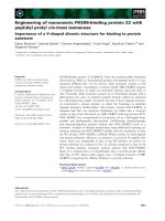
Percentage of reports reporting IHC and grading
100
■ 2008–2010
■ 2011–2012
80
*
60
*
**
*
40
20
20 (4.6)
48 (11.0)
15 (3.4)
71 (16.3)
32 (7.3)
70 (16.1)
180 (41.3)
89 (20.4)
168 (38.5)
179 (41.1)
(85.1%) patients (Fig. 2). Of 398 (93.9%) tumours at least
one neuroendocrine marker was reported. Grade was reported in smaller amounts of patients: Ki-67 was reported
in 185 (43.6%) patients and mitotic rate in 180 (42.5%).
Sixty-nine percent of cases could be graded because either
Ki-67 (n = 107, 25.2%), mitotic rate (n = 112, 26.4%) or
Neuroendocrinology
DOI: 10.1159/000505920
ng
IH
C
an
An
y
d
gr
gr
ad
i
ad
i
ng
sis
ito
M
7
Ki
-6
C
rin
e
oc
100
Overall survival, % of patients
66.7±12.6
245 (56.2)
Characteristics of 436 cases with a conclusion of neuroendocrine carcinoma in their local pathology reports. Age: mean ± SD
4
oe
nd
ur
ne
y
An
Table 1. Demographic and disease characteristics
Age, years
Male, n (%)
Primary tumour, n (%)
Oesophagus
Gastroduodenal
Small intestine
Colon
Rectum
Pancreas
Unknown
Extent of disease, n (%)
Localized
Advanced
Unknown
IH
ni
n
ra
og
m
ro
Ch
ap
Sy
n
M
or
ph
ol
Fig. 2. Completeness of pathology reports
of neuroendocrine carcinomas from 2008
to 2012 (percentage). IHC, immunohistochemistry; any neuroendocrine IHC, either
synaptophysin or chromogranin was reported. * p < 0.05, ** p < 0.001.
to
og
ph
ys
in
y
0
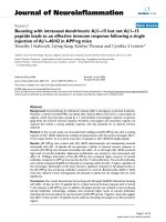
– Large-cell carcinoma NOS (n = 78)
– NET (n = 57)
– NEN, no grade (n = 93)
– NEC (n = 196)
80
60
40
20
0
0
1
2
3
Years since diagnosis
4
5
Fig. 3. Overall survival stratified for diagnosis. Results of the Kaplan-Meier analysis of overall survival (p = 0.02).
both (n = 73, 17.2%) were reported. Altogether, in 268
(63.2%) patients all necessary biomarkers for diagnosis
and grading could be assessed. In 2008, 50.0% of reports
included IHC and grading, increasing to 70.6% in 2012
(p = 0.01). This was mainly determined by the reporting
of grade (mitosis or Ki-67), increasing from 63.7% in
Zandee/van der Zwan/de Herder/
van Velthuysen
Table 2. Diagnosis per year
2008
2009
2010
2011
Neuroendocrine carcinoma, n (%)
Low-grade neuroendocrine tumour, n (%)
Neuroendocrine neoplasia, no grade, n (%)
Large-cell carcinoma NOS, n (%)
19 (31.7)
10 (16.7)
15 (25.0)
16 (26.7)
35 (42.7)
19 (23.2)
18 (22.0)
10 (12.2)
35 (43.2)
8 (9.9)
21 (25.9)
17 (21.0)
48 (48.5)
12 (12.1)
23 (23.2)
16 (16.2)
Total
60
82
81
99
2012
59 (57.8)
8 (7.8)
16 (15.7)
19 (18.6)
102
Total
196
58
93
77
424
Reproducibility of diagnosis with parameters of WHO 2010. NOS, not otherwise specified.
2008–2010 to 74.6% in 2011–2012 (Fig. 2, p = 0.01). The
increase in Ki-67 reporting alone was higher: from 34.5%
in 2008–2010 to 53.7% for 2011–2012 (p < 0.001). Necrosis and differentiation were seldom reported (25.2 and
24.3%).
Classification
Reviewing conclusions and description of morphology in the pathology reports demonstrated that 196 (46.2%)
patients were diagnosed with positive neuroendocrine
markers and either mitosis or Ki-67 compatible with a
NEC in accordance with WHO 2010 guidelines (Fig. 1).
Other diagnoses included low-grade NETs (n = 57,
13.4%), and 78 (18.4%) had a large-cell carcinoma but the
neuroendocrine differentiation could not be confirmed
with IHC (LCC NOS), because IHC was negative (n = 23);
IHC was only described as weakly positive (n = 29), or no
neuroendocrine markers were described at all (n = 26).
However, CD56 was positive in 64.1% of the LCC NOS
(50/78), possibly explaining why these tumours were (incorrectly) classified as neuroendocrine. In 93 patients
with a NEN (21.3%), grading was not possible due to
missing Ki-67 or mitotic count.
Introduction of the WHO 2010 resulted in a clear increase in reproducibility of NEC diagnoses. From 2008 to
2010, 31.7–40.7% of patients could be classified as NEC
with positive IHC and grading. This increased to 48.0 and
56.7% in 2011 and 2012 (p = 0.02, Table 2).
Only low-grade NET was a significant predictor in a
univariate analysis with an HR of 0.60 (95% CI: 0.44–
0.82). LCC NOS (HR 1.01, 95% CI: 0.77–1.33) and NEN,
no grade (HR 1.06; 95% CI: 0.82–1.38), showed similar
overall survival when compared to NEC.
Discussion
Overall Survival and Prognostic Factors
Overall survival patterns are in accordance with the
histopathological classifications (Fig. 3). Low-grade NETs
were associated with the longest survival (mean survival
2.2 years) while the mean survival of NEC was 1.2 years
(p = 0.02). Mean Ki-67 in the group with low-grade NET
was 11% with more than 70% of patients having a Ki-67
of >10.
Diagnostic standards for NEN are described in the
WHO 2010 and 2017 guideline for the classification
of tumours and the Standard of Care for pathology by
ENETS [5, 9]. The main changes in the WHO 2017 guideline for NETs of the pancreas, which will probably also be
adopted for NETs of the gastrointestinal tract, are the
slight change in cut-off between grade 1 and 2 NET and
the introduction of a further stratification in high-grade
NEN. This study demonstrates that implementation of
new guidelines for reporting is an effective measure. Implementation of the WHO 2010 was associated with an
increase in the use of grading based on Ki-67 or mitotic
count from 63.7 to 74.6%, but still many important parameters are lacking in the pathology reports. Even after
2010, only 43.2–57.8% of NECs were classified with reporting of all the necessary parameters. The importance
of the completeness of the report is highlighted by the fact
that due to this completeness, well-differentiated NETs,
having a different survival, could be recognized.
Incorrect classification in the NCR seemed to be present. In 93 (17.5%) patients the conclusion in the pathology report stated a different diagnosis than NEC. Discrepancies in the written information in the patient files
apart from pathology reports are a challenge for NCR
data managers to report these rare types of cancers correctly. Additionally, due to this diversity it is difficult to
build on expertise for the 168 NCR data managers as NEN
can be diagnosed in all Dutch hospitals.
Pertinence of Complete Pathology
Reporting for NEC
Neuroendocrinology
DOI: 10.1159/000505920
5
Secondly, a large number of pathology reports did not
describe all necessary parameters to diagnose a NEC. This
has also been observed in several other cancers. Completeness of pathology reports varies between 10 and
100% but is often around 30% [10]. For example, in a recent Italian study, pathology reports for cutaneous melanoma were complete in 77.8% of cases [11]. Thyroid cancer pathology reports were complete in an Australian
study in only 36.4% of cases [12]. In that perspective, adherence to pathology guidelines for NEN seems comparable with other cancers, and as such the result of the current study is not unique for NEN. The low adherence to
pathology guidelines for diagnosis is probably not only
caused by the rarity of NENs.
As the diagnosis of a NEC is based on morphology,
neuroendocrine IHC and thereafter the demonstration
of a high proliferation rate using the Ki-67 index. These
parameters have major implications for the treatment of
NEN patients. This is demonstrated by the different
therapies in ENETS guidelines for high-grade NEC and
low-grade NET [8, 13]. It is therefore essential that the
pathologist reports the morphology, IHC and grading in
a uniform fashion and that the treating physician can
recognize these parameters to verify the diagnosis of the
patient. The pathology reports in this study were all
written as a narrative, but this way of reporting is famed
for missing parameters and thus misinterpretation [14].
Since 2010, in the Netherlands, synoptic reporting is
available and widely used since 2013. These synoptic reports contain standardized reporting language and
mandatory parameters. This style of reporting has been
shown to significantly increase completeness of pathology reports to nearly 100% for various cancers [10, 15].
The next Standard of Care could suggest such a standardized report.
A third reason for the incorrect classification could
lie in the previous classification (WHO 2000). This classification defined three groups, based on size, metastases
and differentiation. All metastatic disease was classified
as endocrine carcinoma with a further differentiation
between low- and high-grade malignant behaviour
based on differentiation. This could partly explain the
pathology conclusions of NEC before 2010, while with
the current WHO guideline a low-grade NET would be
diagnosed. However, the mandatory differentiation
(poor vs. well) was only reported in 28.4% of reports before 2010, further underlying the need for a standardized report.
The importance of correct diagnosing is illustrated by
the clear difference in survival between low-grade NET
6
Neuroendocrinology
DOI: 10.1159/000505920
and true NEC (Fig. 3). While tumours were considered to
be a NEC by the local pathologist, we recognized a NET
grade 1 or 2 in 13.7% of patients due to the reported proliferation markers in the report. These NETs had a significantly longer survival confirming the heterogeneity in
the cohort and the importance of uniform reporting and
classification. The current study showed that despite implementation of the WHO 2010 guideline, one or more
items were missing for classification and grading in 26.8%
of the pathology reports.
Although well-differentiated NEN, especially in resection specimens, can be readily diagnosed without IHC as
also stated in the paper of the Delphic consensus process
[16], poorly differentiated NECs lose their neuroendocrine morphology and can often only be recognized with
neuroendocrine markers. Therefore, the reporting of
neuroendocrine markers is essential in this group of patients. Moreover, as these tumours are morphologically
seen in a biological continuum, estimation of the proliferative activity by mitotic count and Ki-67 staining is
again shown to predict survival. Thus, although publication of the WHO 2010 guideline is associated with a significant increase in reporting parameters needed for classification, essential parameters are still lacking in almost
one quarter of reports. Universal application will remain
a utopia in narrative reports. Synoptic reporting might
give an important boost to meet requirements more
quickly.
Statement of Ethics
Medical ethics committee approval was not required in accordance with the Dutch Medical Research Involving Human Subjects Act.
Disclosure Statement
The authors have no conflicts of interest to declare.
Author Contributions
All authors contributed to study design and manuscript writing. W.T.Z., J.M.Z. and M.F.V. contributed to the data collection.
All authors approved the final version of the paper.
Zandee/van der Zwan/de Herder/
van Velthuysen
References
1 Hofland J, Zandee WT, de Herder WW. Role
of biomarker tests for diagnosis of neuroendocrine tumours. Nat Rev Endocrinol. 2018
Nov;14(11):656–69.
2 Williams ED, Sandler M. The classification of
carcinoid tum ours. Lancet. 1963 Feb;1(7275):
238–9.
3 Solcia E. Klöppel Gn, Sobin LH: Histological
typing of endocrine tumours. New York:
Springer; 2000. /> 4 Bosman FT. World Health O: WHO classification of tumours of the digestive system.
Lyon, France: IARC Press; 2010.
5 Lloyd RV, Osamura RY, Klöppel G, Rosai J,
Bosman FT, Jaffe ES, Lakhani SR, Ohgaki H;
World Health Organization; International
Agency for Research on Cancer. WHO classification of tumours of endocrine organs, 4th
ed. Geneva: WHO Press; 2017.
6 Casparie M, Tiebosch AT, Burger G, Blauwgeers H, van de Pol A, van Krieken JH, Meijer
GA. Pathology databanking and biobanking
in the Netherlands, a central role for PALGA,
the nationwide histopathology and cytopathology data network and archive. Cell Oncol.
2007;29:19–24.
7Korse CM, Taal BG, van Velthuysen ML,
Visser O. Incidence and survival of neuroendocrine tumours in the Netherlands accord-
Pertinence of Complete Pathology
Reporting for NEC
ing to histological grade: experience of two
decades of cancer registry. Eur J Cancer. 2013
May;49(8):1975–83.
8Garcia-Carbonero R, Sorbye H, Baudin E,
Raymond E, Wiedenmann B, Niederle B, et al.;
Vienna Consensus Conference participants.
ENETS consensus guidelines for high-grade
gastroenteropancreatic neuroendocrine tumors and neuroendocrine carcinomas. Neuroendocrinology. 2016;103(2):186–94.
9 Perren A, Couvelard A, Scoazec JY, Costa F,
Borbath I, Delle Fave G, et al. Antibes Consensus Conference: ENETS consensus guidelines for the standards of care in neuroendocrine tumors: pathology, diagnosis and prognostic stratification. Neuroendocrinology.
2017;105(3):196–200.
10 Sluijter CE, van Lonkhuijzen LR, van Slooten
HJ, Nagtegaal ID, Overbeek LI. The effects of
implementing synoptic pathology reporting
in cancer diagnosis: a systematic review. Virchows Arch. 2016 Jun;468(6):639–49.
11 Tumino R, Minicozzi P, Frasca G, Allemani
C, Crocetti E, Ferretti S, et al. Populationbased method for investigating adherence to
international recommendations for pathology reporting of primary cutaneous melanoma: results of a EUROCARE-5 high resolution study. Cancer Epidemiol. 2015 Jun;39(3):
424–9.
Neuroendocrinology
DOI: 10.1159/000505920
12 Kahn C, Simonella L, Sywak M, Boyages S,
Ung O, O’Connell D. Postsurgical pathology
reporting of thyroid cancer in New South
Wales, Australia. Thyroid. 2012 Jun; 22(6):
604–10.
13 Pavel M, O’Toole D, Costa F, Capdevila J,
Gross D, Kianmanesh R, et al.; Vienna Consensus Conference participants. ENETS Consensus Guidelines Update for the Management of Distant Metastatic Disease of Intestinal, Pancreatic, Bronchial Neuroendocrine
Neoplasms (NEN) and NEN of Unknown
Primary Site. Neuroendocrinology. 2016;
103(2):172–85.
14 Powsner SM, Costa J, Homer RJ. Clinicians
are from Mars and pathologists are from Venus. Arch Pathol Lab Med. 2000 Jul; 124(7):
1040–6.
15 Srigley JR, McGowan T, Maclean A, Raby M,
Ross J, Kramer S, et al. Standardized synoptic
cancer pathology reporting: a populationbased approach. J Surg Oncol. 2009 Jun;99(8):
517–24.
16 Klimstra DS, Modlin IR, Adsay NV, Chetty R,
Deshpande V, Gönen M, et al. Pathology reporting of neuroendocrine tumors: application of the Delphic consensus process to the
development of a minimum pathology data
set. Am J Surg Pathol. 2010 Mar; 34(3): 300–
13.
7