Effect of preparation conditions on optical properties of CdTe quantum dot dispersed in water
Bạn đang xem bản rút gọn của tài liệu. Xem và tải ngay bản đầy đủ của tài liệu tại đây (405.07 KB, 7 trang )
<span class='text_page_counter'>(1)</span><div class='page_container' data-page=1>
39
Effect of preparation conditions on optical properties of CdTe
quantum dot dispersed in water
Nguyen Van Hung
1,*, Duong Anh Tuan
1, Trinh Duc Thien
1, Nguyen Dang Phu
1,
Danh Bich Do
1, Pham Van Vinh
1<i>1</i>
<i>Hanoi National University of Education </i>
<i>136 Xuan Thuy Road, Cau Giay District, Hanoi, Vietnam </i>
Received 12 August 2012, received in revised form 03 November 2012
<b>Abstract. CdTe quantum dots dispersed in water were prepared successfully by the microwave </b>
irradiation method. The influence of the pH value of the solution precursor, irradiation time
and microwave power on the structure, size and optical properties of CdTe quantum dots was
investigated. The fluorescence of CdTe quantum dots studies were illustrated that the samples
which were synthesized at pH value of 7 exhibited the best fluorescence. The absorption,
fluorescence spectra and TEM images showed that the microwave power and the irradiation time
influenced significantly on the size of CdTe quantum dot as well as their optical properties. The
<b>fluorescence spectra were red-shift with the increase of the irradiation time and microwave power. </b>
<i>Keywords:</i> CdTe, Quantum dots, optical properties.
<b>1. Introduction</b>∗∗∗∗
The study and synthesis of quantum dot (QDs) have been the attractive subject in past few decades
because of their applications in many fields. Quantum dots belonging to AIIBVI group, such as CdS,
CdSe, CdTe have potential application to photo devices [1-3], bio sensors [4], bio-label [5-7] and solar
cells [8]. In particular, the conjugation of QDs with biomolecules became an increasingly attractive
research area after waterdispersed nanocrystals were successfully applied for biological labeling
[9-10]. Obviously, water-dispersed nanocrystals with good spectral properties play a critical role in
<b>biological applications such as fluorescent labeling [11]. Although nanocrystals with a high </b>
photoluminescence quantum yield (PLQY) were obtained through the TOP/TOPO organometallic
synthetic approach [12-14], their hydrophobic character rendered them unsuitable for direct use in
_______
∗
</div>
<span class='text_page_counter'>(2)</span><div class='page_container' data-page=2>
<b>biological systems. Therefore the search for simple procedures and synthesis in aqueous solution of </b>
QDs materials has attracted increasing interest due to the innovative technological applications offered
by these materials. Many procedures have been developed with the aim of optimizing the spectral
properties of QDs directly prepared in aqueous phase [15-17]. For example, the hydrothermal method
providing higher temperatures was employed to synthesize QDs, which were successfully used as
biological labels [17]. However, the synthesis of QDs is carried out in an autoclave at high pressure
and temperature. This process also takes much time.
Recently, microwave irradiation is an attractive method for synthesis of QDs. The synthesis of
QDs by microwave irradiation was first introduced by the Kotov group [18]. Some studies showed that
the synthesis of nanocrystals by microwave irradiation was generally quite faster, simpler, and very
energy efficient as compared to conventional hydrothermal synthesis [19]. The combination of using
water as a solvent to synthesize nanocrystals and applying microwave irradiation as an efficient
heating source is a very desirable way to make the synthesis of nanocrystals cleaner and more
environment-friendly [20]. In this paper, this microwave irradiation method was used to synthesize
CdTe QDs in aqueous phase. According to the grown conditions, CdTe QDs with different particle
size and fluorescence energy were obtained. We found the significant effects of microwave irradiation
and experimental conditions on the size and optical properties of as-prepared CdTe QDs.
<b>2. Experiment </b>
All chemicals were used without further purification. Sodium borohydride (NaBH4, 99.9%),
tellurium powder (99.8%) was purchased from Andrich. Cadmium bromide (CdBr2, 99.9%),
3-mercaptopropionic acid (MPA, 99%) and NaOH 1M were obtained from Shanghai Chemical Reagents
Company.
In our experiments, the CdTe precursor solution was prepared by adding freshly prepared NaHTe
solution to a N2-saturated CdBr2 solution at different pH values in the presence of
3-mercaptopropionic acid (MPA) as a stabilizer. The pH of solution was controlled by NaOH. Typically,
a 4 mL volume of CdTe precursor solution was injected into the vitreous vessel with a volume of 10
mL. A series of high-quality CdTe QDs were prepared under microwave irradiation process. After
microwave irradiation, the CdTe QDs sample was taken when the temperature cooled to room
temperature naturally.
</div>
<span class='text_page_counter'>(3)</span><div class='page_container' data-page=3>
<b>3. Result and discussion </b>
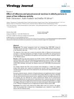
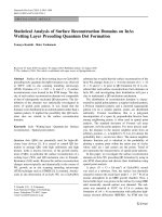
Figure 1 shows XRD patterns obtained from powdered precipitated fractions of CdTe QDs
synthesized through microwave irradiation at pH=7, 480 min, 900W . The XRD pattern of the
nanocrystalline CdTe powder shows the (1 1 1), (2 2 0) and (3 1 1) planes of the cubic CdTe phase. It
is clearly shown that the as-prepared CdTe QDs belonged to the cubic (zinc blende) structure, which
was also the dominant crystal phase of bulk CdTe
Figure 1. XRD diffractogram of CdTe quantum dots under microwave irradiation for 420 minute at pH=7, 900W
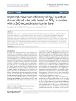
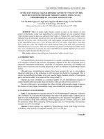
Figure 2 is the TEM images of typical sample irradiated in 5 min and 480 min at pH=7 and power
900W. Particle size of the 5 min and 480 min samples were corresponding to about 2 - 3 nm, 8 – 10
nm, respectively. When irradiation time increased the particle size increased significantly due to the
agglomeration and particle growth. In addition, the size distribution of the long time particles was
larger than that of the short time samples. Irradiation time was used to change particles size than
changing pH in solution and power of microwave
</div>
<span class='text_page_counter'>(4)</span><div class='page_container' data-page=4>
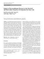
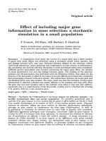
Fig 3 and Fig 4 presents the UV – Vis absorption and fluorescence spectra of CdTe quantum dots
in the range pH from 7 to 12 with the same of the power and time irradiation. In the UV – Vis
absorption, the peak position shifted from 520 nm to 536 nm when pH changed from 7 to 12.
However, we saw that the peak position was shightly shifted to the longer wavelength. This was the
same with the fluorescence spectra in Fig.4 and its intensity also decreased. The highest intensity was
found on the samples synthesized at pH value of 7. Therefore, the pH value of 7 was chosen for all
experiments.
Figure 3. Exciton absorption spectrum of CdTe
quantum dots with different pH
Figure 4. fluorescence spectrum of CdTe
quantum dots with different pH
The absorption spectra in Fig.3 showed that the exciton absorption peaks were red-shift from 521
<b>nm to 531 nm. The red-shift was believed due to the increase of QDs size. Indeed, because the </b>
bonding between Cd2+ and the stabilizer decreases with the increase of pH, the Cd2+ and Te2- in
solution were easy to bond with the ions in the surface of QDs resulting in the QDs size increase.
</div>
<span class='text_page_counter'>(5)</span><div class='page_container' data-page=5>
Figure 5. Absorption spectra of CdTe quantum
dots with different microwave powers
Figure 6. Fluorescent spectra of CdTe quantum
dots with different microwave powers
In order to investigate the influence of irradiation time on properties of CdTe QDs, the irradiation
time was varied while the pH value and microwave power were fixed at 7 and 300 W, respectively.
Fig. 7 and Fig. 8 show the effect of irradiation time on the optical properties of the QDs.
Figure 7. Absorption spectra of CdTe quantum dots
with different irradiation time
Figure 8. fluorescence spectra of CdTe quantum dots
with different irradiation time
</div>
<span class='text_page_counter'>(6)</span><div class='page_container' data-page=6>
synthesis rate. The crystalline growing depended on the concentration of the micro crystal present in
solution. and the amount of micro crystal decreased with reaction time of ‘Ostwald ripening’ [21].
Beside red-shift, the broader fluorescent peaks were also observed when rising the irradiation
time. This indicated the QDs size was not uniform. The optimum time for QDs to crystallize is
100 min.
<b>4. Conclusion </b>
The water-dispersed CdTe quantum dots were synthesized successfully by using the micr0wave
irradiation method. The pH, microwave power and irradiation time influenced significantly on the
QDs size as well as the optical properties. The fluorescent spectra were red-shift while their peaks
intensity decreased and FWHM broadened when the microwave power increased from 300 W to 900
W. The fluorescent spectra were strongly red-shift with the irradiation time in the range from 5 min to
360 min. Further increasing irradiation time, the red-shift increased slowly.
<b>Acknowledgments. </b>
This work was supported by Ministerial-level project of MOET, No B2010-17-237 and the basic
research project of Hanoi National University of Education No. SPHN 11-9.
<b>References </b>
[1] N. C. Greenham, X. Peng, and A. P. Alivisatos, “Charge separation and transport in conjugated–
polymer/semiconductor–nanocrystal composites studied by photoluminescence quenching and
photoconductivity,” Phys. Rev. B 54 (1996) 17628–17637.
[2] L. Li, T. J. Daou, I. Texier, T. T. K. Chi, N. Q. Liem, and P. Reiss, “Highly Luminescent CuInS2/ZnS Core/Shell
anocrystals: Cadmium–Free Quantum Dots for In Vivo Imaging,” Chem. Mater 21 (2009) 2422–2429.
[3] M. Shim and P. G. Sionest, “Permanent dipole moment and charges in colloidal semiconductor quantum dots,” J.
Chem. Phys. 111 (1999) 6955–6964.
[4] M. Frasco and N. Chaniotakis, “Semiconductor Quantum Dots in Chemical Sensors and Biosensors,” Sensors 9
(2009) 7266-7286.
[5] S. K. Mahto, C. Park, T. H. Yoon, and S. W. Rhee, “Assessment of cytocompatibility of surface-modified
CdSe/ZnSe quantum dots for BALB/3T3 fibroblast cells,” Toxicology in Vitro 24 (2010) 1070-1077.
[6] T. Yu, J. S. Shen, H. H. Bai, L. Guo, J. J. Tang, Y. B. Jiang, and J. W. Xie, “A photoluminescent
nanocrystal-based signaling protocol highly sensitive to nerve agents and highly toxic organophosphate pesticides,” Analyst
134 (2009) 2153-2157.
[7] C. Zhou, H. Shen, Y. Guo, L. Xu, J. Niu, Z. Zhang, Z. Du, J. Chen, and L. S. Li, “A versatile method for the
preparation of water-soluble amphiphilic oligomer-coated semiconductor quantum dots with high fluorescence
and stability,” Journal of Colloid and Interface Science 344 (2010) 279-285.
</div>
<span class='text_page_counter'>(7)</span><div class='page_container' data-page=7>
[9] M. P. Bruchez, M. Moronne, P. Gin, S. Weiss, and A. P. Alivisatos, “,Semiconductor nanocrystals as fluorescent
biological labels,” Science 281 (1998) 2013-2016.
[10] W. C. W. Chan, S. Nie, <i>"Quantum dot bioconjugates for ultrasensitive nonisotopic detection,"</i>
<b>Science 281 (1998) 2016-2018. </b>
[11] X. Michalet, F. F. Pinaud, L. A. Bentolila, J. M. Tsay, S. Doose, J. J. Li, G. Sundaresan, A. M. Wu, S. S.
Gambhir, and S. Weiss, “Quantum Dots for Live Cells, in Vivo Imaging, and Diagnostics,” Science 307 (2005)
538-544.
[12] D. V. Talapin, S. Haubold, A. L. Rogach, A. Kornowski, M. Haase, and H. Weller, “A Novel Organometallic
<b>Synthesis of Highly Luminescent CdTe Nanocrystals,” J. Phys. Chem. B 105 (2001) 2260-2263. </b>
[13] L. Qu and X. Peng, “Control of Photoluminescence Properties of CdSe Nanocrystals in Growth,” J. Am. Chem.
<b>Soc. 124 (2002) 2049-2055. </b>
[14] W. W. Yu, Y. A. Wang, X. Peng, “Formation and Stability of Size-, Shape-, and Structure-Controlled CdTe
<b>Nanocrystals: Ligand Effects on Monomers and Nanocrystals,” Chem. Mater. 15 (2003) 4300-4308. </b>
[15] T. Rajh, O. Micic, and A. Nozik, “Synthesis and characterization of surface-modified colloidal CdTe qunatum
<b>dots,” J. Phys. Chem. B 97 (1993) 11999-12003. </b>
[16] N. Gaponik, D. Talapin, A. L. Rogach, K. Hoppe, E. Shevchenko, A. Kornowski, A. Eychmuller, and H. Weller,
<b>“Thiol-capping of CdTe nanocrystals: an alternative to organometallic synthetic routes,” J. Phys. Chem. B 106 </b>
<b>(2002) 7177-7185. </b>
[17] H. Zhang, L. Wang, H. Xiong, L. Hu, B. Yang, and W. Li, “Hydrothermal synthesis to high quality CdTe
<b>nanocrystals” Adv.Mater. 15 (2003) 1712-1715. </b>
[18] M. A. Correa-Duarte, M. Giersig, N. A. Kotov, and L. M. Liz-Marzan, “Control of Packing Order of
Self-Assembled Monolayers of Magnetite Nanoparticles with and without SiO2 Coating by Microwave Irradiation,”
Langmuir 14 (1998) 6430-6435.
[19] H. Grisaru, O. Palchik, A. Gedanken, M. A. Slifkin, A. M. Weiss, V. Palchik, “Microwave-Assisted Polyol
Synthesis of CulnTe2 and CulnSe2 Nanoparticles,” Inorg. Chem. 42 (2003) 7148.
[20] N. E. Leadbeater, “Fast, easy, clean chemistry by using water as a solvent and microwave heating: the Suzuki
<b>coupling as an illustration,” Chem. Commun. 23 (2005) 2881-2902. </b>
</div>
<!--links-->