Hinh CT bung
Bạn đang xem bản rút gọn của tài liệu. Xem và tải ngay bản đầy đủ của tài liệu tại đây (3.24 MB, 131 trang )
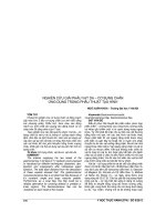
Nam 28 tuổi với đau HC(P)
Đại tràng lên
Khối cạnh ĐT kèm thậm nhiểm
Thậm nhiễm quanh ĐT
1
Viêm ruột thừa lạc chổ.
Brief review of Epiploic Appendagitis Rare inflammatory and
ischemic condition Results from torsion or spontaneous venous
thrombosis of one of the appendices epiploicae ischemia or
infarction of the appendix epiploica & localized inflammation
Sudden, severe, focal abdominal pain, mimic other conditions
such as appendicitis.
Can be managed conservatively CT: 1- 4-cm, oval, fatty pericolic
lesion with surrounding mesenteric inflammation Adjacent cecal
wall thickening and compression Rarely, a central highattenuation "dot" within the inflamed appendage; corresponds to
the thrombosed vein (17).
2
3
Diffuse largeB-cell lymphoma
Brief review of round solid
mesenteric masses Malignant
solid tumors have a tendency to
be located near root of mesentery
benign solid tumors in periphery
near bowel! 1. Metastases
especially from colon, ovary (most
frequent neoplasm of mesentery)
2. Lymphoma 3.
Leiomyosarcoma (more frequent
than leiomyoma) 4. Neural tumor
(neurofibroma, ganglioneuroma)
5. Lipoma (uncommon),
lipomatosis, liposarcoma 6.
Fibrous histiocytoma 7.
Hemangioma 8. Desmoid tumor
(most common primary)
4
5
Figure(s)
60/M
Chief complaint: jaundice, fever and
chill
*not hach
*day thanh
6
Gallbladder carcinoma
Brief review of gallbladder carcinoma Most common biliary
cancer Associated with: (1) Gallstones in 64 - 98%
Gallbladder carcinoma occurs in only 1% of all patients with
gallstones! (2) Porcelain gallbladder (in 4 - 60%) (3)
Inflammatory bowel disease (predominantly ulcerative colitis)
(4) Familial polyposis coli (5) Chronic cholecystitis Growth
types: replacement of gallbladder by mass (37 - 70%) focal
/ diffuse asymmetric irregular thickening of GB wall (15 - 47%)
polypoid / fungating intraluminal mass with wide base (14 25%) Differential diagnosis see note below
7
8
Figure(s)
45/M
Chief complaint: general weakness
9
Addison disease caused by adrenal tuberculosis
Brief review of addison disease
= Primary adrenal insufficiency 90% of adrenal cortex must be destroyed!
Cause:
1. Idiopathic adrenal atrophy (60 - 70%): likely autoimmune disorder
2. Granulomatous disease: tuberculosis, sarcoidosis
3. Fungal infection: histoplasmosis, blastomycosis, coccidioidomycosis 4.
Adrenal hemorrhage: anticoagulation therapy, bleeding, coagulation disorders,
sepsis, shock
5. Bilateral metastatic disease (rare) Diminutive glands (in idiopathic atrophy
+ chronic inflammation) Enlarged glands (acute inflammation, acute
hemorrhage, metastasis
10
11
These are images from contrastenhanced abdomen CT. There is a
large, round mass between the
right hepatic lobe and the
duodenum. The mass is well
encapsulated. Majority of the mass
shows fat attenuation and
geographic or tread-like areas with
soft tissue attenuation are scattered
between them. The duodenum and
the pancreas are displaced by the
mass but look clearly separated
from the mass. What are the
differential diagnoses?
12
AnswerMyxoid liposarcoma
Brief review of myxoid liposarcoma most common type of
liposarcoma varying degrees of mucinous
+ fibrous tissue
+ relatively little lipid intermediate differentiation CT solid
pattern: inhomogeneous poorly marginated infiltrating
mass mixed pattern: focal fatty areas
+ areas of higher density pseudocystic pattern: waterdensity mass calcifications in up to 12% DDx: malignant
fibrous histiocytoma, leiomyosarcoma, desmoid tumor
13
14
M/40
chief complaint:
jaundice
PTC
15
Percutaneous transhepatic cholangiography shows
multiple ovoid filling defects in dilated intrahepatic
bile ducts. Focal stricture is noted in right main IHD.
What are the differential diagnoses?
16
Clonorchiasis of the liver
Brief review of clonorchiasis of the liver Endemic Country: Japan,
Korea, China, Taiwan, Indochina Organism: Chinese liver fluke =
Clonorchis sinensis Pathology (a) desquamation of epithelial bile duct
lining with adenomatous proliferation of ducts + thickening of duct
walls (inflammation, necrosis, fibrosis) (b) bacterial superinfection
with formation of liver abscess Remittent incomplete obstruction +
bacterial superinfection Multiple crescent- / stiletto-shaped filling
defects within bile ducts Complication (1) Bile duct obstruction
(conglomerate of worms / adenomatous proliferation (2) Calculus
formation (stasis / dead worms / epithelial debris) (3) Jaundice in 8%
(stone / stricture / tumor) (4) Generalized dilatation of bile ducts (2%)
17
18
M/49
Chief complaint: fever,chill
19
Explanation for figure(s)
Air in anterior pararenal space
Infiltrations adjacent to the duodenum and thickened renal fasciae & septi
20
neumoperitoneum due to perforated duodenal ulcer Radiologic findings of
neumoperitoneum air lesser peritoneal sac gas in scrotum (through open
rocessus vaginalis) Large collection of gas: abdominal distension, no gastric
r-fluid level "wall sign" = "Rigler sign" = "bas-relief sign" =air on both sides of
owel as intraluminal gas + free air outside (usually requires >1,000 mL of gas)
ootball sign" = large pneumoperitoneum outlining entire abdominal cavity
utline of falciform ligament (medial RUQ); most common structure outlined
elltale triangle sign" = triangular air pocket between 3 loops of bowel
nverted V sign" = outline of both lateral umbilical ligaments "urachus sign" =
utline of middle umbilical ligament
21
Figure(s): CT
M/57
Chief complaint: fever and chill
Past medical history: went through
whipple’s operation due to
pancreatic cancer
22
Afferent loop syndrome caused by recurred pancreatic cancer Brief review of
afferent loop syndrome Complication of subtotal gastrectomy with Billoth II
gastrojejunostomy Cause internal hernia, kinking of anastomosis, adhesive band,
stomal stenosis, neoplasm, inflammation Abdominal radiographs often normal
because the afferent loop is fluid filled as a result of distal obstruction Barium study
non-filling of the afferent loop or preferential filling of dilated proximal loop with
stasis CT , US two or more thinly marginated, round, cystic structures adjacent to
pancreas anterior displacement of the superior mesenteric artery
23
24
Figure(s)F/59
Chief complaint: went
through extended left
hepatic lobectomy
and radiation therapy
for klatskin tumor
25