Electrochemical Sensors
Bạn đang xem bản rút gọn của tài liệu. Xem và tải ngay bản đầy đủ của tài liệu tại đây (503.03 KB, 49 trang )
201
Analytical Electrochemistry, Third Edition, by Joseph Wang
Copyright © 2006 John Wiley & Sons, Inc.
6
ELECTROCHEMICAL SENSORS
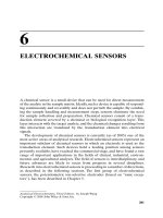
A chemical sensor is a small device that can be used for direct measurement
of the analyte in the sample matrix. Ideally, such a device is capable of respond-
ing continuously and reversibly and does not perturb the sample. By combin-
ing the sample handling and measurement steps, sensors eliminate the need
for sample collection and preparation. Chemical sensors consist of a trans-
duction element covered by a chemical or biological recognition layer. This
layer interacts with the target analyte, and the chemical changes resulting from
this interaction are translated by the transduction element into electrical
signals.
The development of chemical sensors is currently (as of 2005) one of the
most active areas of analytical research. Electrochemical sensors represent an
important subclass of chemical sensors in which an electrode is used as the
transduction element. Such devices hold a leading position among sensors
presently available, have reached the commercial stage, and have found a vast
range of important applications in the fields of clinical, industrial, environ-
mental, and agricultural analyses. The field of sensors is interdisciplinary, and
future advances are likely to occur from progress in several disciplines.
Research into electrochemical sensors is proceeding in a number of directions,
as described in the following sections. The first group of electrochemical
sensors, the potentiometric ion-selective electrodes (based on “ionic recep-
tors”), has been described in Chapter 5.
6.1 ELECTROCHEMICAL BIOSENSORS
Electrochemical biosensors combine the analytical power of electrochemical
techniques with the specificity of biological recognition processes. The aim is
to biologically produce an electrical signal that relates to the concentration of
an analyte. For this purpose, a biospecific reagent is either immobilized or
retained at a suitable electrode, which converts the biological recognition
event into a quantitative amperometric or potentiometric response. Such bio-
component–electrode combinations offer new powerful analytical tools that
are applicable to many challenging problems. A level of sophistication and
state-of-the art technology are commonly employed to produce easy-to-use,
compact, and inexpensive devices.Advances in electrochemical biosensors are
progressing in different directions. Two general categories of electrochemical
biosensors may be distinguished, depending on the nature of the biological
recognition process: biocatalytic devices (utilizing enzymes, cells, or tissues as
immobilized biocomponents) and affinity sensors (based on antibodies, mem-
brane receptors, or nucleic acids).
6.1.1 Enzyme-Based Electrodes
Enzymes are proteins that catalyze chemical reactions in living systems. Such
catalysts are not only efficient but also extremely selective. Hence, enzymes
combine the recognition and amplification steps, as needed, for many sensing
applications.
Enzyme electrodes are based on the coupling of a layer of an enzyme with
an appropriate electrode. Such electrodes combine the specificity of the
enzyme for its substrate with the analytical power of electrochemical devices.
As a result of such coupling, enzyme electrodes have been shown to be
extremely useful for monitoring a wide variety of substrates of analytical
importance in clinical, environmental, and food samples.
6.1.1.1 Practical and Theoretical Considerations The operation of an
enzyme electrode is illustrated in Figure 6.1. The immobilized enzyme layer is
chosen to catalyze a reaction, which generates or consumes a detectable
species:
(6.1)
where S and C are the substrate and coreactant (cofactor), and P and C′ are
the corresponding products. The choice of the sensing electrode depends pri-
marily on the enzymatic system employed. For example, amperometric probes
are highly suitable when oxidase or dehydrogenase enzymes (generating elec-
trooxidizable hydrogen peroxide or NADH species) are employed, pH–glass
electrodes for enzymatic pathways which result in a change in pH, while gas
SC PC
enzyme
+ → +
′
202
ELECTROCHEMICAL SENSORS
(carbon dioxide) potentiometric devices will be the choice when decarboxy-
lase enzymes are used.
The success of the enzyme electrode depends, in part, on the immobiliza-
tion of the enzyme layer. The objective is to provide an intimate contact
between the enzyme and the sensing surface while maintaining (and even
improving) the enzyme stability. Several physical and chemical schemes can
thus be used to immobilize the enzyme onto the electrode (Fig. 6.2). The sim-
plest approach is to entrap a solution of the enzyme between the electrode
and a dialysis membrane. Alternately, polymeric films (e.g., polypyrrole,
Nafion) may be used to entrap the enzyme (via casting or electropolymeriza-
tion). Additional improvements can be achieved by combining several mem-
branes and/or coatings. Figure 6.3 displays a useful, and yet simple,
immobilization based on trapping the enzyme between an inner cellulose
acetate film and a collagen or polycarbonate membrane, cast at the tip of an
amperometric transducer. Such coverage with a membrane/coating serves also
to extend the linear range (via reduction of the local substrate concentration)
ELECTROCHEMICAL BIOSENSORS
203
Biocatalytic
layer
Electrode
S+C
Bulk solution
P+C′
Figure 6.1 Enzyme electrode based on a biocatalytic layer immobilized on an elec-
trode transducer.
and to reject potential interferences (e.g., coexisting electroactive species or
proteins). In chemical immobilization methods the enzyme is attached to the
surface by means of a covalent coupling through a cross-linking agent (e.g.,
glutaraldehyde, amide). Covalent coupling may be combined with the use
of functionalized thiolated monolayers for assembling multilayer enzyme
networks on electrode surfaces (2). Biotin–avidin interactions can also be
employed using streptavidin-coated surfaces and biotinylated enzymes (e.g.,
see Fig. 6.2). Other useful enzyme immobilization schemes include entrapment
within a thick gel layer, low-temperature encapsulation onto sol-gel films,
adsorption onto a graphite surface, incorporation (by mixing) within the bulk
of three-dimensional carbon-paste or graphite–epoxy matrices (3,4), or elec-
trochemical codeposition of the enzyme and catalytic metal particles (e.g., Pt,
Rh). Such codeposition, as well as electropolymerization processes, are par-
ticularly suited for localizing the enzyme onto miniaturized sensor surfaces
(5,6). The electropolymerization route can be accomplished by entrapping the
enzyme within the growing film or anchoring it covalently to the monomer
prior to the film deposition. Such an avenue can also reduce interferences and
fouling of the resulting biosensors. The mixed-enzyme/carbon paste immobi-
lization strategy is attractive for many routine applications, as it couples
the advantages of versatility (controlled doping of several modifiers, e.g.,
enzyme, cofactor mediator), speed (due to close proximity of biocatalytic and
sensing sites, and absence of membrane barriers), ease of fabrication, and
renewability.
204
ELECTROCHEMICAL SENSORS
++++++++++++
S
S
S
S
S
S
S
S
S
S
S
S
S
S
Polymer entrapment
Covalent binding
(nondirected)
Defined covalent
binding
Surface adsorption
Electrostatic
Biospecific interaction
(e.g., biotin–avidin)
Figure 6.2 Methods for immobilizing enzymes onto electrode surfaces.
The immobilization procedure may alter the behavior of the enzyme (com-
pared to its behavior in homogeneous solution). For example, the apparent
parameters of an enzyme-catalyzed reaction (optimum temperature or pH,
maximum velocity, etc.) may all be changed when an enzyme is immobilized.
Improved stability may also accrue from the minimization of enzyme unfold-
ing associated with the immobilization step. Overall, careful engineering of the
enzyme microenvironment (on the surface) can be used to greatly enhance
the sensor performance. More information on enzyme immobilization
schemes can be found in several reviews (7, 8).
ELECTROCHEMICAL BIOSENSORS
205
Platinum anode
Silver cathode
Electrode body
Thin CA
layer
Drop of
enzyme solution
Outer
membrane
CA solution
Figure 6.3 Steps in preparation of an amperometric enzyme electrode, with a simple
enzyme immobilization by trapping between an inner cellulose acetate and outer
collagen membrane, cast on the electrode body. (Reproduced with permission from
Ref. 1.)
The response characteristics of enzyme electrodes depend on many vari-
ables, and an understanding of the theoretical basis of their function would
help to improve their performance. Enzymatic reactions involving a single sub-
strate can be formulated in a general way as
(6.2)
In this mechanism, the substrate S combines with the enzyme E to form an
intermediate complex ES, which subsequently breaks down into products P
and liberates the enzyme. At a fixed enzyme concentration, the rate of the
enzyme-catalyzed reaction V is given by the Michaelis-Menten equation:
(6.3)
where K
m
is the Michaelis-Menten constant and V
m
is the maximum rate of
the reaction. The term K
m
corresponds to the substrate concentration for
which the rate is equal to half of V
m
. In the construction of enzyme electrodes,
it is desirable to obtain the highest V
m
and lowest K
m
. Figure 6.4 shows the
dependence of the reaction rate on the substrate concentration, together with
the parameters K
m
and V
m
.The initial rate increases with substrate, until a non-
limiting excess of substrate is reached, after which additional substrate causes
no further increase in the rate. Hence, a leveling off of calibration curves is
expected at substrate concentrations above the K
m
of the enzyme.Accordingly,
low K
m
values—while offering higher sensitivity—result in a narrower linear
range (which reflects the saturation of the enzyme). The preceding discussion
assumes that the reaction obeys the Michaelis-Menten kinetics theory. Exper-
imentally, the linear range may exceed the concentration corresponding to K
m
,
VV K=
[]
+
[]
()
mm
SS
k
k
k
1
2
1
ES ES EP
-
+→+
∫
206
ELECTROCHEMICAL SENSORS
Analytically useful
region for substrate
determination S < <K
m
0.5 V
m
Reaction velocity, V (units of Vm)
0
0 K
m
Substrate molarity, S
Analytically useful
region for enzyme
determination S > > K
m
V
m
Figure 6.4 Dependence of the velocity of an enzyme-catalyzed reaction on the sub-
strate concentration (at a constant level of the enzymatic activity).
because the local substrate concentration in the electrode containment region
is often lower than the bulk concentration (as common with amperometric
probes coated with diffusion-limiting membranes).The level of the cosubstrate
may also influence the linear range.
Improved sensitivity and scope can be achieved by coupling two (or more)
enzymatic reactions in a chain, cycling, or catalytic mechanism (9). For
example, a considerable enhancement of the sensitivity of enzyme electrodes
can be achieved by enzymatic recycling of the analyte in two-enzyme systems.
Such an amplification scheme generates more than a stoichiometric amount
of product and hence large analytical signals for low levels of the analyte. In
addition, a second enzyme can be used to generate a detectable (electroac-
tive) species, from a nonelectroactive product of the first reaction.
The most important challenge in amperometric enzyme electrodes is the
establishment of satisfactory electrical communication between the active site
of the enzyme and the electrode surface. Different mechanisms of electron
transfer can be exploited for amperometric biosensing, including the use of
natural secondary substrates, artificial redox mediators, or direct electron
transfer (Fig. 6.5). The latter obviates the need for cosubstrates or mediators,
holds promise for designing reagentless devices, and allows efficient trans-
duction of the biorecognition event. Only a restricted number of enzymes have
shown direct electron transfer reactions between the prosthetic group of the
enzyme and electrodes (10). The challenges in establishing such direct elec-
trical communication between redox enzymes and electrode surfaces have
been reviewed (2,11,12).
ELECTROCHEMICAL BIOSENSORS
207
O
2
Electrode
Substrate Product
(a)
H
2
O
2
Med
ox
Med
red
Electrode
Substrate Product
(b)
Electrode
Substrate Product
(c)
Figure 6.5 Three generations of amperometric enzyme electrodes based on the use
of natural secondary substrate (a), artificial redox mediators (b), or direct electron
transfer between the enzyme and the electrode (c).
6.1.1.2 Enzyme Electrodes of Analytical Significance
6.1.1.2.1 Glucose Sensors The determination of glucose in blood plays a
crucial role in the diagnosis and therapy of diabetes. Electrochemical biosen-
sors for glucose have played a key role in the move toward simplified wide-
scale glucose testing, and have dominated the $5 billion/year diabetes
monitoring market (13). The glucose amperometric sensor, developed by
Updike and Hicks (14), represents the first reported use of an enzyme elec-
trode. The electrode is commonly based on the entrapment of glucose oxidase
(GOx) between polyurathene and permselective membranes on a platinum
working electrode (Fig. 6.6). The liberation of hydrogen peroxide in the enzy-
matic reaction
(6.4)
can be monitored amperometrically at the platinum surface:
(6.5)
The multilayer membrane coverage (of Fig. 6.6) improves the relative surface
availability of oxygen and excludes potential interferences (common at the
potentials used for detecting the peroxide product). Electrocatalytic trans-
ducers based on Prussian Blue layers (15) or metallized carbons (16), which
preferentially accelerate the oxidation of hydrogen peroxide, are also useful
for minimizing potential interferences. The enzymatic reaction can also be fol-
lowed by monitoring the consumption of the oxygen cofactor.
Further improvements can be achieved by replacing the oxygen with a non-
physiological (synthetic) electron acceptor, which is able to shuttle electrons
from the flavin redox center of the enzyme to the surface of the working elec-
trode. Glucose oxidase (and other oxidoreductase enzymes) do not directly
transfer electrons to conventional electrodes because their redox centers are
surrounded by a thick protein layer. Such insulating shell introduces a spatial
separation of the electron donor–acceptor pair, and hence an intrinsic barrier
to direct electron transfer, in accordance to the distance dependence of the
electron transfer (ET) rate (17):
(6.6)
where ∆G and λ correspond to the free and reorganization energies accom-
panying the electron transfer, respectively, and d is the actual electron trans-
fer distance. The interfacial ET rate is thus dependent on the distance between
the enzyme redox center and the electrode surface, that is, on the depth of the
redox group inside the protein shell, and the orientation of the protein on the
surface.
As a result of using artificial (diffusional) electron-carrying mediators,
measurements become insensitive to oxygen fluctuations and can be carried
Kee
dGRT
et
=
−−
()
−+
()
[]
10
13
091 3 4. ∆λ λ
HO O H e
2
electrode
22
22→++
+−
Glucose O gluconic acid H O
glusoce oxidase
+ → +
222
208
ELECTROCHEMICAL SENSORS
out at lower potentials that do not provoke interfering reactions from coex-
isting electroactive species (Fig. 6.7). Many organic and organometallic redox
compounds have been considered for this role of enzyme mediator (18–20).
Some common examples are displayed in Figure 6.8. In particular, ferricyanide
ELECTROCHEMICAL BIOSENSORS
209
Platinum
cathode
Reaction 4
Reaction 2
Reaction 1
H
2
C
2
Reaction 1
Oxidase
enzyme
Immobilized
enzyme
Polycarbonate
membrane
Reaction 3
Cellulose acetate
membrane
O-ring
Glucose
gluconic
acid
Glucose + O
2
H
2
O
2
H
2
O
2
+
AgCl + e
Silver anode
Reaction 2
Platinum
anode
O
2
+ 2H
+
+ 2e
–
Reaction 3
Silver
reference
Ag
0
+
Cl
–
4H
+
+ O
2
Reaction 4
Auxiliary
electrode
2H
2
O – 4e
–
Figure 6.6 Schematic of a “first generation” glucose biosensor (based on a probe
manufactured by YSI Inc.).
Gluconic acid
Glucose
GO
x
(ox)
GO
x
(red)
Mediator
x
(ox)
Mediator (red)
Electrode
Current
signal
Figure 6.7 “Second generation” enzyme electrodes : sequence of events that occur in
a mediated system (ox = oxidation; red = reduction). (Reproduced with permission
from Ref. 19.)
and ferrocene derivatives (e.g., Fig. 6.8a) have been very successful for shut-
tling electrons from glucose oxidase to the electrode by the following scheme:
(6.7)
(6.8)
(6.9)
where M
(ox)
and M
(red)
are the oxidized and reduced forms of the mediator.
This chemistry has led to the development of hand-held battery-operated
meters for personal glucose monitoring in a single drop of blood (21). The
single-use disposable strips used with these devices are usually made of
polyvinyl chloride and a screen-printed carbon electrode containing a mixture
of glucose oxidase and the mediator (Fig. 6.9). The screen-printing technology
used for mass-scale production of this and similar biosensors, along with the
ink-jet localization of the dry reagent layer, are discussed in Section 6.3. The
control meter typically relies on a potential-step (chronoamperometric) oper-
ation. Other classes of promising mediators for glucose oxidase are quinone
derivatives, ruthenium complexes, phenothiazine compounds, and organic
conducting salts [particularly tetrathiafulvalene–tetracyanoquinodimethane
222MMe
red ox
() ()
−
→+
GOx M GOx M H
red ox ox red
( ) () () ( )
+
+→ + +222
Glucose GOx gluconic acid GOx
ox red
+→ +
() ( )
210
ELECTROCHEMICAL SENSORS
Fe
CH
3
CH
3
S
(a)
(b)
(c)
(d)
S
S
S
NC
NC
N
+
CH
3
N
CN
CN
Figure 6.8 Chemical structures of some common redox mediators: (a) dimethyl fer-
rocene; (b) tetrathiafulvalene; (c) tetracyanoquinodimethane; (d) Meldola Blue.
(TTF-TCNQ)]. An elegant nondiffusional route for establishing electrical
communication between GOx and the electrode is to “wire” the enzyme to
the surface with a long polymer having a dense array of electron relays [e.g.,
osmium(bipyridyl) bound to poly(vinyl pyridine), Fig. 6.10a (22)]. Such a poly-
meric chain is flexible enough to fold along the enzyme structure (Fig. 6.10b).
The resulting three-dimensional redox-polymer/enzyme network offers high
current outputs and stabilizes the mediator to the surface. It has been
ELECTROCHEMICAL BIOSENSORS
211
PVC substrate
Contacts
Conductive
carbon track
Conductive
silver track
Working
electrode
Ag/AgCl reference
electrode
Dielectric
layer
Figure 6.9 Schematic representation of a disposable glucose sensor strip. (Repro-
duced with permission from Ref. 20.)
Os
2+/3+
(bpy)
2
Cl
m
(
(
(a)
(b)
no
NN
N
+
CH
2
CH
2
CH
2
Glycoprotein
Electron
relay
FADH
2
R
R
R
R
R
R
FADH
2
Electrode
)
(
))
Figure 6.10 (a) Composition of an electron-relaying redox polymer and (b) use of the
polymer for electrical “wiring” of an enzyme to the electrode surface. (Reproduced
with permission from Ref. 22.)
successfully used in a commercial painless forearm blood glucose monitoring
system. Nanoscale materials, such as gold nanoparticles or carbon nanotubes,
have also shown to be extremely useful for “plugging” an electrode into GOx.
(23,24). An even more elegant possibility is the chemical modification of the
enzyme with the redox-active mediator (25). Glucose electrodes of extremely
efficient electrical communication with the electrode can be generated by the
enzyme reconstitution process (26). For this purpose, the flavin active center
of GOx is removed to allow positioning of the electron-mediating ferrocene
unit prior to reconstitution of the enzyme (Fig. 6.11). Ultimately, these and
similar developments would lead to minimally invasive subcutaneously
implanted (needle-type) and noninvasive devices for continuous real-time
monitoring of glucose (27,28). Such probes would offer a tight control of dia-
betes, in connection with an alarm detecting hypo- or hyperglucemia or for a
future closed-loop insulin release system (i.e., artificial pancreas). In addition
to their biosensing utility, mediated enzyme electrodes (particularly those
relying on electron-conducting redox polymers) have been shown extremely
useful for increasing the power density of energy-producing biofuel cells
(29,30). Such devices exploit the biocatalytic oxidation of biofuels, such as
glucose, coupled to the enzymatic reduction of dissolved oxygen (by bilirubin
oxidase or laccase), to generate electricity.
6.1.1.2.2 Ethanol Electrodes The reliable sensing of ethanol is of great sig-
nificance in various disciplines. The enzymatic reaction of ethanol with the
212
ELECTROCHEMICAL SENSORS
FAD
FAD
Fc
GO
x
-holoenzyme
Reconstituted
GO
x
Glucose
Gluconic acid
e
–
e
–
GO
x
-apoenzyme
FAD
FAD
Fc
Figure 6.11 Electrical contacting of a flavoenzyme by its reconstitution with a relay
FAD semi-synthetic cofactor. (Reproduced with permission from Ref. 2.)
cofactor nicotinamide adenine dinucleotide (NAD
+
), in the presence of
alcohol dehydrogenase (ADH)
(6.10)
serves as a basis of amperometric sensors for ethanol (31). Reagentless devices
based on the coimmobilization of ADH and NAD
+
to various carbon or plati-
num anodes are employed for this task (e.g., Fig. 6.12). NAD
+
is regenerated
electrochemically by oxidation of the NADH, and the resulting anodic current
is measured:
(6.11)
To circumvent high overvoltage and fouling problems encountered with reac-
tion (6.11) at conventional electrodes, much work has been devoted to the
development of modified electrodes with catalytic properties for NADH.
Immobilized redox mediators, such as the phenoxazine Meldola Blue or phe-
nothiazine compounds, have been particularly useful for this task (32) (see
also Fig. 4.17). Such mediation should be useful for many other dehydroge-
nase-based biosensors. High sensitivity and speed are indicated from the flow
injection response illustrated in Figure 3.22. The challenges of NADH detec-
tion and the development of dehydrogenase biosensors have been reviewed
(33). Alcohol biosensing can be accomplished also in the presence of alcohol
oxidase, based on measurements of the liberated peroxide product.
6.1.1.2.3 Urea Electrodes The physiologically important substrate urea can
be sensed on the basis of the following urease-catalyzed reaction:
(6.12)
The electrode is an ammonium ion-selective electrode surrounded by a gel
impregnated with the enzyme urease [Fig. 6.13 (34)]. The generated ammo-
NH CONH H O H NH HCO
urease
222 4 3
22++→+
++−
NADH NAD e H
+
→++
−+
2
C H OH NAD C H O NADH
ADH
25 25
+→+
+
ELECTROCHEMICAL BIOSENSORS
213
Containment
region
NAD
+
Electrode
NADH
ADH
Sample
solution
CH
3
CH
2
OH CH
3
CHO
Figure 6.12 Reagentless ethanol bioelectrode.
nium ions are detected after 30–60s to reach a steady-state potential. Alter-
nately, the changes in the proton concentration can be probed with glass pH
or other pH-sensitive electrodes. As expected for potentiometric probes, the
potential is a linear function of the log[urea] in the sample solution.
Enzyme electrodes for other substrates of analytical significance have been
developed. Representative examples are listed in Table 6.1. Further advances
in enzyme technology, particularly the isolation of new and more stable
214
ELECTROCHEMICAL SENSORS
Internal
reference
electrode
Ion-selective
membrane
Substrate
Enzyme–matrix
layer
Reference
solution
Figure 6.13 Urea electrode, based on the immobilization of urease onto an
ammonium-ion-selective electrode.
TABLE 6.1 Some Common Enzyme Electrodes
Measured
Species Enzyme Detected Species Type of Sensing Ref.
Cholesterol Cholesterol oxidase O
2
Amperometric 35
Creatinine Creatinase NH
3
Potentiometric 36
gas sensing
Amperometric 37
Lactate Lactate dehydrogenase NADH Amperometric 38
Lactate oxidase H
2
O
2
Amperometric 39
Penicillin Penicillinase H
+
Potentiometric 40
Phenol Tyrosinase Quinone Amperometric 41
Salicylate Salicylate hydroxylase CO
2
Potentiometric 42
gas sensing
Uric acid Uricase CO
2
Potentiometric 43
gas sensing
enzymes, should enhance the development of new biocatalytic sensors. New
opportunities (particularly assays of new environments or monitoring of
hydrophobic analytes) accrued from the finding that enzymes can maintain
their biocatalytic activity in organic solvents (44,45).
6.1.1.2.4 Toxin (Enzyme Inhibition) Biosensors Enzyme affectors (inhibitors
and activators), which influence the rate of biocatalytic reactions, can also be
measured. Sensing probes for organophosphate and carbamate pesticides, for
the respiratory poisons cyanide or azide, or for toxic metals have thus been
developed using enzymes such as acetylcholinesterase, horseradish peroxi-
dase, or tyrosinase (46,47). The analytical information is commonly obtained
from the decreased electrochemical response to the corresponding substrate
(associated with the inhibitor–enzyme interaction). Pesticide measurements
with cholinesterase systems often employ a bienzyme cholinesterase/choline
oxidase system, in connection to amperometric monitoring of the liberated
peroxide species. Changes in the substrate response can also be exploited for
measuring the activity of enzymes.
6.1.1.3 Tissue and Bacteria Electrodes The limited stability of isolated
enzymes, and the fact that some enzymes are expensive or even not available
in the pure state, has prompted the use of cellular materials (plant tissues, bac-
terial cells, etc.) as a source of enzymatic activity (48). For example, the banana
tissue (which is rich with polyphenol oxidase) can be incorporated by mixing
within the carbon paste matrix to yield a fast-responding and sensitive
dopamine sensor (Fig. 6.14). These biocatalytic electrodes function in a
manner similar to that for conventional enzyme electrodes (i.e., enzymes
present in the tissue or cell produce or consume a detectable species).
Other useful sensors rely on the coupling of microorganisms and electro-
chemical transducers. Changes in the respiration activity of the microorgan-
ism, induced by the target analyte, result in decreased surface concentration
of electroactive metabolites (e.g., oxygen), which can be detected by the
transducer.
ELECTROCHEMICAL BIOSENSORS
215
Banana
HO
HO
Dopamine Dopamine
quinone
Carbon paste
CH
3
fast
PPO+ O
2
CH
3
NH
2
CH
3
CH
3
NH
2
O
O
1
2
Figure 6.14 The mixed tissue (banana)–carbon paste sensor for dopamine. (Repro-
duced with permission from Ref. 49.)
6.1.2 Affinity Biosensors
Affinity electrochemical biosensors exploit selective binding of certain bio-
molecules (e.g., antibodies, receptors, or oligonucleotides) toward specific
target species for triggering useful electrical signals. The biomolecular recog-
nition process is governed primarily by the shape and size of the receptor
pocket and the ligand of interest (the analyte). Such an associative process is
governed by thermodynamic considerations (in contrast to the kinetic control
exhibited by biocatalytic systems). The high specificity and affinity of bio-
chemical binding reactions (such as DNA hybridization and antibody–antigen
compexation) lead to highly selective and sensitive sensing devices. As will be
shown in the following sections, electrochemical transducers are very suitable
for detecting these molecular recognition events. Such devices rely on meas-
uring the electrochemical signals resulting from the binding process.
6.1.2.1 Immunosensors Immunoassays are among the most specific of the
analytical techniques, provide extremely low detection limits, and can be used
for a wide range of substances. As research moves into the era of proteomic,
such assays become extremely useful for identifying and quantitating proteins.
Immunosensors are based on immunological reactions involving the shape
recognition of the antigen (Ag) by the antibody (Ab) binding site to form the
antibody/antigen (AbAg) complex:
(6.13)
The antibody is a globular protein produced by an organism to bind to foreign
molecules, namely, antigens, and mark them for elimination from the organ-
ism. The remarkable selectivity of antibodies is based on the stereospecificity
of the binding site for the antigen, and is reflected by large binding constants
(ranging from 10
5
to 10
9
L/mol). Antibody preparations may be monoclonal or
polyclonal. The former are produced by a single clone of antibody-producing
cells, and thus have the same affinity. Polyclonal antibodies, in contrast, are
cheaper but possess varying affinities.
Electrochemical immunosensors, combining specific immunoreactions with
an electrochemical transduction, have gained considerable attention (50–55).
Such sensors are based on labeling of the antibody (or antigen) with an
enzyme that acts on a substrate and generate an electroactive product that can
be detected amperometrically. Enzyme immunosensors can employ competi-
tive or sandwich modes of operation (Fig. 6.15). In competitive-type sensors,
the sample antigen (analyte) competes with an enzyme-labeled antigen for
antibody-binding sites on a membrane held on an amperometric or potentio-
metric sensing probe. After the reaction is complete, the sensor is washed to
remove unreacted components. The probe is then placed in a solution
containing the substrate for the enzyme, and the product or reactant of the
biocatalytic reaction is measured. Because of the competitive nature of the
Ab Ag AbAg+ ∫
216
ELECTROCHEMICAL SENSORS
assay, the measurement signal is inversely proportional to the concentration
of the analyte in the sample. Several enzymes, such as alkaline phosphatase,
horseradish peroxidase, glucose oxidase, and catalase, have been particularly
useful for this task.
Sandwich-type sensors are applicable for measuring large antigens that are
capable of binding two antibodies. Such sensors utilize an antibody that binds
the analyte antigen, which then binds the enzyme-labeled antibody. After
removal of the nonspecifically adsorbed label, the probe is placed into the sub-
strate-containing solution, and the extent of the enzymatic reaction is moni-
tored electrochemically. Other types of immunosensors based on labeling the
antigen or antibody with an electroactive tag (e.g., heavy metal or a ferrocene
derivative), metal (gold) nanoparticle tracer, label-free capacitance, imped-
ance or amperometric measurements, immobilizing antigen carrier conjugates
at the tip of potentiometric electrodes, or amplifying the antigen–antibody
complex equilibria by liposome lysis, are also being explored. For example,
antibodies incorporated in conducting polymers have been shown to retain
their affinity properties in connection with label-free pulsed amperometric
measurements (56). Similarly, impedance spectroscopy (described in Section
2.5) offers a label-free electronic detection based on the increased interfacial
electron transfer resistance associated with the formation of bioaffinity
complexes (57). Changes in the conductivity of one-dimensional antibody-
functionalized nanowires conjugated on binding of the target proteins can also
lead to a powerful label-free electrical immunoassays (58).
Instead of immobilizing the antibody onto the transducer, it is possible to
use a bare (amperometric or potentiometric) electrode for probing enzyme
immunoassay reactions (59). In this case, the content of the immunoassay reac-
ELECTROCHEMICAL BIOSENSORS
217
E
Sandwich
Antibody
membrane
Competitive
Antibody
membrane
Labled
antibody
Antigen
Antigen
E
E
S
P
S
P
E
E
E
S
P
Figure 6.15 Enzyme immunosensors based on the competitive or sandwich modes of
operation. (Reproduced with permission from Ref. 53.)
tion vessel is injected into an appropriate flow system containing an electro-
chemical detector, or the electrode can be inserted into the reaction vessel.
Remarkably low (femtomolar) detection limits have been reported in con-
nection to the use of the alkaline phosphatase label (60,61). This enzyme cat-
alyzes the hydrolysis of phosphate esters to liberate easily oxidizable phenolic
products. Even lower detection limits can be achieved by coupling the elec-
trochemical immunoassays with a dual-enzyme substrate regeneration (62).
The use of gold nanoparticles has also been shown useful for highly sensitive
immunoassays with stripping voltammetric detection of the dissolved gold (63).
More recent trends aim in the direction of fabricating electrochemical
protein array systems (for detecting multiple protein targets) and miniatur-
ization of such immunoassays. These include an electrochemical protein chip
with an array of 36 platinum electrodes on a glass substrate (64) and electri-
cal immunoassays using microcavity formats down to the zmol antigen level
(65).
In addition to antibodies, it is possible to use artificial nucleic acids ligands,
known as aptamers, for the selective detection of proteins. The tight binding
properties make aptamers attractive candidates as molecular recognition ele-
ments in a wide range of bioassays and for the development of protein arrays.
Electrochemistry has been shown useful for monitoring aptamer–protein
interactions (66).
6.1.2.2 DNA Hybridization Biosensors
6.1.2.2.1 Background and Principles Nucleic acid recognition layers can be
combined with electrochemical transducers to form new and important types
of affinity biosensors. The use of nucleic acid recognition layers represents an
exciting area in biosensor technology. Electrochemical DNA hybridization
biosensors offer considerable promise for obtaining sequence-specific infor-
mation in a simpler, faster, and cheaper manner, compared to traditional
hybridization assays (67–71). Such strategies hold an enormous potential for
clinical diagnosis of genetic or infectious diseases, for the detection of food-
contaminating organisms, for early warning against biowarfare agents, for
environmental monitoring, or in criminal investigations.
The basis for these devices is the DNA base pairing. Accordingly, these
sensors rely on the immobilization of a relatively short [20–40-bp (basepair)]
single-stranded DNA sequence (the “probe”) on the transducer surface,
which, on hybridization to a specific complementary region of the target DNA,
gives rises to an electrical signal (Fig. 6.16). A wide range of chemistries have
been exploited for monitoring electrochemically the DNA hybridization.
These can be divided into two major principles, involving the use of labels gen-
erating an electrical signal or label-free protocols. The hybridization event can
thus be detected via the increased current signal of an electroactive indicator
(that preferentially binds to the DNA duplex), or due to captured enzyme or
nanoparticle tags, or from other hybridization-induced changes in electro-
218
ELECTROCHEMICAL SENSORS
chemical parameters (e.g., capacitance or conductivity). Control of the probe
immobilization (e.g., linking chemistry, surface coverage) is essential for assur-
ing high reactivity, orientation and/or accessibility, and stability of the surface-
bound probe, as well as for avoiding non-specific binding/adsorption events.
Control of the hybridization conditions (e.g., ionic strength, temperature, time)
is also crucial for attaining high sensitivity and selectivity (including the detec-
tion of point mutations).
6.1.2.2.2 Electrical Transduction of DNA Hybridization Several studies
have demonstrated the utility of electroactive indicators for monitoring the
hybridization event (67). Such redox-active compounds have a much larger
affinity for the resulting duplex (compared to their affinity to the probe alone).
Their association with the surface duplex thus results in an increased electro-
chemical response. Very successful has been the use of a threading intercala-
tor ferrocenyl naphthalene diimide (FND) (72), which binds to the DNA
duplex more tightly than do the usual intercalators and displays a negligible
affinity to the single-stranded probe (Fig. 6.17). It is also possible to employ
metal nanoparticle labels (e.g., colloidal gold), and to quantitate them follow-
ing the hybridization and acid dissolution by a highly sensitive electrochemi-
cal stripping protocol [73].
The use of enzyme labels to generate electrical signals also offers great
promise for ultrasensitive electrochemical detection of DNA hybridization.
This can be accomplished by combining the hybridization step with an
electrochemical measurement of the product of the enzymatic reaction. The
ELECTROCHEMICAL BIOSENSORS
219
G
A
T
G
T
A
C
C
T
G
G = C
A = T
T = A
G = C
T = A
A = T
C = G
C = G
T = A
G = C
Hybridization Signal
Probe
Target
Transducer
Transducer
Figure 6.16 Steps involved in the detection of a specific DNA sequence using an elec-
trochemical DNA hybridization biosensor. (Reproduced with permission from Ref.
71.)