Calcitriol restores antiestrogen responsiveness in estrogen receptor negative breast cancer cells: A potential new therapeutic approach
Bạn đang xem bản rút gọn của tài liệu. Xem và tải ngay bản đầy đủ của tài liệu tại đây (955.17 KB, 10 trang )
Santos-Martínez et al. BMC Cancer 2014, 14:230
/>
RESEARCH ARTICLE
Open Access
Calcitriol restores antiestrogen responsiveness in
estrogen receptor negative breast cancer cells: A
potential new therapeutic approach
Nancy Santos-Martínez1,2†, Lorenza Díaz1†, David Ordaz-Rosado1, Janice García-Quiroz1,2, David Barrera1,
Euclides Avila1, Ali Halhali1, Heriberto Medina-Franco3, María J Ibarra-Sánchez4, José Esparza-López4,
Javier Camacho2, Fernando Larrea1 and Rocío García-Becerra1*
Abstract
Background: Approximately 30% of breast tumors do not express the estrogen receptor (ER) α, which is necessary
for endocrine therapy approaches. Studies are ongoing in order to restore ERα expression in ERα-negative breast
cancer. The aim of the present study was to determine if calcitriol induces ERα expression in ER-negative breast
cancer cells, thus restoring antiestrogen responses.
Methods: Cultured cells derived from ERα-negative breast tumors and an ERα-negative breast cancer cell line
(SUM-229PE) were treated with calcitriol and ERα expression was assessed by real time PCR and western blots. The
ERα functionality was evaluated by prolactin gene expression analysis. In addition, the effects of antiestrogens were
assessed by growth assay using the XTT method. Gene expression of cyclin D1 (CCND1), and Ether-à-go-go 1 (EAG1)
was also evaluated in cells treated with calcitriol alone or in combination with estradiol or ICI-182,780. Statistical
analyses were determined by one-way ANOVA.
Results: Calcitriol was able to induce the expression of a functional ERα in ER-negative breast cancer cells. This
effect was mediated through the vitamin D receptor (VDR), since it was abrogated by a VDR antagonist. Interestingly,
the calcitriol-induced ERα restored the response to antiestrogens by inhibiting cell proliferation. In addition,
calcitriol-treated cells in the presence of ICI-182,780 resulted in a significant reduction of two important cell
proliferation regulators CCND1 and EAG1.
Conclusions: Calcitriol induced the expression of ERα and restored the response to antiestrogens in ERα-negative
breast cancer cells. The combined treatment with calcitriol and antiestrogens could represent a new therapeutic
strategy in ERα-negative breast cancer patients.
Keywords: Estrogen receptor, Breast cancer, Hormonal therapy, Calcitriol, VDR
Background
Breast cancer is a heterogeneous disease, encompassing a number of distinct biological entities that are
associated with a variety of pathological and clinical
features [1]. The gene expression profile of breast
cancer allows to classify this disease in five groups,
two of them estrogen receptor (ER)-positive (luminal
* Correspondence:
†
Equal contributors
1
Departments of Reproductive Biology, Instituto Nacional de Ciencias
Médicas y Nutrición Salvador Zubirán, Vasco de Quiroga No. 15, Tlalpan
14000 México, México
Full list of author information is available at the end of the article
A and B) and three ER-negative (normal breast-like,
human epidermal growth factor receptor- 2 (HER2)
and basal-like) [2]. Approximately 30% of all breast
tumors do not express ER, a protein with both prognostic and predictive values. Indeed, the presence of
ERα correlates with increased disease-free survival
and better prognosis. Importantly, ERα-positive breast
cancers respond appropriately to endocrine therapies
[3-5]. Tamoxifen is the most common and effective
therapy in pre- and postmenopausal patients affected
with ER-positive tumors, since a long-term use of this
compound increases disease-free survival and reduces
© 2014 Santos-Martínez et al.; licensee BioMed Central Ltd. This is an Open Access article distributed under the terms of the
Creative Commons Attribution License ( which permits unrestricted use,
distribution, and reproduction in any medium, provided the original work is properly credited. The Creative Commons Public
Domain Dedication waiver ( applies to the data made available in this
article, unless otherwise stated.
Santos-Martínez et al. BMC Cancer 2014, 14:230
/>
tumor recurrence [6,7]. Unfortunately, up to 50% of
patients bearing ERα-positive primary tumors lose
receptor expression in recurrent tumors, and about
one third of metastatic tumors develop resistance to
tamoxifen and lose ERα expression [8]. The lack of ER
expression has been linked to epigenetic mechanisms or
to others such as hyperactivation of the mitogen-activated
protein kinase (MAPK) signaling pathway or increased
expression of specific microRNAs [9-11]. In fact, knockdown of specific microRNAs or inhibition of MAPK
activity is followed by restoration of a functional
ERα in ER-negative breast cancer cells [9,10]. These
findings indicate that the ERα-negative phenotype
could be reverted for therapeutic purposes.
Calcitriol, the most active metabolite of vitamin D,
elicits significant antiproliferative activity in breast
cancer cells by several vitamin D receptor (VDR) mediated
mechanisms including regulation of growth arrest, cell differentiation, migration, invasion and apoptosis [12-14].
Epidemiological studies have demonstrated an association
between low levels of calcidiol, the precursor of calcitriol,
and increased risk of developing breast cancer [15].
Moreover, low levels of calcitriol are associated with
disease progression and high incidence of ER-negative
and triple-negative breast tumors [16,17], while VDRpositive breast cancer patients had significantly longer
disease-free survival than those with VDR-negative tumors [18]. Indeed, VDR knock-out mice are more
likely to develop ER- and progesterone receptor (PR)negative mammary tumors as compared with their
wild type littermates [17], highlighting calcitriol prodifferentiating properties. Our laboratory and other groups
have demonstrated the potent antipropiferative activity of calcitriol in cells derived from biopsies or in established cell lines from breast cancer [19-21]. Additionally,
other studies have demonstrated the antiproliferative effects of vitamin D compounds in ER-responsive human
breast cancer cells through downregulation of ER and
disruption of estrogen dependent signaling pathways
[20,22,23]. However, calcitriol also inhibited proliferation in ER-negative cell lines, suggesting that growth
inhibition induced by calcitriol is not solely mediated
through the ER [12]. In this regard, ERα regulation
studies in several human breast cancer cell lines
showed that calcitriol treatment decreased or did not modify ER expression [20,22-24]. In contrast, in an ER-negative
breast cancer cell line calcitriol increased estrogen binding
proteins [24].
In order to increase our knowledge concerning the
participation of calcitriol in ER regulation, the aim of the
present study was to investigate if this hormone induces
a functional ER and consequently could restore the
antiproliferative effects of antiestrogens in ER-negative
breast cancer cells.
Page 2 of 10
Methods
Reagents
Estradiol (E2), 4-hydroxytamoxifen and calcipotriol
(MC 903) were purchased from Sigma (St. Louis,
MO, USA). Cell culture medium was obtained from
Life Technologies (Grand Island, NY, USA). Fetal
bovine serum (FBS) was from Hyclone Laboratories Inc.
(Logan, UT, USA) and the antiestrogen ICI-182,780
(Fulvestrant) from Zeneca Pharmaceuticals (Wilmington,
DE, USA). Gefitinib (Iressa, ZD1839) was kindly provided by AstraZeneca (Wilmington, DE, USA). U0126
was from Millipore (MA, USA). Trizol and the oligonucleotides for real time polymerase chain reaction
(qPCR) were from Invitrogen (CA, USA). The TaqMan
Master reaction, probes, capillaries, reverse transcription
(RT) system and the cell proliferation assay (XTT) were
purchased from Roche (Roche Applied Science, IN,
USA). MCF-7 nuclear extract was purchased from
Santa Cruz Biotechnology Inc., (CA, USA). The VDR
antagonist (23S)-25-dehydro-1-hydroxyvitamin D3-26,23lactone (TEI-9647) and 1α,25-dihydroxycholecalciferol
(calcitriol) were kindly donated from Teijin Pharma
Limited (Tokyo, Japan) and Hoffmann-La Roche Ltd.
(Basel, Switzerland), respectively.
Human tissues
The protocol was approved by the Institutional Review
Board “Comité Institucional de Investigación Biomédica en
Humanos (No. 1967, 2009)” of the “Instituto Nacional de
Ciencias Médicas y Nutrición Salvador Zubirán (Mexico
City). Before mammary biopsies donation, all participating
patients signed an informed consent. Biopsies were
obtained from patients with ER-negative breast cancer.
The samples were harvested and processed as described
previously [19]. A total of 5 independent cultured
specimens were used for this study. The ER-negative
SUM-229PE (Asterand, San Francisco, CA) and the
ER-positive BT-474 (ATCC) and MCF-7 (ATCC)
established cell lines were also studied.
Cell culture
Primary tumor cultures were derived from biopsies of
breast cancer patients as described previously [19,25].
The cells were cultured in DMEM-HG medium
supplemented with 5% heat-inactivated-FBS, 100 U/ml
penicillin, 100 μg/ml streptomycin; and incubated in 5%
CO2 at 37°C. After approximately 8 passages cells were
characterized by western blot and immunocytochemistry.
Established cell lines were maintained according to indications from suppliers. All experimental procedures were
performed in DMEM-F12 medium supplemented with 5%
charcoal-stripped-heat-inactivated FBS, 100 U/ml penicillin
and 100 μg/ml streptomycin.
Santos-Martínez et al. BMC Cancer 2014, 14:230
/>
Immunocytochemistry
Cultured cells were grown on glass coverslips and fixed
in 96% ethanol. Antigen retrieval was done by autoclaving
in EDTA decloaker 5× solution (pH 8.4-8.7, Biocare
Medical, CA, USA) during 10 min. Slides were blocked with
immunodetector peroxidase blocker (Bio SB, CA, USA)
and incubated with ERα (1:250, Bio SB) [26] and
VDR antibodies (1:100, Santa Cruz Biotechnology Inc, CA,
USA) [27]. After washing, the slides were sequentially
incubated with immune-Detector Biotin-Link and
Immuno-Detector HRP label (Bio SB) during 10 min each.
Staining was completed with DAB and 0.04% H2O2.
Western blots
Cells were incubated in the presence of calcitriol (1X10⁻⁸ M
and 1X10⁻⁷ M), MAPK inhibitors (U0126; 10 μM, Gefitinib;
0.8 μM) or the vehicle alone during 72 hr. Afterwards, whole-cell protein lysates were prepared using
lysis buffer (50 mM Tris-HCl, 150 mM NaCl, 1%
Nonidet P-40, pH 7.5) in the presence of a protease
inhibitor cocktail. Protein concentrations were determined
using the Protein Assay Dye Reagent Concentrate
(Bio-Rad, Hercules, CA, USA). The proteins were separated
on 10% SDS-PAGE and transferred to nitrocellulose membranes. The membranes were blocked with 5% skim milk
and incubated overnight at 4°C in the presence of mouse
anti-ERα (1:200, Santa Cruz) [28]. The membranes were
washed and incubated with goat anti-mouse HRPconjugated secondary antibody (1:2000, Santa Cruz). For
visualization, membranes were processed with BM chemiluminescence blotting substrate (Roche Applied Science,
IN, USA). For normalization, blots were stripped in boiling stripping buffer (2% w/v SDS, 62.5 mM Tris-HCl
pH 6.8, 100 mM 2- mercapto-ethanol) for 30 min at 50°C
and sequentially incubated with mouse anti-GAPDH
(1:10000, Millipore) [29] and anti-mouse-HRP (1:10000,
Jackson ImmunoResearch Laboratories, Inc., West
Grove, PA, USA). Densitometric analysis of resulting
bands was performed by using ImageJ software (NIH,
USA).
Page 3 of 10
was measured in a microplate reader (BioTek, Winooski,
VT, USA).
Real time RT-PCR
For ERα gene expression analysis the cells were incubated
in the presence of different calcitriol concentrations or the
vehicle alone (0.1% ethanol) during 24 hr. In order to establish the participation of the VDR on calcitriol effects upon
the ERα, the VDR antagonist TEI-9647 (1X10⁻⁶ M) was
coincubated with calcitriol in some experiments. Gene
expression analyses of prolactin (PRL), cyclin D1 (CCND1)
and the potassium channel Ether-à-go-go (EAG1) were also
performed. For this, the cells were treated with calcitriol
(1X10⁻⁸ M) during 48 hr. Afterwards, E2 (1X10⁻⁸ M) or
ICI-182,780 (1X10⁻⁶ M) were added to the culture media
and the incubations proceeded for additional 24 hr. Next,
RNA was extracted with Trizol reagent and then subjected
to reverse transcription using the transcriptor RT system.
Real-time PCR was carried out using the LightCycler 2.0
from Roche (Roche Diagnostics, Mannheim, Germany),
according to the following protocol: activation of Taq DNA
polymerase and DNA denaturation at 95°C for 10 min,
proceeded by 45 amplification cycles consisting of 10 s at
95°C, 30 s at 60°C, and 1 s at 72°C. The following oligonucleotides were used: ERα-F, CCTTCTTCAAGAGAAGTA
TTCAAGG; ERα-R, GTTTTTATCAATGGTGCACTGG;
EAG1-F, CCTGGAGGTGATCCAAGATG; EAG1-R, CCA
AACACGTCTCCTTTTCC; CCND1-F, GAAGATCGTCG
CCACCTG; CCND1-R, GACCTCCTCCTCGCACTTCT;
PRL-F, AAAGGATCGCCATGGAAAG; PRL-R, GCACAG
GAGCAGGTTTGAC. The gene expression of the
housekeeping gene glyceraldehyde-3-phosphate dehydrogenase (GAPDH) GAPDH-F, AGCCACATCGCTGAGA
CAC; GAPDH-R, GCCCAATACGACCAAATCC was used
as an internal control. Stimulatory concentration (EC50)
values were obtained by non-linear regression analysis
using sigmoidal fitting with a dose-response curve by
means of a scientific graphing software (SigmaStat,
Jandel Scientific).
Statistical analyses
Cell proliferation assay
The cells were seeded in 96-well tissue culture plates at
a density of 500-1000 cells/well by sextuplicate. After
incubating for 24 hr, cells were incubated in the presence or
absence of calcitriol (1X10⁻⁸ M) during 48 hr. Afterwards,
culture medium was removed and incubations with E2
(1X10⁻⁸ M), as an ER agonist, or tamoxifen (1X10⁻⁶ M) and
ICI-182,780 (1X10⁻⁶ M), as ER antagonists, or their
combination were performed in the absence or presence
of calcitriol. Plates were incubated at 37°C for 6 days and
cell viability was determined by using the colorimetric
XTT Assay Kit (Roche) according to manufacturer’s
instructions. After 4 hr incubation, absorbance at 492 nm
Data are expressed as the mean ± standard deviation (S.D.).
Statistical analyses were determined by one-way ANOVA
followed by the Holm-Sidak method, using a specialized
software package (SigmaStat, Jandel Scientific). Differences
were considered significant at P ≤ 0.05.
Results
Calcitriol induced ERα expression through a VDRdependent mechanism in ER-negative breast cancer cells
Biopsies from five patients with ER-negative breast
cancer were obtained and used for cell culturing. These
biopsies had a diagnosis of invasive ductal carcinoma and
ranged between 5 and 9 in the Scarff-Bloom-Richardson
Santos-Martínez et al. BMC Cancer 2014, 14:230
/>
system score. All cultured breast tumor-derived cells were
positive for VDR and further confirmed to be negative for
ERα (Figure 1). In addition, the ER-negative SUM-229PE
and ER-positive BT-474 established cell lines were
also studied. All cell lines were incubated in the presence
of calcitriol (1X10⁻⁷ M) during 24 hr and ERα gene
expression was assessed by qPCR. As shown in Figure 2A,
calcitriol significantly induced ERα mRNA expression
in all tumor-derived cultured cells and SUM-229PE cells.
In contrast, calcitriol downregulated ERα mRNA levels in
BT-474 as it has been previously reported [30].
As shown in Figure 2B, calcitriol significantly increased
ERα mRNA in a dose dependent manner with an EC50 of
9.8X10⁻⁹ M. This effect was specifically mediated through
the VDR, since the VDR antagonist TEI-9647 significantly
abolished the stimulatory effect of calcitriol upon ERα
gene expression. The presence of the VDR antagonist by
itself did not modify ERα gene expression (Figure 2C).
In order to assess if calcitriol induced ERα protein
expression, the SUM-229PE cell line was incubated in
the presence of calcitriol and western blot analyses were
performed. Figure 3 shows the results of cells incubated
Page 4 of 10
with two calcitriol concentrations (1X10⁻⁸ and 1X10⁻⁷ M)
during 72 hr. The presence of a 66 KDa band corresponding to ERα, as judged by the positive control in MCF-7
cells, was observed in calcitriol-treated cells. Moreover, a
higher calcitriol concentration further increased the relative
abundance of ERα as shown in Figure 3. Inhibitors of the
MAPK signaling pathway (U0126 and Gefitinib) were used
as controls of ERα induction [10].
Calcitriol induced a functional ERα
In order to determine the functionality of the ERα
induced by calcitriol, we evaluated the effects of E2
and the antiestrogen ICI-182,780 on the expression
of PRL, cathepsin D (CTSD) and trefoil factor 1 (TFF1) as
examples of estrogen inducible genes [31]. Breast
tumor-derived cells were cultured first in the presence
or absence of calcitriol (1X10⁻⁸ M) during 48 hr and
subsequently incubated in the presence of E2 (1X10⁻⁸ M)
or ICI-182,780 (1X10⁻⁶ M) with or without calcitriol for
24 hr (Figure 4). In the absence of calcitriol (black bars),
E2 and ICI-182,780 did not modify PRL mRNA; however,
in calcitriol-treated cells (white bars), E2 significantly
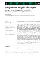
Figure 1 Immunocytochemical analysis of ERα and VDR in primary and established breast cancer cells. Representative images of cultured
tumor-derived (A-C), SUM-229PE (D-F) and BT-474 (G-I) cells are shown. Tumor-derived (A) and SUM-229PE (D) cells were negative for ERα, while
BT-474 was ERα positive (G). All cells were positive for VDR (B, E and H) in the cytoplasmic, nuclear and perinuclear regions (brown staining).
Negative controls were carried out in the absence of primary antibody for each cell line (C, F and I). Representative pictures are displayed (20 ×).
Santos-Martínez et al. BMC Cancer 2014, 14:230
/>
Page 5 of 10
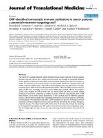
Figure 3 Calcitriol induced ERα protein expression. SUM-229PE
cells were treated with two calcitriol concentrations (Cal, 1X10⁻⁸ M
and 1X10⁻⁷ M) and two MAPK inhibitors: U0126 (U, 10 μM) or Gefitinib
(G, 0.8 μM) used as controls of ERα induction during 72 hr. Control
incubations were done in the presence ethanol (C) or DMSO (D).
Results were analyzed by western blots. MCF-7 (M) nuclear extracts
were used as positive control for ERα and GAPDH was utilized as the
loading control for normalization. Results are representative from two
independent experiments.
upregulated PRL expression. The presence of the antiestrogen alone did not change PRL gene expression.
These data suggest that the calcitriol-induced ERα is
a fully-transcriptionally active receptor. Interestingly,
calcitriol per se significantly stimulated the expression
of both CTSD and TFF1 genes, which may explain
why E2 was not able to further increase gene expression
(data not shown).
Calcitriol restored the antiestrogenic response in
ERα-negative breast cancer cells
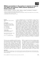
Figure 2 Calcitriol induced ERα mRNA expression through the VDR
in ERα-negative breast cancer cells. A) Cultured tumor-derived cells
from five patients (1-5) with ERα-negative breast cancer and the
ER-negative SUM-229PE (S) and ER-positive BT-474 (B) established
cell lines were incubated with calcitriol (1X10⁻⁷ M) or its vehicle
(C, ethanol) for 24 hr. Subsequently, mRNA was extracted and real time
RT-PCR (qPCR) was performed. B) Cultured breast tumor-derived cells
were treated with increasing calcitriol concentrations (1X10⁻¹⁰ M and
1X10⁻⁷ M) for 24 hr. C) Cells were incubated in the absence (C) or
presence of calcitriol (Cal, 1X10⁻⁸ M), without or with a VDR antagonist
(TEI, 1X10⁻⁶ M). Results shown are the mean ± S.D. of ERα/GAPDH
mRNA normalized ratio of two independent experiments per triplicate.
Data were normalized to 1 for vehicle-treated cells. *P ≤ 0.05 vs.
C. **P ≤ 0.05 vs. calcitriol alone.
In order to assess whether the calcitriol-induced ERα was
sensitive to the antiproliferative effects of the antiestrogens
in ERα-negative breast cancer cells, growth assays
were performed. Breast cancer cells were incubated in
the presence of calcitriol (1X10⁻⁸ M) or the vehicle
alone for 48 hr. Afterwards, cells were incubated with
ER agonist (1X10⁻⁸ M), antagonists (1X10⁻⁶ M) or the
combination of E2 plus antagonists during 6 days.
The results demonstrated that in the absence of calcitriol
(black bars), none of the compounds affected cell growth
in both cultured breast tumor-derived cells (Figure 5A)
and the SUM-229PE cell line (Figure 5B). Interestingly,
in calcitriol-treated tumor-derived cells (white bars), antiestrogens alone or in combination with E2 significantly
inhibited cell proliferation as compared with control cells
Santos-Martínez et al. BMC Cancer 2014, 14:230
/>
Page 6 of 10
Figure 4 Calcitriol induced a fully active ERα. Cultured breast
tumor-derived cells were incubated in the absence (black bars) or
presence of calcitriol 1X10⁻⁸ M (white bars) for 48 h. Subsequently,
cells were coincubated with or without calcitriol plus estradiol
(E2, 1x10⁻⁸ M), ICI-182,780 (ICI, 1x10⁻⁶ M) or vehicle (C) for 24 h. PRL
gene expression was determined by qPCR. Results are shown as the
mean ± S.D. of PRL/GAPDH mRNA normalized ratio. Data were
normalized to 1 for vehicle-treated cells. *P ≤ 0.05 vs. C.
(C, white bar). The presence of E2 at the dose of 1X10⁻⁸ M
did not modify cell growth (Figure 5A); however, higher
E2 concentrations (1X10⁻⁷ M) significantly inhibited cell
growth (data not shown). Similar results were observed in
SUM-229PE cells, but tamoxifen alone or in combination
with E2 did not affect cell growth (Figure 5B). MCF-7
cells were used as control of the inhibitory effect of
the antiestrogens via ERα (Figure 5C). As depicted,
E2 significantly increased cell proliferation in cells not
treated with calcitriol; however, this effect was not
observed in those cells cultured in the presence of
calcitriol, most likely due to its antiproliferative activity.
As expected, antiestrogens and their combination with E2
significantly inhibited cell growth in both treated and
not-treated calcitriol cells.
Antiestrogen treatment downregulated CCND1 and EAG1
gene expression in calcitriol-treated breast cancer cells
One of the molecular mechanisms by which antiestrogens inhibit cell proliferation is by decreasing CCND1
expression and blockage of cell cycle progression via the
ER [32,33]. Thus, we studied the effects of ICI-182,780
and E2 on CCND1 expression in calcitriol-treated
ERα-negative breast tumor-derived cells. As shown in
Figure 6A, only in calcitriol-treated cells the presence
of ICI-182,780 (1X10⁻⁶ M) but not E2 downregulated
CCND1 gene expression.
In breast cancer cell lines the inhibition of EAG1
potassium channel expression is accompanied by a significant reduction of cell proliferation [19,34]. Therefore, we
evaluated the effects of an agonist or antagonist of the
calcitriol-induced ER on EAG1 expression. As shown in
Figure 5 ERα induction restored the response to antiestrogens in
ER-negative breast cancer cells. A) Cultured breast tumor-derived
cells, B) SUM-229PE and C) MCF-7 were incubated in the absence (black
bars) or presence of calcitriol 1X10⁻⁸ M (white bars) for 48 h. Afterwards,
cells were coincubated without (black bars) or with calcitriol (white bars)
plus estradiol (E2, 1X10⁻⁸ M), tamoxifen (Tx, 1X10⁻⁶ M), ICI-182,780
(ICI, 1X10⁻⁶ M), ethanol (C), or combination of antagonists with E2 for
6 days. Cell growth assays by the XTT colorimetric method were
performed. Bars represent the mean ± S.D. Data were normalized to
100% using the activity of vehicle-treated cells. Results are representative
from two independent experiments performed in sextuplicates.*
P ≤ 0.05 vs. control for each group (black bars vs black control or white
bars vs white control).
Santos-Martínez et al. BMC Cancer 2014, 14:230
/>
Page 7 of 10
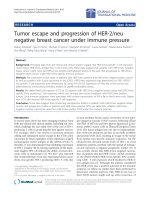
Figure 7 Calcipotriol induced ERα expression in ER-negative
breast cancer cells. SUM-229PE cells were cultured in the presence
of different calcitriol (circles) and calcipotriol (triangles) concentrations
or vehicle alone (C, ethanol) for 24 hr. Afterwards, mRNA was extracted
and qPCR was performed. Results are shown as the mean ± S.D. of
ERα/GAPDH mRNA normalized ratio. Data were normalized setting a
value of 1 for vehicle-treated cells. *P ≤ 0.05 vs. C.
Figure 6 ICI-182,780 downregulated CCND1 and EAG1 gene
expression in calcitriol-treated ERα-negative cells. Cultured breast
tumor-derived cells were incubated in the absence (black bars) or
presence of calcitriol 1X10⁻⁸ M (white bars) for 48 h. Subsequently,
cells were coincubated with or without calcitriol plus estradiol (E2,
1x10⁻⁸ M), ICI-182,780 (ICI, 1X10⁻⁶ M) or its vehicle (C) for 24 hr. A)
CCND1 and B) EAG1 gene expression was determined by qPCR.
Results shown are the mean ± S.D. of CCND1 or EAG1/GAPDH mRNA
normalized ratio. Data were normalized setting a value of 1 for
vehicle-treated cells. *P ≤ 0.05 vs. C.
Figure 6B, neither E2 nor ICI-182,780 altered EAG1
gene expression in non-calcitriol treated cells (black bars);
however, when compared with cells in the presence of
calcitriol, the antiestrogen, in contrast to E2 alone,
significantly decreased EAG1 mRNA levels (white bars).
Calcipotriol, a vitamin D analogue, increased ERα
expression
Calcipotriol, a synthetic low calcemic vitamin D analogue,
has been considered a potent stimulator of cell differentiation
and inhibitor of cell proliferation in cancer cells [35]. Figure 7
shows a comparison between different concentrations of
calcipotriol and calcitriol (1X10⁻¹⁰ to 1X10⁻⁶ M) upon ERα
gene expression in SUM-229PE. As depicted, both compounds increased ERα gene expression in a concentrationdependent manner with similar EC50 values (2.74X10⁻⁸ M
and 2.21X10⁻⁸ M, for calcipotriol and calcitriol, respectively).
Discussion
In breast cancer, the presence of the ERα is considered
as a good indicator of disease-free survival and prognosis
since patients with ERα−positive tumors are candidates
for hormonal therapy [3,4,6]. In contrast, tumors lacking
this receptor have the poorest clinical prognosis [36]. In this
study we demonstrated the ability of calcitriol to induce the
expression of ERα in both primary and established
ERα-negative breast cancer cell lines. This effect was
mediated by a VDR-dependent mechanism. In addition, our
results demonstrated a fully active calcitriol-induced ER by
its ability to increase PRL gene expression. Interestingly,
pretreatment of ER-negative breast tumor-derived
cells with calcitriol and the further incubation with
this secosteroid in combination with tamoxifen or
ICI-182,780 resulted in a significantly lower cell growth
proliferation.
It is noteworthy to mention that, to our knowledge, this
study is the first to demonstrate the ability of calcitriol to
induce the expression of a functional ERα in both primary
and established ERα-negative breast cancer cells, which
we think is of biological importance given its potential for
future treatment strategies to improve prognosis in
ERα-negative breast cancer patients.
Since it has been observed that MAPK inhibitors increase
ERα protein in ER-negative breast tumor cells [10], we
hypothesized that the upregulation of ERα by calcitriol
could be the result of decreased MAPK activity. Although,
in this study we could not demonstrate any change in
this kinase in the presence of calcitriol. An alternative,
mechanism by which calcitriol via its receptor induced
Santos-Martínez et al. BMC Cancer 2014, 14:230
/>
ERα expression might be at the level of promoter-driven
transcriptional regulation. Therefore, in order to identify
putative vitamin D response elements we performed an in
silico analysis with the MatInspector software [37] using a
sequence derived from the human chromosome 6, which
contains the promoter region of ERα [38]. The results
from this analysis showed the presence of several putative
vitamin D response elements of the DR3 and DR4 types,
supporting the idea of a direct transcriptional regulation
of ER promoter by calcitriol.
The observation that tamoxifen and ICI-182,780
inhibited cell growth in calcitriol-treated ER-negative
breast tumor-derived cells indicated the induction of a
functionally active ERα. However, cell growth inhibition by
tamoxifen was not observed in the case of calcitriol-treated
ER-negative SUM-229PE cells. This finding might be
explained as a receptor resistance–like condition resulting
probably from the hyperactivation of the MAPK signaling
pathway due to overexpression of EGFR or HER2 as has
been previously observed in breast cancer cells [10].
It is well known that E2 exhibits proliferative effects
and therefore stimulates tumor growth in breast cancer
[39,40]. However, in the present study, the presence of E2
did not result in increased proliferation of cells pretreated
with calcitriol. It is possible that the lack of mitogenic
activity of E2 through the newly expressed ERα was
due to a priming antiproliferative effect of calcitriol, thus
preventing the expected estradiol-mediated effects on cell
proliferation. This observation agreed with those of Bayliss
et al., [10] who showed that E2 did not increase proliferation in cells where the ERα was reexpressed by MAPK
inhibitors, including in those studies in ER-negative breast
cancer cells transfected with the ER [41].
In this study, the ability of antiestrogens to inhibit cell
growth in an estradiol-depleted condition might require
further investigation; however, some effects of these compounds on the mitogenic activity of growth factors, in the
absence of estrogens have been already demonstrated in
breast cancer [33,42]. In this regard, one of the most common regulators known to be altered and overexpressed in
various cancers including breast is CCND1, which functions as mitogenic sensor and allosteric activator of cyclindependent kinase (CDK)4/6 [43]. It is known that the
inhibitory actions of antiestrogens on breast cancer are in
part exerted through the downregulation of CCND1 [33].
In this study, the results showing that ICI-182,780 significantly decreased CCND1 mRNA only in calcitriol-treated
cells, indicated that these compounds may affect cell cycle
regulation as has already been shown in ER-positive breast
tumors [33]. Furthermore, the demonstration of a significant inhibition of EAG1 gene expression by ICI-182,780 in
calcitriol-treated cells, suggested that the antiproliferative
effects of these compounds involve a number of regulatory
mechanisms which are under the control of ERα activation.
Page 8 of 10
These results suggest that calcitriol in combination with
ICI-182,780, through downregulation of EAG1 and CCND1
affect cell proliferation and tumor progression [34,44].
There are several markers associated with tumor
aggressiveness. Among these, myoepithelial markers, which
are preferentially expressed in ER-negative breast cancer,
suggest that the loss of the steroid receptor is related to the
degree of cellular dedifferentiation occurring in these
tumors [45]. It is known that calcitriol promotes differentiation of several tumor cell types, including human breast
and colon cancers [14,46]. This process involves the action
of calcitriol on a number of events, such as the induction
of adhesion proteins (E-cadherin, claudin, occludin) or by
interfering with some intracellular signaling pathways,
such as the Wnt/b-catenin signaling [14,46]. Our results
revealed that calcitriol induced ERα gene and protein
expression suggesting that calcitriol affects the phenotype
of ERα-negative breast cancer cells by reverting cellular
mechanisms associated with a more aggressive behavior
and poor prognosis.
The development of numerous vitamin D analogues
and intermittent calcitriol dosing have allowed substantial
dose-escalation and reduced calcemic effects [47,48].
Calcipotriol, a synthetic vitamin D analogue with a
significantly lower calcemic effect, is also known as a
potent antiproliferative compound and an inducer of
cell differentiation [35]. In this study, the demonstration
that calcipotriol was also able to upregulate ERα gene
expression in an ER-negative breast cancer cell line,
suggest that treatment options in breast cancer patients
might also include vitamin D analogues with reduced side
calcemic effects.
Our results suggest that the use of calcitriol in
combination with aromatase inhibitors or ER antagonists
might be considered in the future as a new strategy
for the treatment of ERα-negative breast cancer, including
the triple-negative subtypes.
Conclusions
The results presented herein clearly demonstrated the
ability of calcitriol and its synthetic analog calcipotriol to
upregulate ERα expression in a subset of ER-negative
breast cancer cells. These results may offer a therapeutic
alternative, particularly in those patients affected with
ER-negative tumors by sensitizing them to hormone
therapy, with the aim at improving disease prognosis.
Abbreviations
CTSD: Cathepsin D; CCND1: Cyclin D1; E2: Estradiol; EAG1: Ether-à-go-go 1;
EC50: Stimulatory concentration; EGFR: Human epidermal growth factor
receptor- 1; ER: Estrogen receptor; FBS: Fetal bovine serum;
GAPDH: Glyceraldehyde-3-phosphate dehydrogenase; HER2: Human
epidermal growth factor receptor- 2; MAPK: Mitogen-activated protein
kinase; PR: Progesterone receptor; PRL: Prolactin; qPCR: Real time polymerase
chain reaction; RT: Reverse transcription; S.D: Standard deviation; TFF1: Trefoil
factor 1; VDR: Vitamin D receptor.
Santos-Martínez et al. BMC Cancer 2014, 14:230
/>
Competing interests
The authors declare that they have no competing interests.
Authors’ contributions
RGB and LD were involved in the conception, design and coordination of
the study as well as in data analysis, interpretation of results, actively
participated in all experimental procedures, and were involved in drafting
the manuscript. NSM was in charge of all experimental procedures,
participated in data analysis and interpretation, as well as in drafting the
manuscript. DOR, JGQ, DB, MJIS and JEL participated in the experimental
procedures and revised critically the content of the manuscript. HMF
provided breast biopsies, carried out the clinical data collection and retrieved
patients signed informed-consent forms. EA, AH and JC contributed in the
interpretation of data and critically revised the manuscript for important
intellectual content. FL participated in the interpretation of data, made
substantive intellectual contribution to the study and drafting the
manuscript. All authors read and approved the final manuscript.
Acknowledgments
This work was supported by grants 129315 and 153862 from the Consejo
Nacional de Ciencia y Tecnología (CONACyT), México. The authors state that
there are non-financial competing interests. N. Santos-Martínez is a Ph.D,
student from the Centro de Investigación y Estudios Avanzados, Instituto
Politécnico Nacional (CINVESTAV), México, and recipient of a fellowship from
CONACyT. We acknowledge with thanks to Teijin Pharma Limited (Japan),
Hoffmann-La Roche Ltd and AstraZeneca for TEI-9647, calcitriol and Gefitinib
donations, respectively.
Author details
1
Departments of Reproductive Biology, Instituto Nacional de Ciencias
Médicas y Nutrición Salvador Zubirán, Vasco de Quiroga No. 15, Tlalpan
14000 México, México. 2Department of Pharmacology, Centro de
Investigación y de Estudios Avanzados, I.P.N., Av. Instituto Politécnico
Nacional 2508 Gustavo A. Madero, 07360 México, D.F, México. 3Department
of Surgery, Instituto Nacional de Ciencias Médicas y Nutrición Salvador
Zubirán, Vasco de Quiroga No. 15, Tlalpan 14000 México, D.F, México.
4
Biochemistry Unit. Instituto Nacional de Ciencias Médicas y Nutrición
Salvador Zubirán, Vasco de Quiroga No. 15, Tlalpan 14000 México, D.F,
México.
Received: 24 September 2013 Accepted: 25 March 2014
Published: 29 March 2014
References
1. Simpson PT, Reis-Filho JS, Gale T, Lakhani SR: Molecular evolution of breast
cancer. J Pathol 2005, 205(2):248–254.
2. Sorlie T, Perou CM, Tibshirani R, Aas T, Geisler S, Johnsen H, Hastie T, Eisen MB,
van de Rijn M, Jeffrey SS, Thorsen T, Quist H, Matese JC, Brown PO, Botstein D,
Eystein Lonning P, Borresen-Dale AL: Gene expression patterns of breast
carcinomas distinguish tumor subclasses with clinical implications. Proc Natl
Acad Sci U S A 2001, 98(19):10869–10874.
3. Clark GM, McGuire WL: Steroid receptors and other prognostic factors in
primary breast cancer. Semin Oncol 1988, 15(2 Suppl 1):20–25.
4. McGuire WL, Osborne CK, Clark GM, Knight WA 3rd: Steroid hormone
receptors and carcinoma of the breast. Am J Physiol 1982,
243(2):E99–E102.
5. Nadji M, Gomez-Fernandez C, Ganjei-Azar P, Morales AR: Immunohistochemistry
of estrogen and progesterone receptors reconsidered: experience with 5,993
breast cancers. Am J Clin Pathol 2005, 123(1):21–27.
6. Powles TJ, Ashley S, Tidy A, Smith IE, Dowsett M: Twenty-year follow-up of
the Royal Marsden randomized, double-blinded tamoxifen breast cancer
prevention trial. J Natl Cancer Inst 2007, 99(4):283–290.
7. Clarke MJ: WITHDRAWN: Tamoxifen for early breast cancer. Cochrane
Database Syst Rev 2008, 4, CD000486.
8. Johnston SR: Acquired tamoxifen resistance in human breast
cancer–potential mechanisms and clinical implications. Anticancer
Drugs 1997, 8(10):911–930.
9. Zhao JJ, Lin J, Yang H, Kong W, He L, Ma X, Coppola D, Cheng JQ:
MicroRNA-221/222 negatively regulates estrogen receptor alpha and is
associated with tamoxifen resistance in breast cancer. J Biol Chem 2008,
283(45):31079–31086.
Page 9 of 10
10. Bayliss J, Hilger A, Vishnu P, Diehl K, El-Ashry D: Reversal of the estrogen
receptor negative phenotype in breast cancer and restoration of
antiestrogen response. Clin Cancer Res 2007, 13(23):7029–7036.
11. Oh AS, Lorant LA, Holloway JN, Miller DL, Kern FG, El-Ashry D: Hyperactivation
of MAPK induces loss of ERalpha expression in breast cancer cells.
Mol Endocrinol 2001, 15(8):1344–1359.
12. Fife RS, Sledge GW Jr, Proctor C: Effects of vitamin D3 on proliferation of
cancer cells in vitro. Cancer Lett 1997, 120(1):65–69.
13. Simboli-Campbell M, Narvaez CJ, Tenniswood M, Welsh J:
1,25-Dihydroxyvitamin D3 induces morphological and biochemical
markers of apoptosis in MCF-7 breast cancer cells. J Steroid Biochem Mol
Biol 1996, 58(4):367–376.
14. Pendas-Franco N, Gonzalez-Sancho JM, Suarez Y, Aguilera O, Steinmeyer A,
Gamallo C, Berciano MT, Lafarga M, Munoz A: Vitamin D regulates the
phenotype of human breast cancer cells. Differentiation 2007, 75(3):193–207.
15. Janowsky EC, Lester GE, Weinberg CR, Millikan RC, Schildkraut JM, Garrett
PA, Hulka BS: Association between low levels of 1,25-dihydroxyvitamin D
and breast cancer risk. Public Health Nutr 1999, 2(3):283–291.
16. Mawer EB, Walls J, Howell A, Davies M, Ratcliffe WA, Bundred NJ: Serum
1,25-dihydroxyvitamin D may be related inversely to disease activity in
breast cancer patients with bone metastases. J Clin Endocrinol Metab
1997, 82(1):118–122.
17. Yao S, Ambrosone CB: Associations between vitamin D deficiency and
risk of aggressive breast cancer in African-American women. J Steroid
Biochem Mol Biol 2012, 136:337–341.
18. Berger U, McClelland RA, Wilson P, Greene GL, Haussler MR, Pike JW, Colston K,
Easton D, Coombes RC: Immunocytochemical determination of estrogen
receptor, progesterone receptor, and 1,25-dihydroxyvitamin D3 receptor in
breast cancer and relationship to prognosis. Cancer Res 1991, 51(1):239–244.
19. Garcia-Becerra R, Diaz L, Camacho J, Barrera D, Ordaz-Rosado D, Morales A,
Ortiz CS, Avila E, Bargallo E, Arrecillas M, Halhali A, Larrea F: Calcitriol
inhibits Ether-a go-go potassium channel expression and cell
proliferation in human breast cancer cells. Exp Cell Res 2010.
20. Swami S, Krishnan AV, Feldman D: 1alpha,25-Dihydroxyvitamin D3
down-regulates estrogen receptor abundance and suppresses estrogen
actions in MCF-7 human breast cancer cells. Clin Cancer Res 2000,
6(8):3371–3379.
21. Garcia-Quiroz J, Garcia-Becerra R, Barrera D, Santos N, Avila E, Ordaz-Rosado D,
Rivas-Suarez M, Halhali A, Rodriguez P, Gamboa-Dominguez A, Medina-Franco H,
Camacho J, Larrea F, Diaz L: Astemizole synergizes calcitriol antiproliferative
activity by inhibiting CYP24A1 and upregulating VDR: a novel approach for
breast cancer therapy. PLoS One 2012, 7(9):e45063.
22. Simboli-Campbell M, Narvaez CJ, van Weelden K, Tenniswood M, Welsh J:
Comparative effects of 1,25(OH)2D3 and EB1089 on cell cycle kinetics
and apoptosis in MCF-7 breast cancer cells. Breast Cancer Res Treat 1997,
42(1):31–41.
23. Stoica A, Saceda M, Fakhro A, Solomon HB, Fenster BD, Martin MB: Regulation
of estrogen receptor-alpha gene expression by 1, 25-dihydroxyvitamin D in
MCF-7 cells. J Cell Biochem 1999, 75(4):640–651.
24. Davoodi F, Brenner RV, Evans SR, Schumaker LM, Shabahang M, Nauta RJ,
Buras RR: Modulation of vitamin D receptor and estrogen receptor by
1,25(OH)2-vitamin D3 in T-47D human breast cancer cells. J Steroid
Biochem Mol Biol 1995, 54(3–4):147–153.
25. Li Z, Bustos V, Miner J, Paulo E, Meng ZH, Zlotnikov G, Ljung BM, Dairkee SH:
Propagation of genetically altered tumor cells derived from fine-needle
aspirates of primary breast carcinoma. Cancer Res 1998, 58(23):5271–5274.
26. Tesch M, Shawwa A, Henderson R: Immunohistochemical determination
of estrogen and progesterone receptor status in breast cancer. Am J Clin
Pathol 1993, 99(1):8–12.
27. Maurer U, Jehan F, Englert C, Hubinger G, Weidmann E, DeLuca HF,
Bergmann L: The Wilms’ tumor gene product (WT1) modulates the
response to 1,25-dihydroxyvitamin D3 by induction of the vitamin D
receptor. Biol Chem 2001, 276(6):3727–3732.
28. Lappano R, Recchia AG, De Francesco EM, Angelone T, Cerra MC, Picard D,
Maggiolini M: The cholesterol metabolite 25-hydroxycholesterol activates
estrogen receptor alpha-mediated signaling in cancer cells and in
cardiomyocytes. PloS one 2011, 6(1):e16631.
29. Almeras L, Eyles D, Benech P, Laffite D, Villard C, Patatian A, Boucraut J,
Mackay-Sim A, McGrath J, Feron F: Developmental vitamin D deficiency
alters brain protein expression in the adult rat: implications for
neuropsychiatric disorders. Proteomics 2007, 7(5):769–780.
Santos-Martínez et al. BMC Cancer 2014, 14:230
/>
30. Hussain-Hakimjee EA, Mehta RG: Regulation of steroid receptor expression
by 1alpha-hydroxyvitamin D5 in hormone-responsive breast cancer cells.
Anticancer Res 2009, 29(9):3555–3561.
31. Duan R, Ginsburg E, Vonderhaar BK: Estrogen stimulates transcription from
the human prolactin distal promoter through AP1 and estrogen
responsive elements in T47D human breast cancer cells. Mol Cell
Endocrinol 2008, 281(1–2):9–18.
32. Musgrove EA, Hamilton JA, Lee CS, Sweeney KJ, Watts CK, Sutherland RL:
Growth factor, steroid, and steroid antagonist regulation of cyclin gene
expression associated with changes in T-47D human breast cancer cell
cycle progression. Mol Cell Biol 1993, 13(6):3577–3587.
33. Watts CK, Sweeney KJ, Warlters A, Musgrove EA, Sutherland RL:
Antiestrogen regulation of cell cycle progression and cyclin D1 gene
expression in MCF-7 human breast cancer cells. Breast Cancer Res Treat
1994, 31(1):95–105.
34. Pardo LA, Suhmer W: Eag1 as a cancer target. Expert Opin Ther Targets
2008, 12(7):837–843.
35. Binderup L, Bramm E: Effects of a novel vitamin D analogue MC903 on
cell proliferation and differentiation in vitro and on calcium metabolism
in vivo. Biochem Pharmacol 1988, 37(5):889–895.
36. Osborne CK, Yochmowitz MG, Knight WA 3rd, McGuire WL: The value of
estrogen and progesterone receptors in the treatment of breast cancer.
Cancer 1980, 46(12 Suppl):2884–2888.
37. Cartharius K, Frech K, Grote K, Klocke B, Haltmeier M, Klingenhoff A, Frisch M,
Bayerlein M, Werner T: MatInspector and beyond: promoter analysis based
on transcription factor binding sites. Bioinformatics 2005, 21(13):2933–2942.
38. Kos M, Reid G, Denger S, Gannon F: Minireview: genomic organization of
the human ERalpha gene promoter region. Mol Endocrinol 2001,
15(12):2057–2063.
39. Doisneau-Sixou SF, Sergio CM, Carroll JS, Hui R, Musgrove EA, Sutherland RL:
Estrogen and antiestrogen regulation of cell cycle progression in breast
cancer cells. Endocr Relat Canc 2003, 10(2):179–186.
40. Dickson RB, Lippman ME: Growth factors in breast cancer. Endocrine reviews
1995, 16(5):559–589.
41. Jiang SY, Jordan VC: Growth regulation of estrogen receptor-negative
breast cancer cells transfected with complementary DNAs for estrogen
receptor. J Natl Cancer Inst 1992, 84(8):580–591.
42. Vignon F, Bouton MM, Rochefort H: Antiestrogens inhibit the mitogenic
effect of growth factors on breast cancer cells in the total absence of
estrogens. Biochem Biophys Res Commun 1987, 146(3):1502–1508.
43. Barnes DM, Gillett CE: Cyclin D1 in breast cancer. Breast Cancer Res Treat
1998, 52(1–3):1–15.
44. Ouadid-Ahidouch H, Ahidouch A: K + channel expression in human breast
cancer cells: involvement in cell cycle regulation and carcinogenesis.
J Membr Biol 2008, 221(1):1–6.
45. Gordon LA, Mulligan KT, Maxwell-Jones H, Adams M, Walker RA, Jones JL:
Breast cell invasive potential relates to the myoepithelial phenotype.
Int J Cancer 2003, 106(1):8–16.
46. Palmer HG, Gonzalez-Sancho JM, Espada J, Berciano MT, Puig I, Baulida J,
Quintanilla M, Cano A, de Herreros AG, Lafarga M, Munoz A: Vitamin D (3)
promotes the differentiation of colon carcinoma cells by the induction
of E-cadherin and the inhibition of beta-catenin signaling. J Cell Biol 2001,
154(2):369–387.
47. Beer TM, Munar M, Henner WD: A Phase I trial of pulse calcitriol in
patients with refractory malignancies: pulse dosing permits substantial
dose escalation. Cancer 2001, 91(12):2431–2439.
48. Masuda S, Jones G: Promise of vitamin D analogues in the treatment of
hyperproliferative conditions. Mol Cancer Ther 2006, 5(4):797–808.
doi:10.1186/1471-2407-14-230
Cite this article as: Santos-Martínez et al.: Calcitriol restores antiestrogen
responsiveness in estrogen receptor negative breast cancer cells: A
potential new therapeutic approach. BMC Cancer 2014 14:230.
Page 10 of 10
Submit your next manuscript to BioMed Central
and take full advantage of:
• Convenient online submission
• Thorough peer review
• No space constraints or color figure charges
• Immediate publication on acceptance
• Inclusion in PubMed, CAS, Scopus and Google Scholar
• Research which is freely available for redistribution
Submit your manuscript at
www.biomedcentral.com/submit