Báo cáo y học: "HIV DNA and Dementia in Treatment-Naïve HIV-1-Infected Individuals in Bangkok, Thailand"
Bạn đang xem bản rút gọn của tài liệu. Xem và tải ngay bản đầy đủ của tài liệu tại đây (402.5 KB, 6 trang )
Int. J. Med. Sci. 2007, 4
13
International Journal of Medical Sciences
ISSN 1449-1907 www.medsci.org 2007 4(1):13-18
© Ivyspring International Publisher. All rights reserved
Short Research Communication
HIV DNA and Dementia in Treatment-Naïve HIV-1-Infected Individuals in
Bangkok, Thailand
Bruce Shiramizu
1
, Silvia Ratto-Kim
1 2
, Pasiri Sithinamsuwan
3
, Samart Nidhinandana
3
, Sataporn Thitivi-
chianlert
3
, George Watt
1
, Mark deSouza
2
, Thippawan Chuenchitra
2
, Suchitra Sukwit
2
, Suwicha Chitpatima
4
,
Kevin Robertson
5
, Robert Paul
6
, Cecilia Shikuma
1
, Victor Valcour
1
1. Hawaii AIDS Clinical Research Program, University of Hawaii, Honolulu, HI, USA; 2. Armed Forces Research Inst. Med.
Sciences, Bangkok, Thailand; 3. Phramongkutklao Hosp., Bangkok, Thailand; 4. Royal Thai Army Med. Dept., Bangkok,
Thailand; 5. Univ. North Carolina, Chapel Hill, NC, USA; 6. Univ. Missouri, Dept. Psychology, St. Louis, MO, USA - for the
South East Asia Research Collaboration with the Univ. of Hawaii Protocol 001 Team.
Correspondence to: B. Shiramizu, MD; 3675 Kilauea Ave.; Young Bldg., 5th Floor; Honolulu, Hawaii, USA, 96816; Phone: 808-737-2751;
Fax: 808-735-7047;
Received: 2006.11.16; Accepted: 2006.12.05; Published: 2006.12.06
High HIV-1 DNA (HIV DNA) levels in peripheral blood mononuclear cells (PBMC) correlate with
HIV-1-associated dementia (HAD) in patients on highly active antiretroviral therapy (HAART). If this relation-
ship also exists among HAART-naïve patients, then HIV DNA may be implicated in the pathogenesis of HAD.
In this study, we evaluated the relationship between HIV DNA and cognition in subjects naïve to HAART in a
neuroAIDS cohort in Bangkok, Thailand. Subjects with and without HAD were recruited and matched for age,
gender, education, and CD4 cell count. PBMC and cellular subsets were analyzed for HIV DNA using real-time
PCR. The median log
10
HIV DNA copies per 10
6
PBMC for subjects with HAD (n=15) was 4.27, which was
higher than that found in subjects without dementia (ND; n=15), 2.28, p<0.001. This finding was unchanged in a
multivariate model adjusting for plasma HIV-1 RNA levels. From a small subset of individuals, in which ade-
quate number of cells were available, more HIV DNA was in monocytes/macrophages from those with HAD
compared to those with ND. These results are consistent with a previous report among HAART-experienced
subjects, thus further implicating HIV DNA in the pathogenesis of HAD.
Key words: human immunodeficiency virus type 1; dementia; cognition; HIV DNA
1. INTRODUCTION
Complete eradication of the human immunode-
ficiency virus, type 1 (HIV-1) from infected individu-
als is not currently possible due, in part, to continuing
presence of virus in lymphocytes and cells of the
macrophage lineage [1-3]. Monocytes/macrophages
(M/MΦ) are cellular sanctuaries for HIV-1, which re-
main present even in patients with suppressed plasma
viremia on highly active antiretroviral therapy
(HAART) [4, 5]. These cells may be particularly suited
as sanctuaries for virus because HIV-1 DNA (HIV
DNA), compared to HIV RNA, is less affected by cur-
rent treatment regimens [6-9]. Additionally, these
nondividing cells differ in many respects from that of
CD4 lymphocytes making them unique entities for
long-term persistence of HIV DNA [4, 10]. For in-
stance, mitosis of M/MΦ is not required for nuclear
import or integration of viral DNA; and M/MΦ not
only contribute to establishment and persistence of
HIV-1 infection, they also activate surrounding T-cells
thus favoring their infection.
These circulating monocytes traffic through tis-
sue and to the central nervous system (CNS) and dif-
ferentiate into tissue macrophages. This provides a
basis for theorizing that M/MΦ may contribute to the
ongoing persistence of HIV-1 in these sites [11].
Monocyte trafficking to the CNS is hypothesized to be
an underlying event in neuropathogenesis of
HIV-1-associated dementia (HAD) [12-14]. Our recent
observation of high HIV DNA levels in subjects with
HAD, even among those with undetectable plasma
HIV-1 RNA levels (VL), highlights the significance of
PBMC HIV DNA in the pathogenesis of HAD [15]. We
performed the prior study on subjects who were on
HAART, therefore the question remains regarding the
significance of HIV DNA in HAD pathogenesis before
beginning therapy. In the prior study, our data sug-
gested that HIV DNA was predominantly in
CD14/CD16 M/MФ [15]. Therefore to further assess
the importance of CD14/CD16 phenotype, the current
study attempts to address the question whether a spe-
cific PBMC subset (M/MФ or CD4 lymphocytes) har-
bors HIV DNA.
We undertook the current study to test the hy-
pothesis that HIV DNA levels would be elevated in
cognitively-impaired individuals naïve to HAART;
and that HIV DNA levels in M/MФ are higher than in
CD4 lymphocytes in subjects with HAD compared to
those with no dementia (ND). We hypothesize that an
Int. J. Med. Sci. 2007, 4
14
association of HIV DNA with HAD before starting
HAART would further implicate HIV DNA in the
neuropathogenesis of HAD likely due to the presence
of virus in M/MФ that traffic to the CNS. The work
was completed using a cohort in Bangkok, Thailand,
which was established to characterize cognition
among individuals initiating HAART for the first time
as the country rapidly escalated access to antiretrovi-
ral drugs.
2. METHODS
Subjects and Clinical Data.
We established a longitudinal neuroAIDS cohort
within the Southeast Asia Research Collaboration with
the University of Hawaii (SEARCH) to characterize
HIV-1-related cognitive dysfunction among individu-
als in Bangkok infected with the most commonly
identified subtype in Thailand, recombinant circulat-
ing form (CRF) 01_AE. The protocol and consent
forms were approved by the Ethical Review Commit-
tee and Institutional Review Board of the participating
institutions. The SEARCH institutions involved in this
project included the University of Hawaii,
Phramongkutklao Hospital (PMK), and the Armed
Forces Research Institute of Medical Sciences
(AFRIMS), the latter two located on the same campus
in Bangkok, Thailand. Study volunteers were enrolled
at PMK, a large, tertiary care teaching hospital that is
administered by the Royal Thai Army, which provides
care for all Thai nationals regardless of military affilia-
tion. The study enrolled Thai individuals living in
Bangkok with HAD, ND, and HIV-1-seronegative
controls matched for age, education, and gender.
HIV-1-infected subjects were also matched for CD4
cell counts. The seronegative controls were enrolled
and completed identical neuropsychological tests as
the HIV-1-infected individuals because no Thai nor-
mative data were available to analyze the results. All
individuals had minimal/distant or no exposure to
illicit drug use with negative urine toxicology screens
on two occasions within 30 days prior to enrollment.
Subjects were all seronegative for hepatitis C virus,
free of neurological or psychiatric illnesses including
major depression, and did not have central nervous
system opportunistic infection, active opportunistic
infection in any organ system, pre-existing or known
learning disability, or past brain trauma. Individuals
thought to have cognitive impairment were referred
for study participation from the outpatient neurology
and infectious diseases clinics at PMK or from other
hospitals/clinics. Matched HIV-1-infected individuals
without HAD were then recruited.
The protocol neurologist (P. S.) established a di-
agnosis of HAD using standard-of-care approaches for
Thailand. In general, the evaluation included partici-
pant and proxy informant reports of symptoms and
function, the HIV macroneurological examination as
used in the Adult AIDS Clinical Trails Group (AACTG,
NIAID), bedside cognitive testing (including assess-
ment of orientation, motor and psychomotor speed,
memory, executive functioning, and visuospatial skill),
and the international HIV-1 Dementia Scale [16]. All
participants with HAD were further evaluated to
rule-out other causes of cognitive impairment includ-
ing gadolinium-enhanced brain MRI. If clinically in-
dicated, individuals underwent a lumbar puncture to
exclude opportunistic brain infection, however of the
eight lumbar punctures that were performed, no op-
portunistic infections were found. Similarly, even
though HIV-1-infected subjects had advanced immu-
nosuppression, there were no individuals who had
any history of any opportunistic infections, including
in the CNS. After enrollment, all participants were
evaluated with a modified version of the WHO Inter-
national HIV-1 neuropsychological battery [17]. We
selected this battery as it was designed to minimize
cultural bias and was utilized in a prior study con-
ducted in Bangkok; therefore feasibility was estab-
lished [17]. We substituted the Brief Visual Memory
Test-revised for the Picture Memory Test for logistical
reasons as the latter required immediate and consis-
tent computer and internet access. We assessed de-
pressive symptoms with the Thai Depression Inven-
tory (TDI) which was previously validated in Thailand
[18]. The assessment was a clinical assessment made
by the protocol neurologist (P. S.) at the time of clini-
cal evaluation using the TDI, patient interview, and
patient and proxy information to assist in this assess-
ment.
We validated the diagnosis of HAD by reviewing
the first 27 HIV-1 cases enrolled in a consensus panel
consisting of an HIV neurologist, the study HIV neu-
ropsychologist (R. P.) and the principal investigator of
the cohort (V. V.). We prepared case summaries con-
sisting of all clinical and neurological data. Individual
raw neuropsychological scores were then plotted over
3 box plot distributions of seronegative controls, indi-
viduals with HAD, and those with ND. In a blinded
fashion, we determined a consensus diagnosis of HAD
or ND and reached consensus with the diagnosis de-
termined by our Thai colleague on 100% of the ND
cases. Among the HAD cases, the consensus panel
was congruent in 70% of the cases with the remaining
cases felt to be either mild dementia or minor cogni-
tive motor disorder, with an overall congruence ex-
ceeding 85%. The consensus panel was convened to
validate the diagnosis by the Thai neurologist and not
to substitute it. Therefore, since an excellent congru-
ence was achieved, we completed the analysis using
the original diagnoses for the purpose of this evalua-
tion.
Viral load and CD4 lymphocyte counts were
performed at AFRIMS, which maintains a Certificate
of American Pathologists for these tests. Viral sub-
types were determined by ELISA serotyping using
V3-CM237 (Thai subtype B) and V3 CM242 (subtype E)
peptides, which distinguishes HIV-1 subtype B and E
infection in Thai individuals and confirmed by se-
quencing, when indicated [19, 20].
Specimens and HIV DNA Assay.
At entry into the cohort, PBMC were isolated and
stored frozen in dimethyl sulfoxide from blood
Int. J. Med. Sci. 2007, 4
15
(ethylenediaminetetraacetic acid tube). DNA was ex-
tracted from an aliquot of frozen PBMC (5 X 10
6
cells),
as previously reported [15]. HIV DNA, normalized to
the number of copies of HIV-1 DNA per 10
6
cells, was
then measured using real-time polymerase chain reac-
tion (PCR), as previously described [15]. We per-
formed all real-time PCR assays in triplicate using in-
dependent standard curves generated to measure
relative HIV DNA copy number and cellular equiva-
lent genomic DNA. The plasmid used to generate the
standard curves was designed with a single copy each
of HIV-1 and a housekeeping gene, βglobin. We used
two primer sets to distinguish amplification of the two
genes: HIV gag (conserved 296 base pair product for
subtypes A and B) and βglobin (330 base pair product).
The PCR master mix consisted of either the HIV or
βglobin primers and probe sets, 1x iQ supermix (Bio-
Rad Laboratories, Hercules, CA), 100 ng DNA, and
water (final volume 25 μL) with the following condi-
tions: initial denaturation for 3 min followed by 45
cycles of 95°C/10 seconds, 57°C/30 seconds; with fi-
nal extension of 72°C/2 min. We used non-HIV-1 in-
fected genomic DNA for a negative control and DNA
from three HIV-1 infected cell lines (8E5, OM10.1, and
ACH-2; NIH AIDS Research and Reference Reagent
Program, NIH, Bethesda, MD) as positive controls.
The HIV-1 primers were tested on HIV-1 clades A, E,
and B; and demonstrated equivalent amplification of
the target gene.
Separation of PBMC Subsets and HIV DNA Analay-
sis.
Because HIV DNA is present in both lympho-
cytes and monocytes, we were interested in assessing
whether more HIV DNA was in one particular PBMC
subset versus the other. We previously measured HIV
DNA in PBMC subsets from individuals from a dif-
ferent cohort and showed that there were higher levels
in M/MΦ compared to CD14
-
cells in those diagnosed
with HAD versus those with ND [15, 21]. The same
procedures were performed on the specimens from
HIV-1-positive subjects for the current study from
which adequate numbers of cells were available to
recover reasonable quantities of cells in the subsets. To
separate the cells, we used RosetteSep (Stemcell
Technologies, Vancouver, BC, Canada) combined with
magnetic beads. Initially a CD14
-
subset from a small
aliquot of blood, which includes CD4 lymphocytes,
was isolated with beads; with the remaining cells
separated into CD14
+
/CD16
+
by enrichment and bead
separation. An aliquot of the sorted cells was then
analyzed by flow cytometry (FACSCalibur, Becton
Dickinson, San Jose, CA) to verify the phenotype in
each subset. The cells were analyzed using FlowJo
software (Tree Star Inc, San Jose, CA) following stain-
ing with the following antibodies (BD Biosciences, San
Jose, CA): murine anti-human antibodies,
FITC-conjugated anti-CD14, PE-conjugated anti-CD16
(3G8; PharMingen), PerCP-conjugated anti-HLA-DR,
and isotype controls. Total DNA was isolated from
each subset and HIV DNA measured as described
above.
Statistical Analysis.
We used logistic regression models to examine
the independent effect of HIV DNA on HAD vs. ND
with the Likelihood-Ratio test on the odds-ratio.
Analyses were conducted using SAS 9.0 (SAS Institute,
Cary, N.C.) with a p-value <0.05 interpreted as a sig-
nificant result. A two sample t test for the educa-
tion/age/CD4/VL variables and Fisher's Exact test for
the gender variable were used.
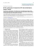
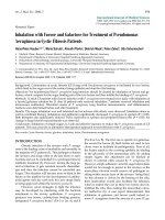
Figure 1. HIV DNA in Subjects with HAD vs. Non-HAD.
Log
10
HIV DNA levels in subjects with HAD (n=15; me-
dian=4.27) are higher than those without HAD (ND, n=15;
median=2.28), p<0.001.
3. RESULTS
Sixty individuals entered the study (n=15 each
for the HAD and ND groups and n=30 for HIV-1
seronegative controls); matched for age, gender, and
years of education, Table 1. All participants were Thai
nationals with the majority of the participants being
female. The HIV-1-seropositive subjects (n=30) were
HAART-naïve initially and had relatively low CD4
cell counts prior to initiation of therapy, Table 1. All of
the HIV-1-infected individuals were infected with
circulating HIV-1 subtype (CRF) 01_AE. In subjects
with HAD, compared to those with ND, the median
(IQR) CD4 cell counts were 21 (6-74) cells/ μL and 39
(16-71) cells/ μL, respectively, with no difference be-
tween the two groups, p=0.775. As would be expected,
in treatment-naïve patients with low CD4 cell counts,
the log
10
HIV-1 plasma RNA levels were relatively
high with no difference in between the two groups
(median 5.28 and 5.33 for HAD and ND, respectively),
p=0.811.
Comparing subjects with and without HAD, we
found significantly higher log
10
HIV DNA copies per
10
6
PBMC in the HAD group [n=15; median 4.27 (2.10
to 5.28)] versus the ND group [n=15; 2.28 (0.69 to
4.30)], p<0.001, Table 1. The calculated log
10
HIV
DNA/10
6
PBMC and medians for ND and HAD indi-
viduals is shown in Figure 1. In an unadjusted logistic
regression model, we identified an association of HIV
DNA to HAD resulting in an odds ratio of 1.841 (95%
confidence interval, CI, 1.286-2.635), p<0.001, with the
odds ratio representing a one unit increase in log
10
HIV DNA copies per 10
6
cells. This effect was un-
changed in a multivarate model adjusting for plasma
Int. J. Med. Sci. 2007, 4
16
HIV RNA levels (odds ratio 1.867, 95% CI 1.297-2.688).
As expected, HIV DNA was not detected in any of the
HIV-1 seronegative control subjects.
Table 1. Demographic and Laboratory Parameters
HIV-1-Seronegative (n=30) HAD (n=15) ND (n=15) p
Age (years) [mean (SD)] 34.1 (9.6) 33.1 (8.6) 33.7 (8.0) 0.947
Years of education [Mean (SD)] 7.6 (1.8) 6.9 (2.3) 6.6 (1.7) 0.186
Female/Male 18/12 9/6 10/5 0.904
CD4 cell count (cells/μL)
Median (IQR) 797.6 (679-1012) 21 (6-74) 39 (16-71)
Log
10
HIV-1 RNA (copies/mL)
Median (IQR)
Not applicable
5.28 (5.04-5.54)
5.33 (5.08 to 5.53)
0.811
Log
10
HIV DNA (copies/10
6
PBMC)
Median (IQR)
Not applicable
4.27 (2.10 to 5.28)
2.28 (0.69 to 4.30)
<0.001
HAD: HIV-1-associated dementia; ND: no dementia
Table 2. HIV DNA Copy and Total Burden in PBMC and Subsets
HIV DNA Copy per Total HIV DNA Copy Calculated From Diagnosis
PBMC CD14/CD16 CD4 PBMC CD14/CD16 CD4
Ratio*
HAD 4.10X10
-2
1.27X10
-2
1.92X10
-4
2.50X10
8
1.48X10
6
3.5X10
4
>1
HAD 1.34X10
-2
1.25X10
-2
1.06X10
-4
9.91X10
7
6.18X10
5
6.28X10
4
>1
HAD 2.09X10
-2
2.16X10
-2
1.59X10
-4
1.23X10
8
3.15X10
6
9.38X10
3
>1
HAD 2.00X10
-2
1.24X10
-2
5.30X10
-4
1.08X10
8
1.79X10
6
5.72X10
4
>1
HAD 3.35X10
-2
5.75X10
-2
2.16X10
-4
2.41X10
8
4.85X10
7
7.78X10
4
>1
Non-HAD 1.89X10
-4
9.10X10
-6
1.56X10
-4
1.25X10
6
7.46X10
2
1.03X10
6
<1
Non-HAD 4.45X10
-3
3.77X10
-4
4.29X10
-3
2.83X10
7
2.70X10
4
1.62X10
6
<1
Non-HAD 4.68X10
-3
3.13X10
-4
4.08X10
-3
1.54X10
7
2.41X10
4
5.39X10
5
<1
Non-HAD 2.00X10
-2
1.06X10
-3
1.94X10
-2
1.10X10
8
6.76X10
4
6.40X10
6
<1
*Ratio of CD14/CD16 to CD4 > 1.00 denotes total higher HIV DNA levels in CD14/CD16 compared to CD4 subsets
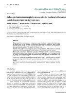
Figure 2. Phenotypic Expression of CD14
+
and CD14
-
Sorted Subsets. Cells from sorted fractions were stained for CD14. A,
B) Two examples of CD14-stained sorted cell populations from monocyte fractions from two different subjects demonstrating
the majority of cells isolated were CD14
+
(82.3% and 92.3%); C) CD14-negative subset showing low CD14-staining (0.49%).
A limited number of individuals (HAD n=5; ND
n=4) had analyses of PBMC subsets in which an ade-
quate number of cells was available for separation.
Using flow cytometry (monocyte & CD4/CD8 per-
centages) and data from sorted cells, we estimated the
total HIV DNA copies from CD14/CD16 and CD4
subsets. An assumption was made whereby HIV DNA
measurements from the CD14
-
subsets were primarily
from CD4 lymphocytes. The efficiency of our sorting
procedure is depicted in Figures 2A & 2B, where
greater than 80-90% of isolated monocyte subsets were
Int. J. Med. Sci. 2007, 4
17
CD14
+
. Less than 1% of cells from the CD14
-
subset
were positive for CD14, Figure 2C. By extrapolating
from the calculated values, HIV DNA levels in PBMC
were relatively high in all individuals diagnosed with
HAD with the highest in the CD14/CD16 subsets
compared to CD4 subsets, Table 2. We initially esti-
mated HIV DNA copy per PBMC; per CD14/CD16
cell; and per CD4 lymphocyte, Table 2. We then esti-
mated the total HIV DNA contribution from each
subset using the flow data and complete blood counts
obtained at the same time the blood was collected. In
this analysis, the total HIV DNA contribution from
CD14/CD16 cells was higher than the HIV DNA con-
tribution from CD4 cells in subjects with HAD, Table 2,
which was not apparent in subjects without HAD. In
order to estimate differences in the HIV DNA contri-
bution from the two subsets, the ratios of HIV DNA
from CD14/CD16 subsets to HIV DNA from CD4
subsets were calculated. This resulted in ratios sig-
nificantly higher from those with HAD (n=5; me-
dian=188.5) compared to those with ND (n=4; me-
dian=0.0059), p<0.029, Table 2.
4. DISCUSSION
Current antiretroviral therapy for HIV-1 focuses
on eradication of the virus from plasma. In contrast to
the cytotoxic effects of HIV-1 on lymphocytes,
HIV-1-infection usually leads to chronic infection in
M/MΦ. Recent studies suggest that PBMC HIV DNA
may be a marker for HIV-1 disease progression [22-25].
Our laboratory previously reported the presence of
high HIV DNA in PBMC as a risk for HAD in
HAART-experienced individuals; and preliminary
analyses suggest that the majority of this HIV DNA
may be in circulating M/MФ [15]. We demonstrated
that this effect was independent of plasma HIV-1 RNA
levels by a separate analysis of HIV DNA in individu-
als with undetectable plasma VL. We now confirm our
findings in a different cohort who are naïve to
HAART and hypothesize that high HIV DNA levels
are an important factor in HAD pathogenesis.
In the current study, we found the effect of HIV
DNA on HAD was independent of age and current
CD4 count at the time of recruitment, which is similar
to what was found previously in patients on effective
antiretroviral therapy. The HIV DNA data suggesting
a higher contribution from the monocyte/macrophage
subsets in patients with HAD are limited by the small
number of specimens available. The cohort established
in Thailand provided a unique opportunity to test our
hypothesis of the role of HIV DNA in HAD. We were
able to enroll age-, education-, and gender-match
HIV-1 seronegative individuals as controls to establish
normative data for the current study, which have not
previously been established in Thailand. Additional
data in PBMC subsets are needed to assess the impor-
tance of HIV DNA in the pathogenesis of HAD. Other
limitations of the current study include the assump-
tion that the CD14
-
subsets were composed mainly of
CD4 lymphocytes. The calculations of HIV DNA cop-
ies in the PBMC subsets are based on extrapolated
values. To confirm the findings, future experiments
are planned to use a cell sorter to isolate specific cell
populations.
While the mechanism by which HIV DNA leads
to neurocognitive problems remains unclear, we pro-
pose that our results demonstrating an association in
HAART-naïve patients strengthens the relationship of
HIV DNA to HAD neuropathogenesis. Even though
the mechanisms linking HIV DNA to HAD patho-
genesis are not fully known, studying HIV DNA in
PBMC subsets such as memory and naïve CD4
T-lymphocytes, and CD14
+
monocytes may provide
clues to HIV-1-associated neuropathogenesis [6]. Oth-
ers have shown that HIV DNA was detected in both T
lymphocytes and monocytes in severely immuno-
compromised subjects on HAART, but with higher
levels in monocytes [26]. In another study, monocytes
were identified as the predominant cellular reservoir
of virus in the majority of subjects who had been on
HAART for longer than 2 years [24]. Calcaterra et al.
found higher levels of HIV DNA in monocytes than in
CD4
+
lymphocytes in a subset of non-viremic patients
[24]. Pertinent to our results was the finding that three
patients in the Calcaterra analyses had HIV DNA
titers in monocytes that were at least six-fold higher
than in CD4+ lymphocytes [24].
Activated CD4
+
lymphocytes, once infected, are
rapidly killed by HIV-1 while M/MФ are less affected
by the cytopathic effect of the virus [27-29]. Several
studies demonstrated the presence of HIV-1 in M/MФ
in HAART-treated patients, even among those with
consistently undetectable viral loads [15, 30-32]. The
presence of elevated HIV DNA levels in PBMC in
HAART-naïve and HAART-treated individuals with
HAD relative to ND suggests a critical need to identify
the interrelationship among M/MΦ, HIV DNA, and
HAD. This may expose underlying mechanisms to
explain the continued prevalence of HAD in the era of
HAART. Since HIV DNA in M/MФ persists while
individuals are on HAART and since monocytes likely
play a critical role in HIV-1 neuropathogenesis, these
M/MФ may be important cellular reservoirs of virus
[33, 34]. Future studies are planned to assess other
markers of monocyte/macrophage activation other
than CD14/CD16 to determine the importance of HIV
DNA in M/MФ in the pathogenesis of HAD.
In summary, our findings confirm the association
between HIV DNA and dementia in HIV-1-infected
patients even prior to instituting HAART. We also
demonstrate that this effect does not appear to relate
to age, CD4 count, or plasma HIV-1 RNA levels. The
current study provides new evidence supporting the
hypothesis that HIV DNA may be an important factor
in HIV-1 neuropathogenesis. Further research is nec-
essary to understand the mechanisms underlying this
relationship, and particularly, to evaluate longitudinal
cohorts to determine the prognostic significance of
HIV DNA and its relationship to HAD incidence.
ACKNOWLEDGEMENTS
The work was presented, in part, at the 13th