Hypermethylation of the GATA binding protein 4 (GATA4) promoter in Chinese pediatric acute myeloid leukemia
Bạn đang xem bản rút gọn của tài liệu. Xem và tải ngay bản đầy đủ của tài liệu tại đây (2.47 MB, 13 trang )
Tao et al. BMC Cancer (2015) 15:756
DOI 10.1186/s12885-015-1760-5
RESEARCH ARTICLE
Open Access
Hypermethylation of the GATA binding
protein 4 (GATA4) promoter in Chinese
pediatric acute myeloid leukemia
Yan-Fang Tao1†, Fang Fang1†, Shao-Yan Hu1†, Jun Lu1, Lan Cao1, Wen-Li Zhao1, Pei-Fang Xiao1, Zhi-Heng Li1,
Na-Na Wang1, Li-Xiao Xu1, Xiao-Juan Du2, Li-Chao Sun3, Yan-Hong Li1, Yi-Ping Li1, Yun-Yun Xu1, Jian Ni4,
Jian Wang1, Xing Feng1* and Jian Pan1*
Abstract
Background: Acute myeloid leukemia (AML) is the second-most common form of leukemia in children. Aberrant
DNA methylation patterns are a characteristic feature of AML. GATA4 has been suggested to be a tumor suppressor
gene regulated by promoter hypermethylation in various types of human cancers although the expression and
promoter methylation of GATA4 in pediatric AML is still unclear.
Methods: Transcriptional expression levels of GATA4 were evaluated by semi-quantitative and real-time PCR.
Methylation status was investigated by methylation-specific PCR (MSP) and bisulfate genomic sequencing (BGS).
The prognostic significance of GATA4 expression and promoter methylation was assessed in 105 cases of Chinese
pediatric acute myeloid leukemia patients with clinical follow-up records.
Results: MSP and BGS analysis showed that the GATA4 gene promoter is hypermethylated in AML cells, such as
the HL-60 and MV4-11 human myeloid leukemia cell lines. 5-Aza treatment significantly upregulated GATA4
expression in HL-60 and MV4-11 cells. Aberrant methylation of GATA4 was observed in 15.0 % (3/20) of the normal
bone marrow control samples compared to 56.2 % (59/105) of the pediatric AML samples. GATA4 transcript levels
were significantly decreased in AML patients (33.06 ± 70.94; P = 0.011) compared to normal bone marrow/idiopathic
thrombocytopenic purpura controls (116.76 ± 105.39). GATA4 promoter methylation was correlated with patient
leukocyte counts (WBC, white blood cells) (P = 0.035) and minimal residual disease MRD (P = 0.031). Kaplan-Meier
survival analysis revealed significantly shorter overall survival time in patients with GATA4 promoter
methylation (P = 0.014).
Conclusions: Epigenetic inactivation of GATA4 by promoter hypermethylation was observed in both AML cell lines and
pediatric AML samples; our study implicates GATA4 as a putative tumor suppressor gene in pediatric AML. In addition,
our findings imply that GATA4 promoter methylation is correlated with WBC and MRD. Kaplan-Meier survival analysis
revealed significantly shorter overall survival in pediatric AML with GATA4 promoter methylation but multivariate analysis
shows that it is not an independent factor. However, further research focusing on the mechanism of GATA4 in pediatric
leukemia is required.
Keywords: GATA binding protein 4, Pediatric acute myeloid leukemia, Methylation, Tumor suppressor
* Correspondence: ;
†
Equal contributors
1
Department of Hematology and Oncology, Childrens Hospital of Soochow
University, Suzhou, China
Full list of author information is available at the end of the article
© 2015 Tao et al. Open Access This article is distributed under the terms of the Creative Commons Attribution 4.0
International License ( which permits unrestricted use, distribution, and
reproduction in any medium, provided you give appropriate credit to the original author(s) and the source, provide a link to
the Creative Commons license, and indicate if changes were made. The Creative Commons Public Domain Dedication waiver
( applies to the data made available in this article, unless otherwise stated.
Tao et al. BMC Cancer (2015) 15:756
Background
Acute myeloid leukemia (AML) is a heterogeneous
clonal disorder of hematopoietic progenitor cells,
which lose the ability to differentiate normally and
to respond to normal regulators of proliferation [1].
Pediatric AML comprises up to 20 % of all childhood leukemias. Epigenetic disturbances have been
implicated in the development and pathogenesis of
leukemia [2]. These include aberrations in methylation, which is a key epigenetic event responsible for
enhanced proliferation and self-renewal, differentiation arrest, and impaired apoptosis of leukemic cells
[3]. Several studies have evaluated genome-wide
methylation in acute myeloid leukemia [4]. In AML,
the presence of common methylation patterns in a
few genes such as p15 and E-cadherin has been
described independently by several groups across
large patient cohorts [5, 6]. Progression from myelodysplastic syndrome to AML has also been associated with increased aberrant DNA methylation [7].
Identifying these aberrantly methylated genes may
provide a better understanding of AML, thereby paving the way for the development of novel tumor
markers and therapeutic targets.
In vertebrates, the existence of a covalent modification of the base cytosine in the context of CpG dinucleotides by addition of a methyl group to C-5 has
been appreciated since the mid-70s [8]. The promoter
regions of approximately 50 % of human genes contain regions of DNA with a cytosine and guanine
content greater than expected (so-called CpG islands)
that, once hypermethylated, mediate transcriptional
silencing. The human genome consists of approximately 28 million CpGs, of which 60–80 % are normally 5-C methylated [9]. Approximately 10 % of
CpGs occur in the context of CpG islands: CpG-rich
regions which are on average 1 kilobase in size [9].
The following distinct roles in genomic methylation
have been reported for DNMT isoforms: DNMT1
preferentially replicates already existing methylation
patterns; DNMT3A and 3B are responsible for establishing de novo methylation. Abnormal expression of
these methylation-related enzymes may disturb DNA
methylation in pediatric AML. In cancer, aberrantly
occurring DNA hypermethylation of these CpG
islands, especially in tumor suppressor genes, is a
well-established phenomenon, which occurs alongside
a global loss of methylation, which in turn is associated with genomic instability [10, 11]. A common
approach to the study of DNA methylation is to treat
cells with 5-aza-2'-deoxycytidine (5-Aza) demethylation reagent. This epigenetic modifier inhibits DNA
methyltransferase activity, resulting in DNA demethylation (hypomethylation); as such, treatment with 5-
Page 2 of 13
Aza can identify the genes that are inactivated by
methylation.
Transcription factors of the GATA family are essential
regulators of the specification and differentiation of numerous tissues. GATA factors typically bind to the element A/T GATA A/G to coordinate cellular maturation
with proliferation arrest and cell survival. GATA4 is a
member of the GATA family of zinc finger transcription
factor, which regulates gene transcription by binding to
GATA elements [12]. GATA4 was originally discovered
as a regulator of cardiac development and subsequently
identified as a major regulator of adult cardiac hypertrophy. GATA4 works in combination with other essential cardiac transcription factors as well, such as Nkx2-5
and Tbx5 [13]. GATA4 is expressed in both embryo and
adult cardiomyocytes where it functions as a transcriptional regulator for many cardiac genes, and also regulates hypertrophic growth of the heart [14]. Mutations
or defects in the GATA4 gene can lead to a variety of
cardiac problems including congenital heart disease,
abnormal ventral folding, and defects in the cardiac
septum separating the atria and ventricles, and hypoplasia
of the ventricular myocardium [15]. In addition to the
heart, GATA4 plays important roles in the reproductive
system, gastrointestinal system, respiratory system and
cancer [16].
Numerous studies gave implicated GATA4 as a
tumor suppressor gene involved in tumorigenesis in
various types of human cancers. A previous investigation
of the methylation status of GATA4 promoters by
methylation-specific PCR in 99 glioblastoma patients
showed that GATA4 was aberrantly methylated in 23.2 %
of glioblastoma tumors, but not in normal brain [17]. In
endometrioid carcinoma, GATA4 promoter methylation
was detected in 81.5 % (44/54) of the carcinoma group
and in none of the control group [18]. In ovarian cancer,
methylation-specific PCR revealed GATA4 promoter
methylation in 31.3 % (21/67) of specimens with ovarian
cancer, and in none of the control ovarian tissue samples
[19]. Furthermore, methylation of GATA4 is significantly
higher in the ovarian cancer group compared with the
control group [20]. Methylation of GATA4 was found in
human gastric mucosa samples, including normal gastric
biopsies, gastric dysplasia (low-grade gastric intraepithelial
neoplasia) and paired sporadic gastric carcinomas (SGC)
as well as the adjacent non-neoplastic gastric
tissues.GATA4 methylation was frequently observed in
SGCs (53.8 %) by MSP. Moreover, a high frequency of
GATA-4 methylation was found in both gastric low-grade
GIN (57.1 %) and indefinite for dysplasia (42.9 %). However,GATA4 methylation was detected only in 4/32
(12.5 %) of normal gastric biopsies. Epigenetic inactivation
of GATA4 by methylation of CpG islands is an early
frequent event during gastric carcinogenesis and is
Tao et al. BMC Cancer (2015) 15:756
significantly correlated with H. pylori infection [21]. Promoter methylation of GATA4 was analyzed in colorectal
tissue and fecal DNA from colorectal cancer patients and
healthy controls using methylation-specific PCR. GATA4
methylation was observed in 70 % (63/90) of colorectal
carcinomas and was independent of clinicopathologic features [22]. In glioblastoma multiforme (GBM), loss of
GATA4 was observed in 58 % (94/163) of GBM operative
samples and was found to be a negative survival
prognostic marker [23]. Furthermore,GATA4 promoter methylation was detected in 67 % (42/63) of
primary lung cancers [24]. In diffuse large B-cell
lymphoma (DLBCL) GATA4 showed significant
methylation in over 85 % of tumors [25].
Currently, the expression of GATA4 and the methylation status of its promoter in pediatric acute myeloid leukemia have not been reported. In this study,
we have provided the first evidence of GATA4 methylation in two AML cell lines and pediatric myeloid
leukemia samples. These data suggest that GATA4
may function as a tumor suppressor in pediatric acute
myeloid leukemia.
Methods
Cell lines
Leukemia cell lines HL-60, MV4-11, U937, DAMI and
K562 were obtained from the American Type Culture
Collection (ATCC). CCRF, Raji, Jurkat, 697 and SHI-1
cell lines (gifts from Professor Wang Jian-Rong, The
Cyrus Tang Hematology center of Soochow University).
The entire cell lines were maintained at 37 °C in the
RPMI 1640 (GibcoR, Life Technologies, Carlsbad, CA)
supplemented with 10 % fetal bovine serum (Invitrogen,
Life Technologies, Carlsbad, CA).
Patients and samples
Bone marrow specimens were obtained at the time of
diagnosis during routine clinical assessment of 105
pediatric patients with AML, who presented at the
Department of Hematology and Oncology, Children's
Hospital of Soochow University between 2006 and
2011. Research involving human subjects, human material, or human data, have been performed in accordance
with the Declaration of Helsinki. Ethical approval was
provided by the Children's Hospital of Soochow
University Ethics Committee (No.SUEC2006-011 and
No.SUEC2000-021), and informed consent was obtained from the parents or guardians. AML diagnosis
was made in accordance with the revised French–
American–British (FAB) classification. Additionally,
bone marrow samples from 12 healthy donors and 8
patients with Idiopathic thrombocytopenic purpura
(ITP) were analyzed as controls. Bone marrow mononuclear cells (BMNCs) were isolated using Ficoll
Page 3 of 13
solution within 2 h after bone marrow samples harvested and immediately subjected for the extraction
of total RNA and genomic DNA.
CD34 + cell purification
For CD34+cell selection, the Miltenyi immunoaffinity
device (VarioMACS 130-046-703) was used according to
the manufacturer’s instructions (Miltenyi Biotech,
Auburn, CA). Briefly, the CD34+ cells are magnetically
labeled with CD34 MicroBeads. Then, the cell suspension is loaded onto a MACSR Column which is placed
in the magnetic field of a MACS Separator. The magnetically labeled CD34+ cells are retained within the
column. The unlabeled cells run through; this cell fraction
is thus depleted of CD34+ cells. After removing the column from the magnetic field, the magnetically retained
CD34+ cells can be eluted as the positively selected cell
fraction.
Analysis of promoter methylation in pediatric AML by
NimbleGen Human DNA Methylation arrays
Analysis of the methylation status of genes in five
pediatric AML samples (M1, M2, M3, M4 and M5) and
three NBM samples (N1, N2, and N3) using NimbleGen
Human DNA Methylation arrays. NimbleGen Human
DNA Methylation arrays Protocol: Step 1, Genomic
DNA Extraction and Fragmentation, Genomic DNA
(gDNA) was extracted from 8 samples using a DNeasy
Blood & Tissue Kit (Qiagen, Fremont, CA). The purified
gDNA was then quantified and quality assessed by nanodrop ND-1000. Step 2, Immunoprecipitation, 1 μg of
sonicated genomic DNA was used for immunoprecipitation using a mouse monoclonal anti-5-methylcytosine
antibody (Diagenode). For this, DNA was heatdenatured at 94 °C for 10 min, rapidly cooled on ice,
and immunoprecipitated with 1 μL primary antibody
overnight at 4 °C with rocking agitation in 400 μL
immunoprecipitation buffer (0.5 % BSA in PBS). To recover the immunoprecipitated DNA fragments, 200 μL
of anti-mouse IgG magnetic beads were added and incubated for an additional 2 h at 4 °C with agitation. After
immunoprecipitation, a total of five immunoprecipitation washes were performed with ice-cold immunoprecipitation buffer. Washed beads were resuspended in TE
buffer with 0.25 % SDS and 0.25 mg/mL proteinase K
for 2 h at 65 °C and then allowed to cool down to room
temperature. MeDIP DNA were purified using Qiagen
MinElute columns (Qiagen). Step 3, Whole Genome
Amplification (WGA). Step 4, DNA Labelling and Array
Hybridization, the purified DNA was quantified using a
nanodrop ND-1000. For DNA labelling, the NimbleGen
Dual-Color DNA Labeling Kit was used according to the
manufacturer’s guideline detailed in the NimbleGen
MeDIP-chip protocol (Nimblegen Systems, Inc.,
Tao et al. BMC Cancer (2015) 15:756
Fig. 1 (See legend on next page.)
Page 4 of 13
Tao et al. BMC Cancer (2015) 15:756
Page 5 of 13
(See figure on previous page.)
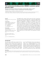
Fig. 1 Promoter methylation analysis of pediatric AML with NimbleGen Human DNA Methylation Arrays. a Analysis of the methylation status of
genes in four pediatric AML samples (M1, M2, M3, M4 and M5) and three NBM samples (N1, N2, and N3) using NimbleGen Human DNA
Methylation Arrays. Each red box represents the number of methylation peaks (PeakScore) overlapping the promoter region for the corresponding
miRNA. The PeakScore is defined as the average -log10 (P-value) from probes within the peak. The scores reflect the probability of positive
methylation enrichment. b DNA methylation array analysis showing significant methylation of the GATA4 promoter in AML samples (4/5), and
unmethylated in NBM samples (0/3)
Madison, WI, USA). Microarrays were hybridized at 42 °
C during 16 to 20 h with Cy3/5 labelled DNA in
Nimblegen hybridization buffer/ hybridization component A in a hybridization chamber (Hybridization
System - Nimblegen Systems, Inc., Madison, WI,
USA). For array hybridization, Roche NimbleGen's
Promoter plus CpG Island array was used, which is a
385 k format array design containing 28,226 CpG
Islands and all well-characterized Promoter regions
(from about -800 bp to +200 bp of the TSSs) totally
covered by ~385,000 probes. This NimbleGen Human
DNA Methylation array analysis was performed by
KangChen Bio-tech, Shanghai P.R. China.
Sodium bisulphite modification of genomic DNA
High-molecular-weight genomic DNA was extracted
from cell lines and biopsies by a conventional phenol/
chloroform method. The sodium bisulphite modification
procedure was as described with slight modification
[26–28]. In brief, 600 ng of genomic DNA was denatured
in 3 M NaOH for 15 min at 37 °C, then mixed with 2 volumes of 2 % low-melting-point agarose. Agarose/DNA
mixtures were then pipetted into chilled mineral oil to
form agarose beads. Aliquots of 200 μl of 5 M bisulphite
solution (2.5 M sodium metabisulphite, 100 mM hydroquinone, both Sigma, USA) were added into each tube
containing a single bead. The bisulphite reaction was then
carried out by incubating the reaction mixture for 4 h at
50 °C in the dark. Treatments were stopped by equilibration against 1 ml of TE buffer, followed by desulphonation
in 500 μl of 0.2 M NaOH. Finally, the beads were washed
with 1 ml of TE buffer and directly used for PCR.
Methylation-specific PCR
The methylation status of the GATA4 (NCBI Reference
Sequence of GATA4 : NG_008177.2) promoter region
was determined by methylation-specific PCR. Primers
were designed with Methprimer design tool (http://
www.urogene.org/methprimer/). Primers distinguishing
unmethylated (U) and methylated (M) alleles were designed to amplify the sequence: GATA4 B M-forward: 5TTTTTTAATTTTTGTTTGTATATCGT-3; GATA4 B
M-reverse: 5- ACTACCTAACACTACCACCCTACGT3; GATA4 B U-forward: 5- TTTTTTAATTTTTGTTTG
TATATTGT-3; GATA4 B U-reverse: 5- CTACCTAAC
ACTACCACCCTACATC-3.
Each PCR reaction contained 20 ng of sodium
bisulphite-modified DNA, 250 pmol of each primer, 250
pmol deoxynucleoside triphosphate, 1 × PCR buffer, and
one unit of ExTaq HS polymerase (Takara, Tokyo) in a
final reaction volume of 20 μl. Cycling conditions were
initial denaturation at 95 °C for 3 min, 40 cycles of 94 °C
for 30 s, 65 °C (M) or 63 °C (U) for 30 s, and 72 °C for
30 s. For each set of methylation-specific PCR reactions,
in vitro-methylated genomic DNA treated with sodium
bisulphite served as a positive methylation control. PCR
products were separated on 4 % agarose gels, stained
with ethidium bromide and visualized under UV illumination. For cases with borderline results, PCR analyses were repeated.
Bisulfite genomic sequencing
Bisulfite genomic sequencing (BGS) was performed as
previously described. BGS primers were from +682 to
+904 including 17 CpGs. GATA4 F: 5- GGATTGAATG
TTTTTTTGGAAGTT-3 and GATA4 R: 5- CCTCCTT
TCCTCAACCTAATAACA-3. Amplified BGS products
were TA-cloned; and five to six randomly chosen colonies were sequenced. DNA sequences were analyzed
with QUMA Analyzer. ( />Leukemia cell cells treated with 5-aza-2'-deoxycytidine
De-methylation was induced with 5-aza-dC (5-Aza,
Sigma-Aldrich, St Louis, MO, USA) treatment at a concentration that induced de-methylation of the DNA
without killing the cells. Culture media for HL-60 and
MV4-11 cells contained 5 μM 5-Aza. DNA and RNA
were extracted after 72 h of 5-Aza treatment for the
following analysis.
Quantitative reverse-transcription PCR for GATA4
Quantitative real-time PCR was performed to determine
the expression levels of GATA4 genes. Total RNA was
reverse transcribed using the Reverse Transcription Kit,
according to the manufacturer's protocol (Applied
Biosystems Inc., Foster City, CA). The real time PCR
primers used to quantify GAPDH expression were: F:
5′-AGAAGGCTGGGGCTCATTTG-3′ and R: 5′-AGG
GGCCATCCACAGTCTTC-3′ and for GATA4 were: F:
Tao et al. BMC Cancer (2015) 15:756
Fig. 2 (See legend on next page.)
Page 6 of 13
Tao et al. BMC Cancer (2015) 15:756
Page 7 of 13
(See figure on previous page.)
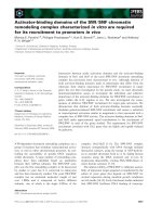
Fig. 2 The GATA4 promoter is methylated in AML cell lines. a Four CpG island regions can be identified in the promoter of GATA4. b MSP
analysis of the methylation status of GATA4 in leukemia cell lines showing hypermethylation in 5/11 cell lines. M and U represent MSP results
using primer sets for methylated and unmethylated GATA4 genes, respectively. c Western blot analysis the expression of GATA4 in 9 NBM
samples and 9 leukemia cell lines. d The GATA4 transcript level is upregulated in cells treated with 5-Aza compared to DMSO: 19.2-fold in HL-60
cells (5-Aza: 19.23 vs. DMSO: 1.00; P = 0.003); 12.5-fold in MV4-11 cells (5-Aza: 29.23 vs. DMSO: 2.33; P = 0.05)
5′- TAGCCCCACAGTTGACACAC-3′ and R: 5′GTCCTGCACAGCCTGCC −3′. Real-time PCR analysis
was according to the MIQE Guidelines and performed
in a total volume of 20 μl including 1 μl of cDNA,
primers (0.2 mM each) and 10 μl of SYBR Green mix
(Roche). Reactions were run on an Lightcycler 480
(Roche) using universal thermal cycling parameters (95 °C
for 5 min, 45 cycles of 10 s at 95 °C, 20 s at 60 °C and 15 s
at 72 °C; followed by a melting curve: 10 s at 95 °C, 60 s at
60 °C and continued melting). The results were obtained
using the sequence detection software of the Lightcycler
480 and analyzed using Microsoft Excel. For quality
control purposes, melting curves were acquired for all
samples. The comparative Ct method was used to quantify
gene expression. The target gene expression level was
normalized to expression of the housekeeping gene glyceraldehyde 3-phosphate dehydrogenase (GAPDH) within
the same sample (−⊿Ct), the relative expression of
GATA4 was calculated with 106 *Log2(-⊿Ct ).
Western blot analysis
Western blot analysis was introduced before [29]. Cellular proteins were extracted in 40 mM Tris–HCl (pH 7.4)
containing 150 mM NaCl and 1 % (v/v) Triton X-100,
supplemented with protease inhibitors. Equal amounts
of protein were resolved on 12 % SDS-PAGE gels, and
then transferred to a PVDF membrane (Millipore,
Bedford, MA). Blots were blocked and then probed with
Polyclonal Goat IgG antibodies against GATA4 (1:1000,
R&D. Minneapolis, MN) and GAPDH (1:5000, Sigma,
St. Louis, MO). After three times’ washing, blots were
then incubated with horseradish peroxidase (HRP) conjugated secondary antibodies and visualized by enhanced
chemiluminescence kit (Pierce, Rockford, IL). Protein
bands were visualized after exposure of the membrane
to Kodak X-ray film.
Statistical analysis
SPSS v11.5 (SPSS Inc., Chicago, IL) was used for statistical analysis. Data are presented as means ± standard
deviation. Group t-test was used to compare the expression of GATA4 between DMSO group and 5-Aza group.
Statistical significance between methylated sample data
and clinical pathological features of AML patients were
analyzed by Pearson chi-square test or Fisher's exact test.
Statistical significance of GATA4 expression among
NBM and pediatric AML groups was determined using
one-way ANOVA. A p <0.05 was considered statistically
significant.
Results and discussion
The GATA4 promoter is hypermethylated in AML cells
The correlation between aberrant methylation and
downregulation of GATA4 has been extensively documented in numerous cancers and cell lines; these are
discussed in the Background. However, the methylation
status of GATA4 in the blood system, particular in
pediatric AML, has not been reported to date. Our analyses of promoter methylation in pediatric AML, using
NimbleGen Human DNA Methylation 385 K Promoter
plus CpG Island arrays, indicated that the GATA4 promoter is hypermethylated in AML (Fig. 1a). The GATA4
promoter was hypermethylated in 80 % (4/5) of pediatric
AML samples and 0 % (0/3) of normal bone marrow
samples (Additional file 1) .
Subsequent analyses of the GATA4 promoter sequence
identified four CpG islands (Fig. 2a). Methylationspecific PCR (MSP) assays were performed to detect the
methylation status of the GATA4 promoter in 11
leukemia cell lines. The MSP primers were designed
using MethPrimer ( />methprimer/methprimer.cgi ) to encompass the CpG
islands of the GATA4 promoter identified in Fig. 2a.
Our results showed that the GATA4 promoter was
hypermethylated in five leukemia cell lines, especially in
SHI-1, HL-60, MV4-11,U937 and K562 cells); and
unmethylated in the other cell lines (Fig. 2b). The results
of RT-PCR analysis of the expression of GATA4 is presented in Fig. 2b; GATA4 expression was detected in
only three cell lines (THP-1, Raji and U937), indicating
that downregulation of GATA4 in AML cells is a common phenomenon. Figure 2c showed that expression of
GATA4 in leukemia cell lines is significantly lower than
NBM. 6/9 NBM samples with obvious expression of
GATA4 and in leukemia cell lines GATA4 only can be
detected in THP-1 and Raji cells. To confirm methylation of the GATA4 promoter, we treated the leukemia
cell lines with the demethylation reagent 5-Aza. Our
results showed that 5-Aza treatment significantly upregulated GATA4 expression. As shown in Fig. 2d, GATA4
expression was upregulated 19.2-fold in HL-60 cells (5Aza: 19.23 vs. DMSO: 1.00; P = 0.003) and 12.5-fold in
MV4-11 cells (5-Aza: 29.23 vs. DMSO: 2.33; P = 0.05).
These results were supported by the MSP analyses,
Tao et al. BMC Cancer (2015) 15:756
Fig. 3 (See legend on next page.)
Page 8 of 13
Tao et al. BMC Cancer (2015) 15:756
Page 9 of 13
(See figure on previous page.)
Fig. 3 GATA4 is inactivated by promoter hypermethylation in pediatric AML. a MSP analysis of the methylation status of GATA4 shows aberrant
methylation in pediatric AML samples compared to NBM/ITP control samples. Aberrant methylation of GATA4 was observed in 15.0 % (3/20) of
the NBM control samples compared to 56.2 % (59/105) of the pediatric AML samples. b Three NBM samples and three AML samples were
analyzed by BSG. The results showed that the CpG islands in the GATA4 promoter were methylated in the AML samples (69.4, 58.8, and 62.4 % in
AML7#, AML10#, and AML11#, respectively). In contrast, the CpG islands of the GATA4 promoter in the NBM samples were unmethylated (24.7,
14.1, and 10.6 % in NBM4#, NBM5#, and NBM9#, respectively). c The transcript levels of GATA4 were examined in 105 pediatric AML
patients by real-time PCR. d GATA4 expression was significantly decreased in 105 AML patients (33.06 ± 70.94; P =0.011) compared to 20
NBM/ITP controls (116.76 ± 105.39); AML patients with GATA4 promoter methylation (16.02 ± 17.59, n = 59) showed lower GATA4 transcript
levels compared to those without GATA4 promoter methylation (54.92 ± 101.80, P <0.001; n = 46)
which also showed a change in the methylation status of
the GATA4 promoter after 5-Aza treatment. In summary, these results showed that the GATA4 promoter
was consistently and significantly methylated in the HL60, MV4-11, SHI-1, U937 and K562 human myeloid
leukemia cell lines. Based on these findings, we hypothesized that the promoter of GATA4 is methylated in
pediatric AML patients.
The GATA4 promoter is methylated in pediatric AML
patients
We next examined the GATA4 promoter methylation
status in pediatric AML samples and NBM/ITP (normal
bone marrow/idiopathicthrombocytopenic purpura)
control samples. Aberrant GATA4 promoter methylation was observed in 15.0 % (3/20) of the NBM control samples compared to 56.2 % (59/105) of the
pediatric AML samples (Fig. 3a). Three NBM samples
and three AML samples were further analyzed by
BSG (Fig. 3b). The results showed that the CpG
islands in the GATA4 promoter were methylated in
the AML samples (69.4, 58.8, and 62.4 % in AML7#,
AML10#, and AML11#, respectively). In contrast, the
CpG islands of the GATA4 promoter in the NBM
samples were unmethylated (24.7, 14.1, and 10.6 % in
NBM4#, NBM5#, and NBM9#, respectively). These
results were supported by MSP assays.
GATA4 transcript levels compared to those in
controls.
The prognostic significance of GATA4 expression
was assessed in 105 cases of Chinese pediatric acute
myeloid leukemia patients with clinical follow-up
records. There was no significant association with
GATA4 expression and patient age, sex, FAB
(French–American–British classification) or cytogenetics (Table 1). Kaplan-Meier survival analysis of 105
pediatric acute myeloid leukemia patients revealed almost identical survival times for patients with GATA4
high or low expressing tumors (P = 0.769, Table 3 and
Fig. 4b). Furthermore, multivariate analysis revealed
that GATA4 expression was not an independent prognostic factor in pediatric AML (P = 0.096, Table 4).
Table 1 Association of GATA4 expression with clinico-pathological
characteristics in 105 pediatric AML samples
Clinical
variables
No. of
patients
GATA4 expression (n)
Low
High
Male
42
24
18
Female
63
29
34
<6
60
32
28
≥6
45
21
24
P
Sex
0.265
Age (years)
0.499
Leukocyte (/μl)
Expression of GATA4 is downregulated with promoter
methylation in Chinese pediatric acute myeloid leukemia
The transcript levels of GATA4 were examined in
105 pediatric AML patients by real-time PCR
(Fig. 3c). As shown in Fig. 3d, GATA4 expression
was significantly decreased in 105 AML patients
(33.06 ± 70.94; P = 0.011) compared to that in 20
NBM/ITP controls (116.76 ± 105.39). Figure 3d shows
that patients with GATA4 promoter methylation
(16.02 ± 17.59, n = 59) exhibited lower GATA4 transcript levels compared to those without GATA4 promoter methylation (54.92 ± 101.80, P < 0.01; n = 46).
Furthermore, AML patients with and without GATA4
promoter methylation showed significantly lower
> 10,000
61
32
29
≤ 10,000
44
21
23
M1–M6
93
49
44
M7
12
4
8
Favorable
50
20
30
Intermediate
27
18
9
Unfavorable
28
15
13
< 0.25 %
49
23
26
≥ 0.25 %
56
30
26
0.632
FAB
0.207
Cytogenetics
0.077
MRD
0.498
Tao et al. BMC Cancer (2015) 15:756
Page 10 of 13
Fig. 4 GATA4 promoter methylation correlates with poor survival in Chinese pediatric acute myeloid leukemia. a Kaplan-Meier survival analysis in
pediatric AML samples with GATA4 promoter methylation status (P = 0.014). b Kaplan-Meier survival analysis in pediatric AML samples with GATA4
expression (P = 0.769)
GATA4 promoter methylation correlates with poor
survival in Chinese pediatric acute myeloid leukemia
The prognostic significance of GATA4 promoter methylation was also assessed in 105 cases of Chinese pediatric
acute myeloid leukemia patients with clinical follow-up
records. Table 2 shows GATA4 promoter methylation
was correlated with leukocyte counts (P = 0.035) and
MRD (P = 0.031). Table 2 also shows there were no significant differences in clinical features, such as sex, age,
FAB or cytogenetics between patients with and without
GATA4 promoter methylation. Kaplan-Meier survival
analysis revealed significantly shorter overall survival
times in patients with GATA4 promoter methylation
(P = 0.014, Table 3 and Fig. 4a). Furthermore, multivariate analysis revealed that GATA4 promoter methylation
was not an independent prognostic factor in pediatric
AML (P = 0.170, Table 4).
In summary, our results showed firstly that the
GATA4 promoter was consistently significantly methylated in leukemia cells, such as HL-60, MV4-11, SHI-1,
U937, and K562 human myeloid leukemia cell lines; the
expression of GATA4 was significantly lower in pediatric
AML compared to NBM control samples, patients with
methylated GATA4 showed lower GATA4 transcript
levels compared to those without methylated; GATA4
promoter methylation was correlated with leukocyte and
MRD, Kaplan-Meier survival analysis revealed a significantly shorter overall survival times in pediatric AML
with GATA4 promoter methylation.
In this study, promoter methylation in Chinese
pediatric AML was analyzed using NimbleGen Human
DNA Methylation 385 K Promoter plus CpG Island
arrays. This approach revealed significant differences in
the methylation status of genes between pediatric AML
and normal bone marrow samples. Previous studies have
demonstrated that promoters of TFPI-2 [30] and miR-
663 [31, 32] were hypermethylated in Chinese pediatric
acute myeloid leukemia. Our results showed significantly
greater GATA4 promoter hypermethylation in pediatric
AML samples and 0/3 (0 %) in normal bone marrow
samples. indicating that the GATA4 promoter is hypermethylated in AML.
GATA4 was suggested to be a tumor suppressor gene
with promoter hypemethylation in various types of
Table 2 Association of GATA4 promoter methylation with
clinico-pathological characteristics in 105 pediatric AML samples
Clinical
variables
No. of
patients
GATA4 methylation (n)
Negative
Positive
Male
42
21
21
Female
63
25
38
<6
60
28
32
≥6
45
18
27
P
Sex
0.297
Age (years)
0.496
Leukocyte (/μl)
> 10,000
61
32
29
≤ 10,000
44
14
30
M1–M6
93
38
55
M7
12
8
4
Favorable
50
16
34
Intermediate
27
14
13
Unfavorable
28
16
12
0.035
FAB
0.090
Cytogenetics
0.062
MRD
< 0.25 %
49
16
33
≥ 0.25 %
56
30
26
0.031
Tao et al. BMC Cancer (2015) 15:756
Page 11 of 13
Table 3 Association of GATA4 expression/promoter methylation
with Kaplan-Meier survival in 105 pediatric AML samples
Variable
No. of
patients
Over survival
Favorable
50
46.664 ± 3.717
Intermediate
27
29.220 ± 3.188
Unfavorable
28
11.161 ± 1.827
P
Median ± SE
Cytogenetics
<0.001
FAB
M1–M6
93
36.113 ± 2.885
M7
12
8.542 ± 1.820
> 10,000
61
30.220 ± 2.974
≤ 10,000
44
33.631 ± 4.063
<0.001
Leukocyte (/μl)
0.803
MRD
< 0.25 %
49
53.627 ± 3.151
≥ 0.25 %
56
18.893 ± 2.425
Low <12.420
53
32.130 ± 3.385
High ≥12.420
52
34.765 ± 3.941
<0.001
GATA4 expression
0.769
GATA4 methylation
Negative
46
39.141 ± 3.554
Positive
59
24.264 ± 3.671
0.014
human cancers. The GATA4 promoter is methylated in
glioblastoma [17], endometrioid carcinoma [18], ovarian
cancer [19], gastric mucosa [21], colorectal carcinomas
[22] and lung cancers [24]. To our knowledge, this is the
first report describing the expression of GATA4 and
promoter methylation status in pediatric AML. In this
study, methylation-specific PCR (MSP) assays showed
that the GATA4 promoter was hypermethylated in five
Table 4 Cox multivariate analysis of GATA4 expression/promoter
methylation and clinico-pathological features in pediatric AML
Variable
Odds ratio
EXP(B) 95 % CI
P
5.894
2.412 (1.185–4.909)
0.015
16.241
5.986 (2.503–14.229)
0.000
0.485
1.225 (0.691–2.172)
0.486
6.645
2.630 (1.261–5.484)
0.010
2.765
1.657 (0.914–3.007)
0.096
1.885
0.661 (0.367–1.193)
0.170
Cytogenetics
Favo vs. Inter and Unfavo
MRD
< 0.25 % vs. ≥0.25 %
Leukocyte (/μl)
> 10,000 vs. ≤10,000
FAB classification
M7 vs. M1–M6
GATA4 Expression
Low vs. High
GATA4 Methylation
Negative vs. Positive
leukemia cell lines, especially in SHI-1, HL-60, MV4-11,
U937 and K562 cells). 5-Aza treatment significantly
upregulated GATA4 expression in HL-60 and MV4-11
cells. Aberrant GATA4 promoter methylation was
observed 15.0 % (3/20) of the NBM control samples
compared to 56.2 % (59/105) of the pediatric AML
samples. BGS analysis also showed that CpG islands in
the GATA4 promoter were methylated in the AML samples and NBM samples were unmethylated. Analysis of
GATA4 transcript levels showed that GATA4 expression
was significantly decreased in AML patients compared
to 20 NBM/ITP control and patients with methylated
GATA4 showed lower GATA4 transcript levels compared to those without methylated GATA4. Taken together, our results show hypermethylation of the GATA4
promoter in Chinese pediatric AML for the first time.
GATA4 promoter hypermethylation is an important
prognostic marker in several tumors. Kaplan-Meier
analysis revealed that high methylation levels of the
GATA4 promoter were significantly correlated with
patient survival in oropharyngeal squamous cell carcinoma (OPSCC) [33]. In high grade serous ovarian
carcinoma (HGSOC), GATA4 promoter methylation
was associated with disease recurrence [34]. In this
study, the prognostic significance of GATA4 promoter
methylation was assessed in 105 cases of Chinese
pediatric AML patients with clinical follow-up
records. GATA4 promoter methylation was correlated
with leukocyte counts and MRD. Kaplan-Meier survival
analysis revealed significantly shorter overall survival in
patients with GATA4 promoter methylation. These observations demonstrate that GATA4 promoter methylation
correlates with poorer survival in Chinese pediatric AML.
The molecular function of GATA4 has been studied in certain tumors. Re-expression of GATA4 in
human glioblastoma multiforme (GBM) cell lines,
primary cultures, and brain tumor-initiating cells
suppressed tumor growth in vitro and in vivo
through direct activation of the cell cycle inhibitor
P21 (CIP1). Re-expression of GATA4 also conferred sensitivity of GBM cells to temozolomide, a DNA alkylating
agent currently used in GBM therapy. GATA4 reduced expression of APNG (alkylpurine-DNA-N-glycosylase), a
DNA repair enzyme which is poorly characterized in
GBM-mediated temozolomide resistance [23]. The potential function of GATA4 as a tumor suppressor was studied
by inducing GATA4-overexpression in human colorectal
cancer cell lines.GATA4 overexpression suppressed
colony formation, proliferation, migration, invasion,
and anchorage-independent growth of colorectal cancer cells [22].GATA4 can control expression of the
anti-apoptotic factor Bcl-2 and the cell cycle regulator
cyclin D2 in normal and neoplastic granulosa cells.GATA4 expression correlated with Bcl-2 and cyclin
Tao et al. BMC Cancer (2015) 15:756
Page 12 of 13
D2 expression in human and murine granulosa cell tumors (GCT). Moreover,GATA4 enhanced Bcl-2 and cyclin
D2 promoter activity in murine GCT cells [35]. To date,
the molecular function of GATA4 in pediatric AML is still
unknown and further investigations are required to elucidate the role of GATA4 in pediatric leukemia.
Biology, Cancer Institute (Hospital), Chinese Academy of Medical Sciences,
Peking Union Medical College, Beijing, China. 4Translational Research Center,
Second Hospital, The Second Clinical School, Nanjing Medical University,
Nanjing, China.
Conclusions
Epigenetic inactivation of GATA4 by promoter hypermethylation was observed in both AML cell lines and
pediatric AML samples. Our study implicates GATA4 as
a putative tumor suppressor gene in pediatric AML. In
addition, our findings indicate that GATA4 promoter
methylation correlates with leukocyte counts, MRD and
significantly shorter overall survival in pediatric AML.
Kaplan-Meier survival analysis revealed significantly
shorter overall survival in pediatric AML with GATA4
promoter methylation but multivariate analysis shows
that it is not an independent factor. However, further research focusing on the molecular mechanism underlying
the role of GATA4 in pediatric leukemia is required.
References
1. Estey E, Dohner H. Acute myeloid leukaemia. Lancet. 2006;368(9550):1894–907.
2. Plass C, Oakes C, Blum W, Marcucci G. Epigenetics in acute myeloid
leukemia. Semin Oncol. 2008;35(4):378–87.
3. Issa JP. CpG island methylator phenotype in cancer. Nat Rev Cancer.
2004;4(12):988–93.
4. Figueroa ME, Lugthart S, Li Y, Erpelinck-Verschueren C, Deng X, Christos PJ,
et al. DNA methylation signatures identify biologically distinct subtypes in
acute myeloid leukemia. Cancer Cell. 2010;17(1):13–27.
5. Bullinger L, Ehrich M, Dohner K, Schlenk RF, Dohner H, Nelson MR, et al.
Quantitative DNA methylation predicts survival in adult acute myeloid
leukemia. Blood. 2010;115(3):636–42.
6. Deneberg S, Grovdal M, Karimi M, Jansson M, Nahi H, Corbacioglu A, et al.
Gene-specific and global methylation patterns predict outcome in patients
with acute myeloid leukemia. Leukemia. 2010;24(5):932–41.
7. Jiang Y, Dunbar A, Gondek LP, Mohan S, Rataul M, O'Keefe C, et al. Aberrant
DNA methylation is a dominant mechanism in MDS progression to AML.
Blood. 2009;113(6):1315–25.
8. Holliday R, Pugh JE. DNA modification mechanisms and gene activity
during development. Science. 1975;187(4173):226–32.
9. Smith ZD, Meissner A. DNA methylation: roles in mammalian development.
Nat Rev Genet. 2013;14(3):204–20.
10. Jones PA. Functions of DNA methylation: islands, start sites, gene bodies
and beyond. Nat Rev Genet. 2012;13(7):484–92.
11. Schoofs T, Muller-Tidow C. DNA methylation as a pathogenic event and as
a therapeutic target in AML. Cancer Treat Rev. 2011;37 Suppl 1:S13–18.
12. White RA, Dowler LL, Pasztor LM, Gatson LL, Adkison LR, Angeloni SV, et al.
Assignment of the transcription factor GATA4 gene to human chromosome
8 and mouse chromosome 14: Gata4 is a candidate gene for Ds
(disorganization). Genomics. 1995;27(1):20–6.
13. Huang WY, Heng HH, Liew CC. Assignment of the human GATA4 gene to
8p23.1–>p22 using fluorescence in situ hybridization analysis. Cytogenet
Cell Genet. 1996;72(2–3):217–8.
14. Perrino C, Rockman HA. GATA4 and the two sides of gene expression
reprogramming. Circ Res. 2006;98(6):715–6.
15. McCulley DJ, Black BL. Transcription factor pathways and congenital heart
disease. Curr Top Dev Biol. 2012;100:253–77.
16. Suzuki YJ. Cell signaling pathways for the regulation of GATA4 transcription
factor: Implications for cell growth and apoptosis. Cell Signal.
2011;23(7):1094–9.
17. Vaitkiene P, Skiriute D, Skauminas K, Tamasauskas A. GATA4 and DcR1
methylation in glioblastomas. Diagn Pathol. 2013;8:7.
18. Chmelarova M, Kos S, Dvorakova E, Spacek J, Laco J, Ruszova E, et al.
Importance of promoter methylation of GATA4 and TP53 genes in
endometrioid carcinoma of endometrium. Clin Chem Lab Med. 2014.
19. Chmelarova M, Dvorakova E, Spacek J, Laco J, Palicka V. Importance of
promoter methylation of GATA4 gene in epithelial ovarian cancer. Biomed
Pap Med Fac Univ Palacky Olomouc Czech Repub. 2013;157(4):294–7.
20. Chmelarova M, Dvorakova E, Spacek J, Laco J, Mzik M, Palicka V. Promoter
methylation of GATA4, WIF1, NTRK1 and other selected tumour suppressor
genes in ovarian cancer. Folia Biol. 2013;59(2):87–92.
21. Wen XZ, Akiyama Y, Pan KF, Liu ZJ, Lu ZM, Zhou J, et al. Methylation of
GATA-4 and GATA-5 and development of sporadic gastric carcinomas.
World J Gastroenterol. 2010;16(10):1201–8.
22. Hellebrekers DM, Lentjes MH, van den Bosch SM, Melotte V, Wouters KA,
Daenen KL, et al. GATA4 and GATA5 are potential tumor suppressors
and biomarkers in colorectal cancer. Clin Cancer Res.
2009;15(12):3990–7.
23. Agnihotri S, Wolf A, Munoz DM, Smith CJ, Gajadhar A, Restrepo A, et al. A
GATA4-regulated tumor suppressor network represses formation of
malignant human astrocytomas. J Exp Med. 2011;208(4):689–702.
Additional file
Additional file 1: Analysis of promoter methylation in pediatric
AML using NimbleGen Human DNA Methylation 385 K Promoter
Plus CpG Island Arrays. (JPEG 777 kb)
Abbreviations
GATA4: GATA binding protein 4; AML: Acute myeloid leukemia;
MSP: Methylation specific PCR; BGS: Bisulfite genomic sequencing;
NBM: Normal bone marrow; ITP: Idiopathic thrombocytopenic purpura.
Competing interests
The authors have no conflicts of interest to disclose.
Authors’ contributions
PJ designed and directed the study. TYF and HSY finished the most of
experiments. ZWL and CL collected the leukemia sample. XPF and LJ
collected the clinical information of samples. DXJ and SLC supported the
design of primer for BGS and MSP analysis. LZH, WNN, FF, LG and LYH
drafted this manuscript. LYP, XYY, WJ, FX and NJ participated in study design
and coordination, data analysis and interpretation and drafted the
manuscript. All authors read and approved the final manuscript.
Acknowledgements
This work was supported by grants from the National Key Basic Research
Program No. 2010CB933902, grants from key medical subjects of Jiangsu
province (XK201120), Innovative team of Jiangsu Province ( LJ201114,
LJ201126 ), Special clinical medical science and technology of Jiangsu
province (BL2012050, BL2013014), Key Laboratory of Suzhou (SZS201108,
SZS201307) , National Natural Science Foundation
(81100371,81370627,81300423,81272143,81170513). Natural Science
Foundation of Jiangsu Province No. BK2011308, Universities Natural Science
Foundation of Jiangsu Province No. 11KJB320014 and Talent’s subsidy
project in science and education of department of public health of Suzhou
City No. SWKQ1020. Major scientific and technological special project for
"significant new drugs creation" No. 2012ZX09103301-040.
Author details
1
Department of Hematology and Oncology, Childrens Hospital of Soochow
University, Suzhou, China. 2Department of Gastroenterology, the 5th Hospital
of Chinese PLA, Yin chuan, China. 3Department of Cell and Molecular
Received: 1 May 2014 Accepted: 9 October 2015
Tao et al. BMC Cancer (2015) 15:756
Page 13 of 13
24. Guo M, Akiyama Y, House MG, Hooker CM, Heath E, Gabrielson E, et al.
Hypermethylation of the GATA genes in lung cancer. Clin Cancer Res.
2004;10(23):7917–24.
25. Pike BL, Greiner TC, Wang X, Weisenburger DD, Hsu YH, Renaud G, et al.
DNA methylation profiles in diffuse large B-cell lymphoma and their
relationship to gene expression status. Leukemia. 2008;22(5):1035–43.
26. Olek A, Oswald J, Walter J. A modified and improved method for bisulphite
based cytosine methylation analysis. Nucleic Acids Res. 1996;24(24):5064–6.
27. Tao YF, Hu SY, Lu J, Cao L, Zhao WL, Xiao PF, et al. Zinc finger protein 382 is
downregulated by promoter hypermethylation in pediatric acute myeloid
leukemia patients. Int J Mol Med. 2014;34(6):1505–15.
28. Tao YF, Xu LX, Lu J, Cao L, Li ZH, Hu SY, et al. Metallothionein III (MT3) is a
putative tumor suppressor gene that is frequently inactivated in pediatric
acute myeloid leukemia by promoter hypermethylation. J Transl Med.
2014;12:182.
29. Tao YF, Lu J, Du XJ, Sun LC, Zhao X, Peng L, et al. Survivin selective inhibitor
YM155 induce apoptosis in SK-NEP-1 Wilms tumor cells. BMC Cancer.
2012;12:619.
30. Jian P, Yan WS, Chao SL, Liang P, Zhen L, Ling QB, et al. Promoter of TFPI-2
is hypermethylated in Chinese pediatric acute myeloid leukemia. J Pediatr
Hematol Oncol. 2012;34(1):43–6.
31. Yan-Fang T, Jian N, Jun L, Na W, Pei-Fang X, Wen-Li Z, et al. The promoter
of miR-663 is hypermethylated in Chinese pediatric acute myeloid leukemia
(AML). BMC Med Genet. 2013;14:74.
32. Jian P, Li ZW, Fang TY, Jian W, Zhuan Z, Mei LX, et al. Retinoic acid induces
HL-60 cell differentiation via the upregulation of miR-663. J Hematol
Oncol. 2011;4:20.
33. Kostareli E, Holzinger D, Bogatyrova O, Hielscher T, Wichmann G, Keck M, et
al. HPV-related methylation signature predicts survival in oropharyngeal
squamous cell carcinomas. J Clin Invest. 2013;123(6):2488–501.
34. Montavon C, Gloss BS, Warton K, Barton CA, Statham AL, Scurry JP, et al.
Prognostic and diagnostic significance of DNA methylation patterns in high
grade serous ovarian cancer. Gynecol Oncol. 2012;124(3):582–8.
35. Kyronlahti A, Ramo M, Tamminen M, Unkila-Kallio L, Butzow R, Leminen A,
et al. GATA-4 regulates Bcl-2 expression in ovarian granulosa cell tumors.
Endocrinology. 2008;149(11):5635–42.
Submit your next manuscript to BioMed Central
and take full advantage of:
• Convenient online submission
• Thorough peer review
• No space constraints or color figure charges
• Immediate publication on acceptance
• Inclusion in PubMed, CAS, Scopus and Google Scholar
• Research which is freely available for redistribution
Submit your manuscript at
www.biomedcentral.com/submit