TNF-alpha promotes lymphangiogenesis and lymphatic metastasis of gallbladder cancer through the ERK1/2/AP-1/VEGF-D pathway
Bạn đang xem bản rút gọn của tài liệu. Xem và tải ngay bản đầy đủ của tài liệu tại đây (2.92 MB, 14 trang )
Hong et al. BMC Cancer (2016) 16:240
DOI 10.1186/s12885-016-2259-4
RESEARCH ARTICLE
Open Access
TNF-alpha promotes lymphangiogenesis
and lymphatic metastasis of gallbladder
cancer through the ERK1/2/AP-1/VEGF-D
pathway
HaiJie Hong1,2†, Lei Jiang1,2†, YanFei Lin1,2, CaiLong He1,2, GuangWei Zhu1,2, Qiang Du1,2, XiaoQian Wang1,
FeiFei She2,3* and YanLing Chen1,2*
Abstract
Background: Tumor necrosis factor-alpha (TNF-α), a key player in cancer-related inflammation, was recently
demonstrated to be involved in the lymphatic metastasis of gallbladder cancer (GBC). Vascular endothelial growth
factor D (VEGF-D) is a key lymphangiogenic factor that is associated with lymphangiogenesis and lymph node
metastasis in GBC. However, whether VEGF-D is involved in TNF-α-induced lymphatic metastasis of GBC remains
undetermined.
Methods: The expression of VEGF-D in patient specimens was detected by immunohistochemistry and the
relationship between VEGF-D in the tissue and TNF-α in the bile of the matching patients was analyzed. The
VEGF-D mRNA and protein levels after treatment with exogenous TNF-α in NOZ, GBC-SD and SGC-996 cell lines
were measured by real-time PCR and ELISA. The promoter activity and transcriptional regulation of VEGF-D were
analyzed with the relative luciferase reporter assay, mutant constructs, electrophoretic mobility shift assay (EMSA),
chromatin immunoprecipitation (ChIP) assay, RNA interference and Western blotting. Inhibitors of JNK, p38 MAPK
and ERK1/2 were used to explore the upstream signaling effector of AP-1. We used lentiviral vector expressing a
VEGF-D shRNA construct to knockdown VEGF-D gene in NOZ and GBC-SD cells. The role of the TNF-α-VEGF-D axis
in the tube formation of human dermal lymphatic endothelial cells (HDLECs) was determined using a threedimensional coculture system. The role of the TNF-α - VEGF-D axis in lymphangiogenesis and lymph node
metastasis was studied via animal experiment.
Results: TNF-α levels in the bile of GBC patients were positively correlated with VEGF-D expression in the clinical
specimens. TNF-α can upregulate the protein expression and promoter activity of VEGF-D through the ERK1/2 - AP1 pathway. Moreover, TNF-α can promote tube formation of HDLECs, lymphangiogenesis and lymph node
metastasis of GBC by upregulation of VEGF-D in vitro and in vivo.
Conclusion: Taken together, our data suggest that TNF-α can promote lymphangiogenesis and lymphatic
metastasis of GBC through the ERK1/2/AP-1/VEGF-D pathway.
Keyword: Gallbladder cancer, TNF-α, VEGF-D, Lymphatic metastasis
* Correspondence: ;
†
Equal contributors
2
Key Laboratory of Ministry of Education for Gastrointestinal Cancer, Fujian
Medical University, 1 Xueyuan Road, Minhou, Fuzhou 350108, China
1
Department of Hepatobiliary Surgery and Fujian Institute of Hepatobiliary
Surgery, Fujian Medical University Union Hospital, 29 Xinquan Road, Fuzhou
350001, China
Full list of author information is available at the end of the article
© 2016 Hong et al. Open Access This article is distributed under the terms of the Creative Commons Attribution 4.0
International License ( which permits unrestricted use, distribution, and
reproduction in any medium, provided you give appropriate credit to the original author(s) and the source, provide a link to
the Creative Commons license, and indicate if changes were made. The Creative Commons Public Domain Dedication waiver
( applies to the data made available in this article, unless otherwise stated.
Hong et al. BMC Cancer (2016) 16:240
Background
Gallbladder cancer (GBC) is rare but represents the
most common cancer of the biliary tract, accounting for
80–95 % of biliary tract malignancies [1, 2]. GBC is a
highly aggressive disease with very poor prognosis (5year survival rate < 5 % [3, 4]), due to its tendency to
metastasize to the lymph nodes in early stages. More
than 50 % of all patients with GBC exhibit lymph node
metastases (LNM) [5]. Therefore, understanding the
mechanism underlying lymphatic metastasis in GBC is
helpful to improve patient treatment and prognosis.
However, the specific mechanisms underlying lymphatic
metastasis in GBC are largely unknown.
In 1863, Virchow first observed that inflammatory cells
can be found in tumors [6]. Since then, many studies
have examined the relationship between inflammation
and cancer. It has been generally accepted that chronic
inflammation promotes cancer [7], including some cancers of the liver [8], intestine [9, 10] and lung [11]. Cytokines secreted by inflammatory cells, including TNF-α,
IL-1, and IL-6, play important roles in cancer-related inflammation [7, 12–15]. Tumor necrosis factor alpha
(TNF-α), a key pro-inflammatory cytokine that was first
identified as a mediator of tumor cell death, is now also
known to promote the tumor progression, proliferation,
epidermal-mesenchymal transition (EMT), angiogenesis,
invasion and metastasis [16–19]. Lymphatic metastasis is
one of the major forms of tumor metastasis. However,
the relationship between TNF-α and lymphatic metastasis requires further research.
Recently, we confirmed that TNF-α can promote lymphangiogenesis and lymph node metastasis of GBC
through upregulation of vascular endothelial growth factor C (VEGF-C) downstream of NF-κB [20]. Furthermore, we determined that vascular endothelial growth
factor D (VEGF-D), another key lymphangiogenic factor
similar to VEGF-C, is associated with lymphangiogenesis
and lymph node metastasis of GBC [21]. Thus, we aimed
to further explore whether VEGF-D is involved in TNFα-induced lymphatic metastasis of GBC and the underlying mechanisms.
In this study, we first analyzed the relationship between TNF-α levels and VEGF-D expression in clinical specimens and demonstrated that TNF-α can
upregulate VEGF-D expression in the NOZ and GBCSD cell lines. Previous studies have demonstrated that
TNF-α promotes the expression of target genes
mainly through NF-κB and (or) AP-1 signaling pathways [22]. We further sought to determine whether
TNF-α upregulates VEGF-D expression and enhances
its promoter activity through these two pathways.
Furthermore, we determined that TNF-α can promote
tube formation of human dermal lymphatic endothelial cells (HDLECs), lymphangiogenesis and lymph
Page 2 of 14
node metastasis of GBC by upregulation of VEGF-D
in vitro and in vivo.
Methods
Patient samples and cell culture
20 GBC tissues and the matching bile used in present
study were obtained from the patients admitted to Fujian Medical University Union Hospital in China. The
informed consents of agreement to use the samples for
further study were signed pre-operation. The samples
were collected according to the protocol approved by
the Ethics Committee of the Medical Faculty of Fujian
Medical University, according to the Declaration of
Helsinki. The details of the patients including the age
and sex of the patient, clinical stage, grade of the tumor
and lymph node metastasis (LNM) had been described
in [20]. The human GBC cell lines: NOZ (obtained from
Health Science Research Resources Bank in Japan),
GBC-SD (purchased from Shanghai Institutes for BiologicalSciences in China) and SGC-996 (provided by the
Tumor Cytology Research Unit, Medical College, Tongji
University, China) were maintained in Dulbecco’s Modified Eagle’s Medium (Gibco, USA) supplemented with
10 % fetal bovine serum (Gibco). Human dermal lymphatic endothelial cells (HDLECs, purchased from Sciencell, San Diego, California, USA) were incubated in
endothelial cell medium (Sciencell). All of the cells were
incubated at 37 °C under 95 % air and 5 % CO2.
Immunohistochemistry
The VEGF-D expression and lymphatic vessels of GBC
specimens were detected by immunohistochemistry as
previously described [21]. The primary antibodies were
VEGF-D (ab155288, Abcam) at a 1:80 dilution and
LYVE-1 (AF2125, R&D Systems) at a 1:150 dilution. The
method used to measure the VEGF-D expression has
been described previously [23]. The density of LYVE-1positive vessels (lymphatic vessels density, LVD) was
assessed according to the method described by Qiang
Du [24].
Quantitative real-time polymerase chain reaction (qRTPCR)
Total RNA was extracted from GBC cells with TRIzol
reagent (Invitrogen). RNA was reverse transcribed using
the RevertAid First Strand cDNA Synthesis Kit
(Thermo). PCR reactions were performed with Fast Start
Universal SYBR Green Master Mix (Roche), and fluorescence was measured using the 7500 quantitative realtime thermocycler (Applied Biosystems). GAPDH served
as an internal control. All procedures followed the manufacturer’s instructions.
Hong et al. BMC Cancer (2016) 16:240
Enzyme-linked immunosorbent assay (ELISA)
VEGF-D levels in cell culture media were measured by
double antibody sandwich enzyme-linked immunosorbent assay using Quantikine ELISA Kits from R&D Systems following the manufacturer’s instructions. VEGF-D
Standards for drawing standard curve were prepared before the antibody reaction. 100 μL of Assay Diluent
RD1X was added to each well, and then 50 μL of Standard, sample or control were added to each well and incubated for 2 h at room temperature. Wash each well
with wash buffer (400 μL) for four times. Add 200 μL of
VEGF-D Conjugate to each well and incubate for 2 h at
room temperature. Wash each well again and add
200 μL of Substrate Solution to each well. Add 50 μL of
Stop Solution to each well after incubation for 30 min
(protect from light). The wells were read at 450 nm with
a Model 550 Microplate Reader (Bio-Rad, Hercules, CA,
USA). Each reaction was run in triplicate.
Construction of VEGF-D promoter luciferase reporter plasmids and dual-luciferase reporter assay
A series of 5′-deletion DNA fragments of the VEGF-D
gene promoter were amplified by PCR with primers containing an XhoI or BglII (Thermo) restriction site, which
were connected to the pGL4.10-Basic vector (Promega)
carrying a firefly luciferase report gene. These recombinant VEGF-D promoter luciferase reporter plasmids were
named PGL4-2148 (−2148 to +117, relative to the transcription start site “ATG”), PGL4-1621 (−1621 to +117),
PGL4-988 (−988 to +117), PGL4-717 (−717 to +117),
PGL4-444 (−444 to +117), PGL4-325 (−325 to +117),
PGL4-154 (−154 to +117), and PGL4-57 (−57 to +117).
Forty-eight hours after transfection with promoter vector, cells were lysed and the intracellular luciferase activity of the lysates was measured using the DualLuciferase Reporter Assay System (Promega) according
to the manufacturer’s instructions. The relative luciferase
units were obtained by comparison with the luciferase
activity of the pRL-TK plasmid (plasmid carrying a
renilla luciferase report gene as an internal reference).
Identification of putative transcription factor binding sites
The websites TFbind ( and Promoter
Scan ( were
used to search for potential transcription factor binding site
motifs.
Site-directed mutagenesis
The site-directed mutagenesis was performed by overlap extension PCR as previously described [20, 25]. The primers
targeting the two mutation sites of the AP-1 binding sites
were as follows: AP-1mut1 (−401 to -393 nt), (forward),
5′-CATCTGCTGCCAATGCTACACAGAAAGCAATC-3′
(reverse); AP-1mut2 (−345 to -337 nt), 5′-CTTAAGCAA
Page 3 of 14
TCCCACCGAGATACAAAGGTC-3′ (forward), 5′-GACC
TTTGTATCTCGGTGGGATTGCTTAAG-3′ (reverse).
Nuclear extraction and electrophoretic mobility shift
assay (EMSA)
Nuclear proteins were extracted from NOZ cells using
the Nuclear and Cytoplasmic Protein Extraction Kit
(Beyotime, JiangSu, China), and electrophoretic mobility
shift assay (EMSA) was performed with the LightShift
Chemiluminescent EMSA kit (Thermo Scientific, Inc.)
according to the manufacturers’ recommendations. Two
biotin-labeled oligonucleotide probes (5′biotin-CTTTC
TGTGTGTCATTGGCAG-3′, which contained −401
to −393 nt, and 5′biotin-ATCCCACTGAGATACAAA
GGT-3′, which contained −345 to −337 nt) were used
to confirm the DNA binding of AP-1. For competition analysis, we used 100-fold excess of unlabeled
competitive probes, including cold probes and mutational cold probes (5′-CTTTCTGTGTAGCATTGG
CAG-3′, and 5′-ATCCCACCGAGATACAAAGGT-3′,
mutation sites underlined).
Chromatin immunoprecipitation (ChIP) assay
The ChIP assay was performed according to the manufacturer’s instructions using the EZ-Magna ChIP kit
(Merck Millipore, Darmstadt, Germany). An antibody
against AP-1 (c-Jun, phosphor S63, Abcam), a negative
control normal rabbit IgG, and a positive control antiacetyl histone H3 antibody were used for immunoprecipitation. The primers for PCR were as follows: 5′TTGCATGTATGGATGGATGTTTT-3′ (forward) and
5′-AAGAAGGGACCTCAGATGCTCAT-3′ (reverse); and
5′-GAGCATCTGAGGTCCCTTCTTAA-3′ (forward) and
5′-AAGAAGGGACCTCAGATGCTCAT-3′ (reverse).
AP-1(c-Jun) siRNA oligonucleotide treatment of cells
The AP-1 (c-Jun) siRNA interference sequence has been
described previously [26] (named siAP-1, sense: 5′GAUGGAAACGACCUUCUAUdTdT-3′, anti-sense: 5′AUAGAAGGUCGUUUCCAUCdTdT-3′), and the nontargeting control (named siNC) were synthesized chemically by GenePharma Co., Ltd. (Suzhou, China). The
transient transfection was performed according to the
manufacturer’s instructions.
Western blotting
Western blot analysis was performed as described previously [27]. Cells were washed twice with ice cold PBS
and then incubated on ice with 100 μL of RIPA buffer
with 100 mM PMSF (phenylmethylsulfonyl fluoride) for
15 min. Plates were scraped and lysates were centrifuged
at 13,000 rpm for 5 min at 4 °C. The protein concentrations
of cell lysates were measured in duplicate using a BCA
Protein Assay Kit (Beyotime Institute of Biotechnology,
Hong et al. BMC Cancer (2016) 16:240
Shanghai, China). The appropriate amount of 5× loading
buffer was mixed with the protein lysates and boiled for
5 min at 100 °C. Equal amounts of total protein were resolved by 10 % SDS (sodium dodecyl sulfate)-polyacrylamide gel electrophoresis and transferred to PVDF
(polyvinylidene fluoride) membranes. The PVDF membranes were then blocked with 5 % nonfat milk in Tris
Buffered Saline with Tween (TBST; 10 mM Tris–HCl,
150 mM NaCl, and 0.05 % Tween) for 2.5 h. The appropriate diluted primary antibodies, including anti-VEGF-D,
anti-AP-1 (c-Jun, phospho-S63), anti-phosphorylated AP1 (p-AP-1) antibodies (1:1000, Abcam) and the β-actin
antibody (1:1000, Santa Cruz), were then incubated with
the membranes overnight at 4 °C. The appropriate secondary antibody conjugated with horseradish peroxidase
diluted in TBST was added for 1 h at room temperature.
Immunoreactivity was detected using a chemiluminescence western blot immunodetection kit (Invitrogen) according to the manufacturer’s instructions and recorded
on Hyperfine-ECL detection film. The amounts of each
protein were semiquantified as ratios to β-actin indicated
on each gel.
Page 4 of 14
Establishment of the orthotopic xenograft model
Thirty male athymic BALB⁄c nude mice 4–6 weeks-old
were obtained from Slaccas Laboratory Animal Co.
(Shanghai, China) and raised in the specefic pathogen
free (SPF) laboratory animal room. All experiments in
this part were carried out in accordance with institutional guidelines and were approved by the Ethics Committee of the Medical Faculty of the Fujian Medical
University. The orthotopic xenograft models were established following the method by Qiang Du [20, 24]. Two
weeks later, exogenous TNF-α (2 μg/kg) was injected
into the peritoneal cavity every 3 days for 3 weeks. Five
weeks after injection of cells, the mice were euthanized
by exposure to CO2, and primary tumors were dissected
and excised.
Statistics
Results are presented as the mean ± SD from at least
three independent experiments. Data were analyzed by
Student’s t-test. A two-sided P-value <0.05 was considered statistically significant.
Results
Construction of a stable NOZ cell line with lentiviral
VEGF-D shRNA
VEGF-D expression in human GBC and the relationship
between VEGF-D and TNF-α
We previously identified an siRNA sequence (5′GCUAUGGGAUAGCAACAAAUG-3′) that effectively
knocked down VEGF-D gene expression in NOZ cells
[21]. To establish a stably expressing cell line, we used
lentiviral vector expressing a VEGF-D shRNA construct
(named LV-siVEGF-D) and a control vector containing a
non-targeting sequence (named LV-siNC). Both vectors
were constructed by Genepharma Co., Ltd. (Suzhou,
China) and were used to infect NOZ and GBC-SD cells;
puromycin was used to screen for stably infected cells.
Our previous study demonstrated that the level of TNFα in the bile of GBC patients was significantly higher
than that in patients with cholesterol gallbladder polyps
[20]. To examine the expression of VEGF-D in human
GBC samples and analyze the relationship between
VEGF-D and TNF-α, we used immunohistochemistry to
detect the expression of VEGF-D in 20 GBC samples.
The TNF-α levels in the bile of these patients had been
detected by ELISA in our previous study [20]. As shown
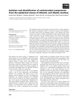
in Fig. 1, The VEGF-D protein was stained as light to
dark brown and is mainly located in the cytoplasm of
GBC cells. As shown in Table 1, VEGF-D was expressed
in 75 % (15/20) of samples. The level of TNF-α in the
bile of GBC patients with positive staining of VEGF-D
was significantly higher than that of patients with negative staining.
Tube formation assay
To assess the role of the TNF-α-VEGF-D axis in the
tube formation of HDLECs, NOZ or GBC-SD cells stably transfected with LV-siVEGF-D were co-cultured with
HDLECs previously labeled by DiI (a cell membrane dye
emitting red fluorescence; Beyotime Institute of Biotechnology, ShangHai, China) in a three dimensional coculture system following the method described by Yiping
Zeng [28]. Briefly, 7.5 × 10³/well of GBC cells and 7.5 ×
103/well of HDLECs were seeded to the same well of
microwell-plate (ibidi) which was previously painted
with matrigel. Tube formation of HDLECs was observed
by inverted fluorescence microscopy (Nikon, Japan), and
images were digitally captured at 1 h, 3 h, 5 h, 8 h and
24 h after cell seeding. The total number of tube-like
structures formed in each well were measured with
Axiovision Rel 4.1 software (Carl Zeiss AG, Jena,
Germany).
TNF-α promotes the expression of VEGF-D in vitro
To determine whether TNF-α could promote the expression of VEGF-D, we measured the expression of
VEGF-D in three GBC cell lines (NOZ, GBC-SD, and
SGC-996) after treatment with exogenous TNF-α. GBC
cells were incubated in 6-well plates and treated with
varying doses of TNF-α (10, 20, 50 and 100 ng⁄mL) for
12 and 24 h; the control samples were not treated with
TNF-α. The relative mRNA of VEGF-D was assayed by
real-time PCR, and VEGF-D protein level in the cell culture supernate was detected by ELISA. As shown in
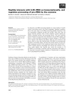
Fig. 2, TNF-α promoted the transcription and protein
Hong et al. BMC Cancer (2016) 16:240
Page 5 of 14
Fig. 1 Representative IHC stainging examples demonstrating VEGF-D expression in GBC specimens: a absent, b weak, c moderate, d strong
expression of VEGF-D in NOZ and GBC-SD cell lines
(but not SGC-996 cells) in a dose- and time-dependent
manner, and the peak effect appeared after 24-h treatment with 50 ng⁄mL TNF-α. So we used NOZ and GBCSD cells to next further study.
Activity analysis of VEGF-D promoter
To further explore the mechanism by which TNF-α
upregulates VEGF-D, we analyzed the promoter of
VEGF-D. Recombinant plasmids carrying a series of
5′-deletion fragments of the VEGF-D gene promoter
and the firefly luciferase report gene (named PGL42148, PGL4-1621, PGL4-988, PGL4-717, PGL4-444,
PGL4-325, PGL4-154, and PGL4-57) were transiently
co-transfected into the NOZ cells with pRL-TK as internal reference. As shown in Fig. 3, cells transfected
with recombinant plasmids PGL4-988, PGL4-444, and
PGL4-154 exhibited higher relative luciferase activities
compared with cells transfected with PGL4-717,
Table 1 The relationship between TNF-α levels in the bile and
VEGF-D expression in the tissues of GBC patients
VEGF-D
Case number
TNF-α (pg/ml)
P value
0.017
Positive
15
666.71 ± 47.26
Negative
5
435.98 ± 49.08
PGL4-325, and PGL4-57, respectively (P < 0.05).
Therefore, we speculated that the three fragments
(−988 to −71 7 nt,-444 to -325 nt,and −154 to
-57 nt) contained sites regulating VEGF-D expression.
Next, we scanned the base sequences of the fragments using the TFbind and Promoter Scan programs
to search for potential binding sites of the transcription factor AP-1 and NF-κB. The region −444 to
-325 nt contained two putative AP-1 binding sites but
no NF-κB site, and neither of the other two regions
contained binding sites. The plasmid PGL4-444 was
therefore selected for further studies.
TNF-α promotes AP-1 binding to the VEGF-D promoter
The sequence of the −444 to −325 nt region of the
VEGF-D promoter is presented in Fig. 4a, and the
two predicted putative AP1-binding sites in the nucleotide region −401 to −393 (AP-1(1)) and −345 to
−337 (AP-1(2)) are underlined. Site-directed mutants
of the putative AP1-binding sites were then generated,
and the promoter activities of the corresponding constructs were measured. As shown in Fig. 4b, both of
the two recombinant plasmids, PGL4-AP-1mut1,
which contains the mutation of the AP-1(1)-binding
site, and PGL4-AP-1mut2, which contains the mutation of the AP-1(2)-binding site, exhibited lower activities than the control non-mutated construct (PGL4-
Hong et al. BMC Cancer (2016) 16:240
A
Page 6 of 14
B
C
Fig. 2 VEGF-D mRNA transcription and protein expression in three GBC cell lines after treatment with TNF-α. GBC cells (NOZ, GBC-SD, and SGC-996) were
treated with varying concentrations of TNF-α (10, 20, 50 and 100 ng⁄ mL) for 12 or 24 h. The VEGF-D mRNA and protein levels were measured by
real-time PCR (a, b) and ELISA (c), respectively, and increased in a dose- and time-dependent manner in NOZ and GBC-SD cell lines but not SGC-996 cells.
(*P < 0.05; **P < 0.01; ***P < 0.001)
444). Furthermore, the activity weakened when the
two sites were mutated simultaneously (AP-1 double
mut), which suggests that both of the AP-1-binding
sites are crucial for the full activity of the VEGF-D
promoter. Upon treatment with TNF-α, the activity of
PGL4-444 increased significantly (P < 0.05), and this
activity was impaired by the mutation of the AP-1binding sites.
The two AP-1 binding sites were further confirmed by
EMSA of nuclear extracts from NOZ cells with and
without TNF-α treatment. As shown in Fig. 4c, the nuclear extracts were combined with a biotin-labeled probe
Fig. 3 Activity analysis of VEGF-D promoter. A series of 5′-deletion fragments of the VEGF-D promoter were amplified by PCR and then inserted into the
firefly luciferase report vector. These constructs (1 μg) were co-transfected into NOZ cells with pRL-TK (0.1 μg) as an internal reference. PGL4-basic served as
the negative control. The constructs PGL4-988, PGL4-444, and PGL4-154 exhibited higher relative luciferase activities (compared with PGL4-717, PGL4-325,
and PGL4-57, respectively (*P < 0.05)). The experiment was repeated three times
Hong et al. BMC Cancer (2016) 16:240
Page 7 of 14
Fig. 4 TNF-α promotes AP-1 binding to the VEGF-D promoter. a. The two predicted putative AP1-binding sites contained in the −444 to −325 nt region
of VEGF-D promoter are underlined (AP-1(1) in the nucleotide region −401 to -393 nt; AP-1(2) in the −345 to -337 nt). b. The effect of mutation of the AP-1
binding sites on the activity of VEGF-D promoter. Both of the two mutated constructs, PGL4-AP-1mut1 and PGL4-AP-1mut2, exhibited lower activities than
the non-mutated construct PGL4-444. Furthermore, the activity weakened when the two sites were mutated simultaneously. The trend persisted upon
treatment with TNF-α (50 ng/ml) (mutants, indicated with the × mark, are depicted schematically on the left; *P < 0.05). c, d. EMSA of AP-1. The nuclear
extracts from NOZ cells could bind the biotin-labeled probes (lane 2). The competition assay revealed that pre-incubation with the cold probes (lane 3) but
not the cold mutated probes (lane 5) diminished the intensity of the bands. TNF-α enhanced the combined effect of the nuclear extracts and the two
AP-1-binding sites (lane 4). e, f. ChIP assay. Chromatin from NOZ or GBC-SD cells was immunoprecipitated with the anti-AP-1 antibody. The total extracted
DNA (Input) and the immunoprecipitated samples were PCR-amplified using primers specific to the regions of the VEGF-D promoter containing the
AP-1(1) binding site (119 bp) and AP-1(2) binding site (150 bp). A normal rabbit IgG and no antibody sample were also included as controls. Another
experiment group was treated with 50 ng⁄ mL of TNF-α (bottom row), and TNF-α enhanced the intensity of the input and anti-AP-1 bands
(lane2). A competition assay revealed that preincubation with a 100-fold molar excess of the cold
probe (lane3) but not the cold mutated probe (lane5) diminished the intensity of the bands. Moreover, TNF-α enhanced the combined effect of the nuclear extract and the
AP-1(1)-binding site (lane 4). The AP-1(2)-binding site had
a similar combined effect (Fig. 4d).
To determine whether the AP-1 transcription factor was associated with the VEGF-D promoter in
vivo, we performed ChIP assays with an AP-1specific antibody and PCR using the primers against
the regulatory elements of the VEGF-D promoter in
NOZ and GBC-SD cell lines. As shown in Fig. 4
(e, f ), DNA fragments covering the two AP-1 binding sites (119 bp for AP-1(1), 150 bp for AP-1(2))
were amplified by chromatin immunoprecipitation
with an anti-AP-1 antibody. The same band was obtained
with the input DNA, whereas the normal IgG control and
no antibody control did not result in the immunoprecipitation of DNA fragments detectable by PCR amplification.
Consistent with the results by EMSA, TNF-α enhanced the
intensity of the anti-AP-1 band.
Taken together, these results demonstrate that the AP1 transcription factor can bind directly to the consensus
binding sites in the VEGF-D promoter region and the
TNF-α can improve the combined effect.
Hong et al. BMC Cancer (2016) 16:240
Upregulation of VEGF-D expression and VEGF-D promoter
activity by the TNF-α/ERK1/2/AP-1 pathway
To determine the effect of the TNF-α⁄AP-1 signaling
pathway on the promoter activity and protein expression
of the VEGF-D gene, we measured the luciferase intensity of the PGL4-444 plasmid and VEGF-D expression in
Page 8 of 14
NOZ (or GBC-SD) cells treated with TNF-α or transfected with AP-1 (c-Jun) siRNA against AP-1 (siAP-1).
The siAP-1 oligos effectively knocked-down the expression of AP-1 and p-AP-1 in NOZ (or GBC-SD) cells
compared with the negative control and siNC groups
(Fig. 5a). As shown in Fig. 5 (a, c), the protein level and
Fig. 5 TNF-α upregulated VEGF-D expression and VEGF-D promoter activity downstream of the ERK1/2/AP-1 pathway. a, c The effect of the TNFα⁄AP-1 signaling pathway on the promoter activity and protein expression of the VEGF-D gene. Transfection with AP-1 siRNA effectively knocked
down the expression of AP-1 and p-AP-1 in both NOZ and GBC-SD cells. The protein level and promoter activity of VEGF-D were accordingly reduced irrespective of treatment with TNF-α. b, d The effect of inhibition of MAPK pathway members on the protein expression and promoter activity of VEGF-D. When treated with SP600125 (10 μM), SB203580 (20 μM) or PD98059 (50 μM), the expression of AP-1 and p-AP-1 in both NOZ
and GBC-SD cells were reduced. However, the protein expression and promoter activity of VEGF-D were significantly reduced only in the
PD98059-treated group. *P < 0.05
Hong et al. BMC Cancer (2016) 16:240
promoter activity of the VEGF-D gene were significantly
reduced after transfection with siAP-1. TNF-α was demonstrated to enhance the expression of AP-1, p-AP-1,
and VEGF-D and to increase the luciferase activity of
the VEGF-D promoter. In contrast, when NOZ (or
GBC-SD) cells were transfected with siAP-1, the ability
of TNF-α to upregulate the luciferase activity and the
protein expression of VEGF-D were blunted.
To explore which member of the MAPK family (JNK,
p38 or ERK1/2) is involved in the TNF-α ⁄AP-1/VEGF-D
Page 9 of 14
signaling pathway, we investigated the effects of MAPK
pathway inhibitors on the protein expression of AP-1, pAP-1, and VEGF-D and the luciferase activity of VEGFD promoter. As shown in Fig. 5 (b, d), treatment of
NOZ (or GBC-SD) cells with SP600125 (10 μM),
SB203580 (20 μM) or PD98059 (50 μM) resulted in reduced expression of AP-1 and p-AP-1. However, the
protein expression and promoter activity of VEGF-D
were significantly reduced in the PD98059-treated group
(compared with control and the TNF-α-treated groups,
Fig. 6 The TNF-α - VEGF-D axis promoted the tube formation of human dermal lymphatic endothelial cells (HDLECs) in vitro. a, b Construction of
a NOZ cell line and a GBC-SD cell line stably expressing lentiviral VEGF-D shRNA and a green fluorescent protein sequence. The cells were observed under a fluorescence microscope with bright or blue light. c, d VEGF-D mRNA and protein expression of NOZ or GBC-SD cells stably transfected with LV-siVEGF-D were analyzed by real-time reverse transcription-polymerase chain reaction (RT-PCR) and enzyme-linked immunosorbent
assay (ELISA), respectively. GAPDH served as an internal control. e, f, g, h DiI-labeled HDLECs (emit red fluorescence) were cocultured with the
three NOZ (or GBC-SD) cell lines and were treated with TNF-α (50 ng⁄ mL) for 5 h. HDLEC tube formation was observed under fluorescence microscopy, and the tube number was counted. (*P < 0.05; **P < 0.01; ***P < 0.001)
Hong et al. BMC Cancer (2016) 16:240
P < 0.05) but not in the SP600125- or SB203580-treated
groups. Therefore, ERK1/2 is involved in the TNF-α/AP1 signaling pathway.
Taken together, these experiments confirm the upregulation of VEGF-D expression and VEGF-D promoter activity by the TNF-α/ERK1/2/AP-1 pathway.
The role of the TNF-α - VEGF-D axis in tube formation of
HDLECs in vitro
After confirming TNF-α-induced expression of VEGF-D
in vitro, we wanted to further analyze the role of the
Page 10 of 14
TNF-α-VEGF-D axis in the tube formation of HDLECs.
We first established a NOZ cell line (Fig. 6a) and a
GBC-SD cell line (Fig. 6b) stably expressing lentiviral
VEGF-D shRNA and employed real-time PCR and
ELISA to measure the efficacy of VEGF-D knockdown at
the mRNA and protein level. As shown in Fig. 6 (c, d),
the mRNA and protein levels of VEGF-D in the LVsiVEGF-D group (NOZ or GBC-SD cells infected with
lentiviral VEGF-D shRNA) were significantly decreased
(**P < 0.01, ***P < 0.001) relative to the control (NOZ or
GBC-SD cells only) and LV-siNC (NOZ or GBC-SD cells
Fig. 7 The TNF-α-VEGF-D axis is involved in lymphangiogenesis and lymph node metastasis (LNM) of GBC in vivo. a. Establishment of orthotopic
xenograft models of GBC in nude mice. After anesthesia, the abdominal cavity of the nude mouse was opened, the gallbladder was exposed, and
one of three NOZ cell lines (NOZ, LV-siNC, or LV-siVEGF-D) was injected into the cavity of gallbladder; the abdominal cavity was subsequently
closed. b, c. After treatment with TNF-α (2 μg⁄ kg) twice a week for 3 weeks, the mice were dissected, and the tumors were excised. Infiltrative
growth (green arrow), LNM (yellow arrow), ascites (red arrow) and hepatic metastasis (white arrow) were observed in the orthotopic xenograft
models. LNM was further confirmed by H-E staining (C-2: 200×, C-3: 400×), and invasive tumor cells (black arrow) could be observed in the lymphoid follicles. d. Detection of lymphatic vessels (marked by LYVE-1 and indicated by blue arrows) in the orthotopic xenograft tumors was achieved
by immunohistochemistry. e. Number of lymphatic vessel in the orthotopic xenograft tumors. TNF-α increased the number of lymphatic vessels
in the NOZ and LV-siNC group, whereas the knockdown of VEGF-D decreased this effect (*P < 0.05)
Hong et al. BMC Cancer (2016) 16:240
Page 11 of 14
infected with empty vector) groups. Subsequently, we
used a three-dimensional coculture system in which the
GBC cells and HDLECs were cultured together to observe the role of TNF-α and VEGF-D in the tube formation of HDLECs. HDLECs labeled by DiI were separately
cocultured with three cell lines (NOZ or GBC-SD, LVsiVEGF-D and LV-siNC) on Matrigel with or without
treatment with TNF-α (50 ng/ml). The phenomenon of
tube formation was observed 1 h, 3 h, 5 h, 8 h, and 24 h
after coculture. As shown in Fig. 6 (e, f, g, h), the greatest number of tubes was observed 5 h after cell seeding,
and the tube-like structures disappeared after 24 h (data
not shown). These data led us to conclude the following:
(1) the tube formation of HDLECs decreased with
knock-down of VEGF-D expression, and (2) the number
tubes formed by HDLECs significantly increased after
treatment with TNF-α, which could be impaired with
knock-down of VEGF-D expression.
The TNF-α-VEGF-D axis promotes the lymphatic metastasis of GBC in vivo
To investigate the role of the TNF-α - VEGF-D axis in
lymphangiogenesis and lymph node metastasis in vivo,
we injected three NOZ cell lines (NOZ, LV-siNC and
LV-siVEGF-D) into the gallbladders of nude mice to
established three orthotopic xenograft models of GBC
(Fig. 7a). Two weeks later, TNF-α (2 μg⁄kg) was injected
into the abdominal cavity twice a week for 3 weeks.
Lymph node metastases were observed with the naked
eye and further confirmed by HE staining (Fig. 7c). The
lymphatic vessels of tumors were detected by immunohistochemistry using an LYVE-1 antibody. As shown in
Fig. 7b, infiltrative growth was observed in most of the
orthotopic xenograft tumors. Lymph node metastases,
ascites or hepatic metastases were observed in some
mice, whereas lung metastases were not observed. Figure 7d demonstrates that TNF-α increased the LVD of
orthotopic xenograft tumors compared with the control
group, and this effect was impaired when the VEGF-D
was knocked down by lentiviral-mediated shRNA (LVsiVEGF-D group). As shown in Table 2, the rates of
lymph node metastasis were increased by TNF-α.
Table 2 Lymphatic vessel density (LVD) and lymph node
metastasis (LNM) of the orthotopic xenograft tumors in nude
mice
TNF-α (2 μg/kg)
Unstimulation
control
LVD
LNM
LVD
11.73 ± 2.28
3/5
23.73 ± 2.17*
LNM
5/5
*
LV-siNC
14.27 ± 1.36
2/5
23.20 ± 2.18
4/5
LV-siVEGF-D
5.67 ± 1.25
1/5
10.07 ± 1.83*
2/5
*P < 0.05
Discussion
As previously mentioned, the relationship between inflammation and cancer was first appreciated by Vichow
in 1863. It is currently estimated that approximately
25 % of the malignancies worldwide are induced by
chronic inflammation [29, 30]. The characteristics of this
chronic inflammation is the infiltration of a large number of inflammatory cells that secrete various cytokines
[31]. TNF-α, mainly secreted by macrophage, is a key
player in cancer-related inflammation. Chronic inflammation induced by gallstones, infection, or other factors
is one of the leading causes of GBC according to epidemiological investigations, [32, 33] and TNF-α has been
detected in the inflammatory environment of the gallbladder [34, 35]. Consistent with these reports, our laboratory recently observed that the level of TNF-α in
the bile of GBC patients was higher than that of patients
with cholecystic polypus (without obvious inflammation)
and demonstrated the ability of TNF-α to promote lymphangiogenesis in GBC [20].
Lymphangiogenesis is thought to be an important step
in cancer metastasis [36]. Our previous study have confirmed that TNF-α can promote lymphangiogenesis of
GBC through upregulation of VEGF-C. Meanwhile, we
found that the effect of TNF-α-induced lymphangiogenesis in GBC was only partially inhibited with knockdown of VEGF-C expression. This interesting
phenomenon promoted us to speculate that there should
be other molecular mechanisms involved in the TNF-αinduced lymphangiogenesis in GBC. Similar to VEGF-C,
VEGF-D is another key lymphangiogenic factor which is
associated with lymphangiogenesis and lymph node metastasis of GBC [21]. Therefore, we hypothesized that
VEGF-D may be involved in the TNF-α-induced lymphatic metastasis of GBC.
In the present study, we found that the level of
TNF-α in the bile of GBC patients was correlated
with the expression of VEGF-D in the tissue. Subsequently, we confirmed in vitro that TNF-α significantly increased the mRNA and protein expression of
VEGF-D in NOZ and GBC-SD cell lines within the
dose range of 10–50 ng⁄mL in a dose- and timedependent manner. We further to reveal that TNF-α
can upregulate the protein expression and promoter
activity of VEGF-D through the ERK1/2 - AP-1 pathway. Moreover, we determined that TNF-α can promote tube formation of HDLECs, lymphangiogenesis
and lymph node metastasis of GBC by upregulation
of VEGF-D in vitro and in vivo. In the tube formation assay, HDLECs were previously labeled by DiI
before co-culture with GBC cells and observed by the
inverted fluorescence microscope after co-culture.
This method can effectively exclude the interference
of GBC cells when observation. In addition, the
Hong et al. BMC Cancer (2016) 16:240
orthotopic xenograft model of GBC in nude mice is
more able to reflect the growth pattern of GBC in
human body.
Many studies have focused on the relationship between VEGF-D and lymphatic metastasis [21, 37–40].
However, few investigations have concentrated on the
regulation of VEGF-D promoter activity. To date, only
two studies have suggested that orphan receptor hepatocyte nuclear factor 4α (HNF-4α), chicken ovalbumin upstream promoter transcription factors 1 and 2 (COUPTF1 and COUP-TF2) and AP-1 bind to the VEGF-D
promoter [41, 42]. A large number of studies have demonstrated that the downstream effector molecules associated with tumor progression are NF-κB or AP-1 [22,
43]. To determine whether TNF-α regulates VEGF-D
promoter activity through these two transcription factors, we used the TFbind and Promoter Scan programs
to search for potential binding sites of NF-κB or AP-1 in
the three fragments of VEGF-D promoter with higher
activities (−988 to -717 nt,-444 to -325 nt,and −154 to
-57 nt), and found that the −444 to -325 nt region contains two putative AP-1 binding sites, whereas NF-κB
sites were not found. Subsequently, we confirmed that
both the AP-1 sites could bind to the VEGF-D promoter
and that TNF-α could enhance the combination by sitedirected mutagenesis, EMSA, and ChIP analysis. Further,
we used siRNA to knock down AP-1, and the protein
level of VEGF-D and the activity of the PGL4-444 plasmid were consequently decreased in the both groups
with or without TNF-α treatment.
It is demonstrated that the multiple effects of TNF-α in
cancers are due to the different downstream signaling pathways activated by the combination of TNF-α and its receptor (mainly through NF-κB and (or) AP-1 pathway). There
are two AP-1 binding sites (no NF-κB site) in the core region of VEGF-D promoter, which revealed that TNF-αinduced upregulation of VEGF-D is mainly through the
AP-1 pathway. Two signaling pathways associated with AP1 have been clarified in previous studies: the TNF-α TNFR1 - signaling complex - MAP3K (ASK1) - JNK or
p38 MAPK - AP-1 pathway and the TNF-α - TNFR1 - Ras
- Raf - MEK1 - ERK1/2 - AP-1 pathway [44]. To further determine which pathway is involved in the TNF-α - VEGF-D
axis, we employed three reagents, SP600125, SB203580 and
PD98059, to selectively inhibit JNK, p38 MAPK and ERK1/
2, respectively. The protein expression of AP-1, p-AP-1,
and VEGF-D and the activity of the PGL4-444 construct
were significantly inhibited in the PD98059 treatment
group, which indicated that TNF-α upregulated VEGF-D
promoter activity and protein expression primarily through
the ERK1/2/AP-1 signaling pathway.
The active Ras proteins combine with the guanosine
triphosphate (GTP) and then activates the downstream
signaling pathways including the MAPK pathway [45].
Page 12 of 14
The alteration of Ras protein conformation caused by
Ras gene mutation makes it lose the GTPase activity and
leads to the continuous activation of the downstream signaling which accordingly promotes cell proliferation and
invasion. K-ras gene is a member of the Ras family and Kras mutation has been reported in various malignancies
including GBC and NOZ cell line [45–48]. As mentioned
above, the Ras protein is an effector between TNFR and
ERK1/2. Thus we can speculate that K-ras mutation could
enhance the activity of the “TNF-α/ERK1/2/AP-1/VEGFD” pathway in NOZ cells which might accordingly enable
the nude mice bearing human GBC in the present study
more prone to appear lymphatic metastasis.
In this study, we first discovered the relationship between
the TNF-α - VEGF-D axis and the lymphangiogenesis and
lymphatic metastasis of GBC. Subsequently, we demonstrated that the regulatory mechanism between TNF-α and
VEGF-D is dependent on the ERK1/2/AP-1 signaling pathway. Furthermore, we determined the core activity region
of the VEGF-D promoter and identified two AP-1 binding
sites in these regions. The regulatory mechanisms of
inflammation-induced tumor metastasis are very complicated, but our work helps elucidate some of these
mechanisms.
Together with our previous study, these results reveal
that TNF-α can promote lymphangigenesis and lymph
node metastasis of GBC at least by two signaling pathways: the NF-κB/VEGF-C pathway and the ERK1/2/AP1/VEGF-D pathway. But, which pathway is dominated
or both are equally important, needs further study.
Conclusions
To our knowledge, our research represents the first report that TNF-α can promote lymphangiogenesis and
lymphatic metastasis of GBC through the ERK1/2/AP-1/
VEGF-D pathway.
Availability of data and materials
The datasets supporting the conclusions of this article
are included within the article.
Abbreviations
TNF-α: Tumor necrosis factor-alpha; VEGF-D: Vascular endothelial growth
factor D; GBC: Gallbladder cancer; HDLECs: Human dermal lymphatic
endothelial cells; LVD: Lymphatic vessel density; LNM: Lymph node
metastases.
Competing interests
The authors declare that they have no competing interests.
Authors’ contributions
CYL, SFF, HHJ and JL contributed to concept design, discussed results. HHJ also
performed immunohistochemistry, relative luciferase reporter assay, mutant
constructs, EMSA, and ChIP assay; participated in cell culture and animal
experiment; and wrote the manuscript. LYF participated in cell culture and tube
formation assay; performed PCR and ELISA. HCL carried out protein isolation and
Western blotting, and also participated in animal experiment. ZGW participated in
the sequence alignment. DQ and TNH performed the statistical analysis. WXQ
Hong et al. BMC Cancer (2016) 16:240
gave assistance with several technical performances. All authors read and
approved the final manuscript.
Acknowledgement
This study was supported by the grants from The National Natural Science
Foundation of China (No. 81272373).
Author details
1
Department of Hepatobiliary Surgery and Fujian Institute of Hepatobiliary
Surgery, Fujian Medical University Union Hospital, 29 Xinquan Road, Fuzhou
350001, China. 2Key Laboratory of Ministry of Education for Gastrointestinal
Cancer, Fujian Medical University, 1 Xueyuan Road, Minhou, Fuzhou 350108,
China. 3Fujian Key Laboratory of Tumor Microbiology, Fujian Medical
University, 1 Xueyuan Road, Minhou, Fuzhou 350108, China.
Received: 16 June 2015 Accepted: 8 March 2016
References
1. Lazcano-Ponce EC, Miquel JF, Munoz N, Herrero R, Ferrecio C, Wistuba II, et
al. Epidemiology and molecular pathology of gallbladder cancer. CA Cancer
J Clin. 2001;51(6):349–64.
2. Misra S, Chaturvedi A, Misra NC, Sharma ID. Carcinoma of the gallbladder.
Lancet Oncol. 2003;4(3):167–76.
3. Levy AD, Murakata LA, Rohrmann Jr CA. Gallbladder carcinoma: radiologicpathologic correlation. Radiographics. 2001;21(2):295–314. doi:10.1148/
radiographics.21.2.g01mr16295. questionnaire, 549–55.
4. Wernberg JA, Lucarelli DD. Gallbladder cancer. Surg Clin North Am. 2014;
94(2):343–60. doi:10.1016/j.suc.2014.01.009.
5. Muratore A, Polastri R, Capussotti L. Radical surgery for gallbladder cancer:
current options. Eur J Surg Oncol. 2000;26(5):438–43.
6. Virchow R. Die krankhaften Geschwülste. Springer Berlin Heidelberg; 1978.
7. Coussens LM, Werb Z. Inflammation and cancer. Nature. 2002;420(6917):
860–7.
8. Bishayee A. The role of inflammation and liver cancer. Adv Exp Med Biol.
2014;816:401–35. doi:10.1007/978-3-0348-0837-8_16.
9. Ekbom A, Helmick C, Zack M, Adami H-O. Ulcerative colitis and colorectal
cancer: a population-based study. N Engl J Med. 1990;323(18):1228–33.
10. Parsonnet J, Hansen S, Rodriguez L, Gelb AB, Warnke RA, Jellum E, et al.
Helicobacter pylori infection and gastric lymphoma. N Engl J Med. 1994;
330(18):1267–71.
11. Gomes M, Teixeira AL, Coelho A, Araujo A, Medeiros R. The role of
inflammation in lung cancer. Adv Exp Med Biol. 2014;816:1–23. doi:10.1007/
978-3-0348-0837-8_1.
12. Mantovani A, Allavena P, Sica A, Balkwill F. Cancer-related inflammation.
Nature. 2008;454(7203):436–44.
13. Szlosarek PW, Balkwill FR. Tumour necrosis factor α: a potential target for
the therapy of solid tumours. Lancet Oncol. 2003;4(9):565–73.
14. Voronov E. IL-1 is required for tumor invasiveness and angiogenesis. Proc
Natl Acad Sci U S A. 2003;100(5):2645–50.
15. Grivennikov S, Karin M. Autocrine IL-6 signaling: a key event in
tumorigenesis? Cancer Cell. 2008;13(1):7–9. doi:10.1016/j.ccr.2007.12.020.
16. Chua HL, Bhat-Nakshatri P, Clare SE, Morimiya A, Badve S, Nakshatri H. NFkappaB represses E-cadherin expression and enhances epithelial to
mesenchymal transition of mammary epithelial cells: potential involvement
of ZEB-1 and ZEB-2. Oncogene. 2007;26(5):711–24. doi:10.1038/sj.onc.
1209808.
17. Johnston DA, Dong B, Hughes CC. TNF induction of jagged-1 in endothelial
cells is NFkappaB-dependent. Gene. 2009;435(1–2):36–44. doi:10.1016/j.gene.
2009.01.003.
18. Katerinaki E, Evans GS, Lorigan PC, MacNeil S. TNF-alpha increases human
melanoma cell invasion and migration in vitro: the role of proteolytic
enzymes. Br J Cancer. 2003;89(6):1123–9. doi:10.1038/sj.bjc.6601257.
19. Zhu G, Du Q, Wang X, Tang N, She F, Chen Y. TNF-alpha promotes
gallbladder cancer cell growth and invasion through autocrine mechanisms.
Int J Mol Med. 2014;33(6):1431–40. doi:10.3892/ijmm.2014.1711.
20. Du Q, Jiang L, Wang X, Wang M, She F, Chen Y. Tumor necrosis factor-α
promotes the lymphangiogenesis of gallbladder carcinoma through nuclear
factor-κB-mediated upregulation of vascular endothelial growth factor-C.
Cancer Sci. 2014;105(10):1261–71.
Page 13 of 14
21. Lin W, Jiang L, Chen Y, She F, Han S, Zhu J, et al. Vascular endothelial
growth factor-D promotes growth, lymphangiogenesis and lymphatic
metastasis in gallbladder cancer. Cancer Lett. 2012;314(2):127–36. doi:10.
1016/j.canlet.2011.09.004.
22. Chu WM. Tumor necrosis factor. Cancer Lett. 2013;328(2):222–5. doi:10.1016/
j.canlet.2012.10.014.
23. Yao X, Zhou L, Han S, Chen Y. High Expression of CXCR4 and CXCR7
Predicts Poor Survival in Gallbladder Cancer. J Int Med Res. 2011;39(4):1253–
64.
24. Du Q, Jiang L, Wang XQ, Pan W, She FF, Chen YL. Establishment of and
comparison between orthotopic xenograft and subcutaneous xenograft
models of gallbladder carcinoma. Asian Pac J Cancer Prev. 2014;15(8):3747–
52.
25. Heckman KL, Pease LR. Gene splicing and mutagenesis by PCR-driven
overlap extension. Nat Protoc. 2007;2(4):924–32. doi:10.1038/nprot.2007.132.
26. Xu Q, Hou W, Zheng Y, Liu C, Gong Z, Lu C, et al. Ultraviolet A-induced
cathepsin K expression is mediated via MAPK/AP-1 pathway in human
dermal fibroblasts. PLoS One. 2014;9(7):e102732. doi:10.1371/journal.pone.
0102732.
27. Chen Y, Jiang L, She F, Tang N, Wang X, Li X, et al. Vascular endothelial
growth factor-C promotes the growth and invasion of gallbladder cancer
via an autocrine mechanism. Mol Cell Biochem. 2010;345(1–2):77–89. doi:10.
1007/s11010-010-0562-y.
28. Zeng Y, Opeskin K, Goad J, Williams ED. Tumor-induced activation of
lymphatic endothelial cells via vascular endothelial growth factor receptor-2
is critical for prostate cancer lymphatic metastasis. Cancer Res. 2006;66(19):
9566–75. doi:10.1158/0008-5472.can-06-1488.
29. Balkwill F, Mantovani A. Inflammation and cancer: back to Virchow? Lancet.
2001;357(9255):539–45.
30. Borroni EM, Buracchi C, Savino B, Pasqualini F, Russo RC, Nebuloni M, et al.
Role of the chemokine scavenger receptor D6 in balancing inflammation
and immune activation. Methods Enzymol. 2009;460:231–43. doi:10.1016/
s0076-6879(09)05211-2.
31. Lu H, Ouyang W, Huang C. Inflammation, a key event in cancer
development. Mol Cancer Res. 2006;4(4):221–33. doi:10.1158/1541-7786.mcr05-0261.
32. Randi G, Malvezzi M, Levi F, Ferlay J, Negri E, Franceschi S, et al.
Epidemiology of biliary tract cancers: an update. Ann Oncol. 2009;20(1):146–
59. doi:10.1093/annonc/mdn533.
33. Li Y, Zhang J, Ma H. Chronic inflammation and gallbladder cancer. Cancer
Lett. 2014;2:242–8.
34. Macarthur M, Hold GL, El-Omar EM. Inflammation and Cancer II. Role of
chronic inflammation and cytokine gene polymorphisms in the
pathogenesis of gastrointestinal malignancy. Am J Physiol Gastrointest Liver
Physiol. 2004;286(4):G515–20. doi:10.1152/ajpgi.00475.2003.
35. Shi JS, Zhou LS, Han Y, Zhu AJ, Sun XJ, Yang YJ. Expression of tumor
necrosis factor and its receptor in gallstone and gallbladder carcinoma
tissue. Hepatobiliary Pancreat Dis Int. 2004;3(3):448–52.
36. Stacker SA, Williams SP, Karnezis T, Shayan R, Fox SB, Achen MG.
Lymphangiogenesis and lymphatic vessel remodelling in cancer. Nat Rev
Cancer. 2014;14(3):159–72. doi:10.1038/nrc3677.
37. Juttner S, Wissmann C, Jons T, Vieth M, Hertel J, Gretschel S, et al. Vascular
endothelial growth factor-D and its receptor VEGFR-3: two novel
independent prognostic markers in gastric adenocarcinoma. J Clin Oncol.
2006;24(2):228–40. doi:10.1200/jco.2004.00.3467.
38. White JD, Hewett PW, Kosuge D, McCulloch T, Enholm BC, Carmichael J, et
al. Vascular endothelial growth factor-D expression is an independent
prognostic marker for survival in colorectal carcinoma. Cancer Res. 2002;
62(6):1669–75.
39. Yokoyama Y, Charnock-Jones DS, Licence D, Yanaihara A, Hastings JM,
Holland CM, et al. Expression of vascular endothelial growth factor (VEGF)-D
and its receptor, VEGF receptor 3, as a prognostic factor in endometrial
carcinoma. Clin Cancer Res. 2003;9(4):1361–9.
40. Du LC, Chen XC, Wang D, Wen YJ, Wang CT, Wang XM, et al. VEGF-Dinduced draining lymphatic enlargement and tumor lymphangiogenesis
promote lymph node metastasis in a xenograft model of ovarian
carcinoma. Reprod Biol Endocrinol. 2014;12:14. doi:10.1186/1477-7827-12-14.
41. Schafer G, Wissmann C, Hertel J, Lunyak V, Hocker M. Regulation of vascular
endothelial growth factor D by orphan receptors hepatocyte nuclear factor4 alpha and chicken ovalbumin upstream promoter transcription factors 1
and 2. Cancer Res. 2008;68(2):457–66. doi:10.1158/0008-5472.can-07-5136.
Hong et al. BMC Cancer (2016) 16:240
Page 14 of 14
42. Ming J, Zhang Q, Qiu X, Wang E. Interleukin 7/interleukin 7 receptor induce
c-Fos/c-Jun-dependent vascular endothelial growth factor-D up-regulation:
a mechanism of lymphangiogenesis in lung cancer. Eur J Cancer. 2009;45(5):
866–73. doi:10.1016/j.ejca.2008.12.006.
43. Parameswaran N, Patial S. Tumor necrosis factor-alpha signaling in
macrophages. Crit Rev Eukaryot Gene Expr. 2010;20(2):87–103.
44. Sethi G, Sung B, Aggarwal BB. TNF: a master switch for inflammation to
cancer. Front Biosci. 2008;13:5094–107.
45. Adjei AA. Blocking oncogenic Ras signaling for cancer therapy. J Natl
Cancer Inst. 2001;93(14):1062–74.
46. Bos JL. ras oncogenes in human cancer: a review. Cancer Res. 1989;49(17):
4682–9.
47. Horiuchi H, Kawamata H, Fujimori T, Kuroda Y. A MEK inhibitor (U0126)
prolongs survival in nude mice bearing human gallbladder cancer cells with
K-ras mutation: analysis in a novel orthotopic inoculation model. Int J
Oncol. 2003;23(4):957–63.
48. Kumari N, Corless CL, Warrick A, Beadling C, Nelson D, Neff T, et al. Mutation
profiling in gallbladder cancer in Indian population. Indian J Pathol
Microbiol. 2014;57(1):9–12. doi:10.4103/0377-4929.130849.
Submit your next manuscript to BioMed Central
and we will help you at every step:
• We accept pre-submission inquiries
• Our selector tool helps you to find the most relevant journal
• We provide round the clock customer support
• Convenient online submission
• Thorough peer review
• Inclusion in PubMed and all major indexing services
• Maximum visibility for your research
Submit your manuscript at
www.biomedcentral.com/submit