Lapatinib sensitivity in nasopharyngeal carcinoma is modulated by SIRT2-mediated FOXO3 deacetylation
Bạn đang xem bản rút gọn của tài liệu. Xem và tải ngay bản đầy đủ của tài liệu tại đây (7.05 MB, 17 trang )
Aimjongjun et al. BMC Cancer
(2019) 19:1106
/>
RESEARCH ARTICLE
Open Access
Lapatinib sensitivity in nasopharyngeal
carcinoma is modulated by SIRT2-mediated
FOXO3 deacetylation
Sathid Aimjongjun1,2†, Zimam Mahmud1†, Yannasittha Jiramongkol1, Glowi Alasiri1, Shang Yao1, Ernesto Yagüe2,
Tavan Janvilisri3 and Eric W.-F. Lam1*
Abstract
Background: Chemoresistance is an obstacle to the successful treatment of nasopharyngeal carcinoma (NPC).
Lapatinib is a targeted tyrosine kinase inhibitor therapeutic drug also used to treat NPC, but high doses are often
required to achieve a result. To investigate the mechanism for the development of Lapatinib resistance, we
characterised a number of NPC cell lines to determine the role of FOXO3 and sirtuins in regulating NPC resistance.
Methods: Sulforhodamine B (SRB) assays, Clonogenic assays, Protein extraction, quantification and western blotting,
RT qPCR, Co-immunoprecipitation assay.
Results: To explore novel treatment strategies, we first characterized the Lapatinib-sensitivity of a panel of NPC cell
lines by SRB and clonogenic cytotoxic assays and found that the metastatic NPC (C666–1 and 5-8F) cells are highly
resistant whereas the poorly metastatic lines (6-10B, TW01 and HK-1) are sensitive to Lapatinib. Western blot analysis
of the Lapatinib-sensitive 6-10B and resistant 5-8F NPC cells showed that the expression of phosphorylated/inactive
FOXO3 (P-FOXO3;T32), its target FOXM1 and its regulator SIRT2 correlate negatively with Lapatinib response and
sensitivity, suggesting that SIRT2 mediates FOXO3 deacetylation to promote Lapatinib resistance. In agreement,
clonogenic cytotoxic assays using wild-type and foxo1/3/4−/− mouse embryonic fibroblasts (MEFs) showed that
FOXO1/3/4-deletion significantly attenuates Lapatinib-induced cytotoxicity, confirming that FOXO proteins are
essential for mediating Lapatinib response. SRB cell viability assays using chemical SIRT inhibitors (i.e. sirtinol, Ex527,
AGK2 and AK1) revealed that all SIRT inhibitors can reduce NPC cell viability, but only the SIRT2-specific inhibitors
AK1 and AGK2 further enhance the Lapatinib cytotoxicity. Consistently, clonogenic assays demonstrated that the
SIRT2 inhibitors AK1 and AGK2 as well as SIRT2-knockdown increase Lapatinib cytotoxicity further in both the
sensitive and resistant NPC cells. Co-immunoprecipitation studies showed that besides Lapatinib treatment, SIRT2pharmaceutical inhibition and silencing also led to an increase in FOXO3 acetylation. Importantly, SIRT2 inhibition
and depletion further enhanced Lapatinib-mediated FOXO3-acetylation in NPC cells.
Conclusion: Collectively, our results suggest the involvement of SIRT2-mediated FOXO3 deacetylation in Lapatinib
response and sensitivity, and that SIRT2 can specifically antagonise the cytotoxicity of Lapatinib through mediating
FOXO3 deacetylation in both sensitive and resistant NPC cells. The present findings also propose that SIRT2 can be
an important biomarker for metastatic and Lapatinib resistant NPC and that targeting the SIRT2-FOXO3 axis may
provide novel strategies for treating NPC and for overcoming chemoresistance.
Keywords: Nasopharyngeal carcinoma, Sirtuin, FOXO3, Chemoresistance, Lapatinib, Acetylation
* Correspondence:
†
Sathid Aimjongjun and Zimam Mahmud are contributed equally and should
be recognised as co-first authors.
1
Department of Surgery and Cancer, Imperial College London, Hammersmith
Hospital Campus, London W12 0NN, UK
Full list of author information is available at the end of the article
© The Author(s). 2019 Open Access This article is distributed under the terms of the Creative Commons Attribution 4.0
International License ( which permits unrestricted use, distribution, and
reproduction in any medium, provided you give appropriate credit to the original author(s) and the source, provide a link to
the Creative Commons license, and indicate if changes were made. The Creative Commons Public Domain Dedication waiver
( applies to the data made available in this article, unless otherwise stated.
Aimjongjun et al. BMC Cancer
(2019) 19:1106
Background
Nasopharyngeal carcinoma (NPC) is a malignancy of
head and neck epithelial cells that originates from
the nasopharyngeal cavity. NPC is an important
health problem in Southern China and Southeast
Asia [1], with about 86,500 NPC new cases recorded
annually, and over 40,000 die from the disease every
year in Asia alone [2]. The most common treatment
for NPC is the combination of radiotherapy and
chemotherapy. Radiotherapy is a mainstay for treatment for early NPC, while chemotherapy is frequently used for the advanced locoregionally NPC. It
has been shown that the combination of chemotherapy and radiation enhances the survival rates [3].
However, advanced NPCs usually fail to respond to
chemotherapy treatment because of the development
of drug resistance that leads to metastasis and relapse, resulting in poor prognosis [4]. In consequence, there is an urgent need for the identification
of novel therapeutic strategies as well as the mechanisms of chemotherapeutic resistance for NPC. To
date, the mechanisms for the development of resistance to conventional therapeutics, such as cisplatin
(CDDP), paclitaxel (PTX), 5-fluorouracil (5-FU), and
vincristine (VCR), for NPC have been extensively
studied [5–8]. These include overexpression of
multidrug-resistant genes, enhanced DNA repair, deregulation of apoptosis, and/or modifications of drug
targets [5–8].
Both the epidermal growth factor receptor (EGFR)
and HER2 (human epidermal growth factor receptor
2; ERRB2) are commonly overexpressed in NPC [9,
10]. Moreover, activation of EGFR/HER2 pathway
has also been shown to promote NPC progression
and invasion [11]. Previous studies have demonstrated that the dual EGFR/HER2 tyrosine kinase inhibitor, Lapatinib, can restrict cell proliferation and
invasion, and promote anoikis in NPC cells [12, 13].
Despite its promising efficacy, Lapatinib has a half
maximal inhibitory concentration (IC50) in the micromolar range in most cell lines [13], and strategies
to sensitize NPC to Lapatinib are currently under
investigation.
FOXO3 and FOXM1 are members of the forkhead
box transcription factor family which play opposite
roles in tumorigenesis, drug resistance and cancer
progression. FOXO3 is a key tumour suppressor
which functions downstream of the PI3K/AKT (protein kinase B) signalling pathway. Conversely,
FOXM1 is an important oncogene that promotes
cell transformation, cancer progression and resistance to chemotherapy. In addition, FOXM1 and
FOXO3 negatively regulate the expression of one
another and compete for same binding sites to
Page 2 of 17
promote or inhibit the transcription of target genes
[14]. Furthermore, FOXO3 has also be shown to be
regulated by post-translational modifications, such
as phosphorylation, methylation, ubiquitination, glycosylation and acetylation. The reversible acetylation/deacetylation of FOXO3 is regulated by class I
histone deacetylases (HDACs) as well as class III
HDACs named sirtuins (SIRT 1–7) and histone
acetyl transferases (HATs e.g. p300/CBP) [15]. The
function of SIRT proteins is to reverse the acetylation of FOXO3 to induce the cell cycle arrest, protection from oxidative stress and to repress the
expression of apoptotic genes [16]. In NPC cells,
FOXM1 is overexpressed and it is associated with
cancer metastasis and chemoresistance [17, 18]. In
NPC patients, low FOXO3 and high HIF-1 expression has been found to be correlated with poor
prognosis in NPC [19]. However, the role and regulation of FOXO3 and FOXM1 in NPC have not
been not fully investigated.
In this study, we investigated the mechanism of action
of a dual tyrosine kinase inhibitor Lapatinib using high
and low metastatic NPC cells and found that the deacetylation of FOXO3 by SIRT2 plays an important role in
the regulation of Lapatinib response and resistance in
NPCs.
Methods
Cell culture
The NPC cell line TW01 is an established cell line
derived from moderately differentiated keratinising
NPC tissues, and was kindly provided by Prof. ChinTarng Lin, National Taiwan University, Taipei [20].
TW01 cells were cultured with DMEM supplemented with 10% v/v foetal bovine serum (FBS), 100
U/mL penicillin, and 100 μg/mL streptomycin. Poorly
differentiated human NPC cell lines SUNE 5–8 F
(highly tumorigenic and metastatic) and 6-10B
(highly tumorigenic, but poorly metastatic) derived
from the parental line SUNE-1, were obtained from
the Prof. Qingling Zhang, Southern Medical University, Guangzhou China [21, 22]. HK1, a welldifferentiated squamous carcinoma line was obtained
from the Queen Elizabeth Hospital, University of
Hong Kong [23]. C666–1 is an undifferentiated NPC
cell line obtained from Prof. Maria L. Lung, Center
for Nasopharyngeal Carcinoma Research, University
of Hong Kong [24]. These last four NPC cell lines
were cultured in RPMI supplemented with 10% FBS,
100 U/mL penicillin, and 100 μg/mL streptomycin.
All NPC cells were maintained in a humidified incubator with 10% CO2 at 37 °C. Wild type mouse embryonic fibroblasts (MEFs) and triple knock-out
foxo1/3/4−/− MEFs were kind gifts from Prof.
Aimjongjun et al. BMC Cancer
(2019) 19:1106
Boudewijn
Burgering,
UMC,
Utrecht,
the
Netherlands, and have been described previously
[25]. MEF cells were cultured in Dulbecco’s modified
eagle’s medium (DMEM) (Sigma Aldrich, Poole, UK)
and supplemented with 10% (v/v) foetal calf serum
(FCS) (First Link Ltd., Birmingham, UK), 100 Unit/
ml penicillin/streptomycin (Sigma-Aldrich, UK) and
2 mM glutamine and maintained at 37 °C in a humidified atmosphere containing 10% CO2. All cell
lines were subjected to DNA fingerprinting analysis
using the AmpF/STR Identifiler PCR Amplification
Kit (Applied Biosystems, Foster City, USA) and are
free from mycoplasma contamination.
siRNA mediated gene knockdown
For gene knockdown, cells were plated in at 60–70%
confluency. The following day, cells were transfected
with ON-TARGET plus siRNA smart pools (GE
Dharmacon) targeting SIRT2 (L− 004826-00-0005) using
oligofectamine (Invitrogen, UK) according to the
manufacturer’s protocol. Non-Targeting siRNA pool
(GE Dharmacon; D-001210-01-05) was used as transfection control.
Sulforhodamine B colorimetric assay
A total of 1000 NPC cells per well were seeded in a 96wells plate. One day after seeding, NPC cells were
treated with increasing concentrations of Lapatinib for
24 and 48 h. The cells were fixed with 40%
trichloroacetic acid at 4 °C for 1 h, washed 3 times with
PBS and stained with 0.4% (w/v) sulforhodamine B
(SRB) solution at room temperature for 1 h. Following
the staining, the cells were washed 5 times with 1%
acetic acid and air-dried overnight. The protein bound
dye was dissolved in 10 mM Tris base solution and the
absorbance was measured at 492 nm using a microplate
reader (Sunrise, Tecan; Männedorf, Switzerland).
Clonogenic assay
A total of 2000–10,000 cells were seeded into 6-well
plates and incubated overnight. The cells were then
treated for 72 h with varying concentrations of
Lapatinib and SIRT inhibitor (SIRT-i). DMSO
(Sigma-Aldrich,) was used as a vehicle and blank.
The drug was removed and surviving cells were left
to form colonies. After 1–2 weeks of incubation,
colonies were fixed with 4% paraformaldehyde for
15 min at room temperature and then washed with
PBS three times. Crystal violet (0.5% w/v) was used
to stain the fixed cells for 30 min, following which
the plates were washed with tap water. Plates were
then left to dry overnight. Quantification was
achieved by solubilising dye with 33% acetic acid
Page 3 of 17
and the absorbance was measured at 592 nm using
Tecan Microplate reader.
Western blotting and antibodies
Western blotting was performed with whole cell extracts prepared by lysing cells with NP40 lysis buffer
[1% NP40, 100 mmol/L NaCl, 20 mmol/L Tris-HCl
(pH 7.4), 10 mmol/L NaF, 1 mmol/L Na orthovanadate, 30 mmol/L Na β-glycerophosphate, and protease inhibitors (“Complete” protease inhibitor
mixture, as instructed by the manufacturer, Roche
Applied Science)] on ice for 15 min. After incubation, the lysates were centrifuged at 13,000×g for 10
min, and the supernatant was collected. Protein
concentrations were determined using a BCA protein assay kit (ThermoFisher Scientific, Waltham,
MA, USA). Ten micrograms of protein were size
fractionated
using
SDS-PAGE
and
electrotransferred onto nitrocellulose membranes (BioRad,
San Diego, CA, USA). Membranes were blocked in
5% (w/v) bovine serum albumin (BSA) in TBS plus
0.5% (v/v) Tween for 1 h at room temperature and
then incubated with specific antibodies overnight at
4 °C. The antibodies against EGFR, P-EGFR, ERBB2,
P-ERBB2, ERBB3, P-ERBB3, JNK, P-JNK, p38, P-p38,
c-Myc, p27Kip1 , P-FOXO3 (T32), FOXO3, cyclin B1,
P-AKT (S473), AKT and β-Tubulin were purchased
from Cell Signalling Technology (New England Biolabs Ltd., Hitchin, UK). The antibody against
FOXM1 (C− 20) was purchased from Santa Cruz Biotechnology (Santa Cruz, CA, USA). Primary antibodies were detected using horseradish peroxidaselinked anti-mouse or anti-rabbit conjugates (Dako,
Glostrup, Denmark) and visualized using the ECL
detection system (Perkin Elmer, Beaconsfield, UK).
Co-Immunoprecipitation assay
NPC cell lysates were prepared in IP buffer (1%
Nonidet P− 40, 150 mM NaCl, 50 mM Tris-HCl [pH
7.4], 10 mM NaF, 1 mM Na3VO4, 10 mM N-ethylamide (NEM) and protease inhibitors [Complete protease inhibitor cocktail; Roche, Lewes, UK]). The
pre-cleared lysate was immunoprecipitated with the
indicated antibodies and with protein A/G-sepharose
for 4 h. After that, the fractionated supernatant was
transferred to the new tube. Samples and incubated
overnight with 0.5 μg of primary antibody-FOXO3
(sc-11,351, Santa Cruz) [1, 26], Anti-acetyl lysine
(Cell-Signalling technology) or IgG negative control
(2729, Cell-Signalling technology) (1500) at 4 °C on a
rotator. The supernatant was discarded on the following day and beads were washed three times with
PBS. Samples were boiled with 2 × SDS sample buffer
Aimjongjun et al. BMC Cancer
(2019) 19:1106
for 5 min at 100 °C and were analysed by SDS-PAGE
followed by western blotting.
Gene expression analysis by RT-qPCR
Total mRNA was extracted from cell pellets using the
RNeasy Mini Kit (Qiagen, UK) according to manufacturer’s instructions. The purity and concentration of
mRNA samples were determined by measuring the spectrophotometric absorption at 260 nm and 280 nm using
a NanoDrop ND-1000. Then, 2 μg of the extracted total
RNA was used as a template for first strand cDNA synthesis reaction, The resulting first-strand cDNA was
then used as template in the real-time PCR. Real time
quantitative PCR (RT-qPCR) was performed on 100 ng
of cDNA with SYBER-Green Master Mix (Applied BioSystems). All RT-qPCR assays were assayed in 96-well
plates in the ABI PRISM® 7900 HT Fast Real-time PCR
System (Applied BioSystems) on a cycling program of
90 °C for 10 min for enzyme activation followed by 40
cycles of denaturation and primer annealing/extension
consisting of 95 °C for 3 s and 60 °C for 30s respectively.
The nonregulated ribosomal housekeeping gene, RPL19
was used as an internal control to normalize gene expression between samples. All samples were done in
triplicates. Primer sequences used for RT-qPCR are,
RPL19-F: 5’GGTGCTTCCGATTCCAGAGT3’,
RPL19-R: 5’CCCATTCCCTGATCGCTTGA3’,
Erbb2-F: 5’GCTCTTTGAGGACAAGTATG3’,
Erbb2-R: 5’AAGATCTCTGTGAGACTTCG3’,
Egfr-F: 5’CTGTCGCAAAGTTTGTAATG3’,
Egfr-R: 5’GAATTTCTAGTTCTCGTGGG3’,
Foxo3-F: 5’CCGGACAAACGGCTCACT3’,
Foxo3-R: 5’GGCACACAGCGCACCAT3’,
Foxo4-F:5’AGGACAAGGGTGACAGCAAC3’,
Foxo4-R: 5’GGTTCAGCATCCACCAAGAG3’,
Foxo1-F: 5’AAGAGCGTGCCCTACTT-CAA3’,
Foxo1-R: 5’TCCTTCA-TTCTGCACTCGAA 3′.
Statistical data analysis
The results were represented as the means ± SEM of at
least 3 separate experiments. For categorical variables,
we used the analysis of variance (ANOVA) which was
followed by a Bonferroni’s post-hoc test for multiple
comparisons. Student’s t test was used to analyse the
statistical significance of differences between the control
groups and the experimental groups. The differences
were considered as significant at p < 0.05. The GraphPad
Prism 5.0 software (version 5.00 for Windows, GraphPad
Software; San Diego, California, USA) was used for the
statistical analyses. ImageJ software was used to analyse
and compare the intensity of the protein bands obtained
from Western blots.
Page 4 of 17
Results
Effects of Lapatinib on cell viability of different
nasopharyngeal carcinoma (NPC) cell lines
In order to identify the cellular mechanisms which
modulate Lapatinib sensitivity, we first analysed its effect
on the proliferation of NPC cell lines with distinct metastatic status. To this end, two highly metastatic (C666–1
and 5-8F) and three poorly metastatic (6-10B, TW01
and HK-1) NPC cell lines were treated with increasing
concentrations of Lapatinib (from 0 μM to 20 μM) for
48 h and their viability measured by the sulforhodamine
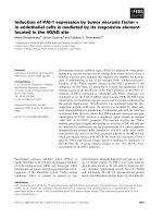
B (SRB) assay. The results showed that Lapatinib caused
a dose-dependent reduction of cell proliferation in all
NPC cells. The results also indicated that there were significant differences in Lapatinib sensitivity between the
NPC cell lines at 48 h (Fig. 1a) (2-way ANOVA, ****P <
0.0001). Moreover, the highly metastatic (C666–1 and 58F) lines were significantly more resistant to Lapatinib
than the three poorly metastatic lines (6-10B, TW01 and
HK-1) (Fig. 1b). These results were further confirmed by
clonogenic assays, in which the surviving cells were
allowed to further proliferate and form clones. The
highly metastatic C666–1 and 5-8F lines were also significantly more resistant to Lapatinib than the three
poorly metastatic lines (6-10B, TW01 and HK-1) in longer term assays (Fig. 1c, d).
Effects of Lapatinib on the expression and activity of
proteins involved in EGFR/ERBB2 signalling in the SUNE
5-8F and 6-10B NPC cells
In order to identify the potential mechanisms involved in modulating Lapatinib sensitivity, we analysed by Western blotting the effects of Lapatinib on
the expression of molecules implicated in Lapatinib
signalling and sensitivity. To this end, the two NPC
cell lines, SUNE 5-8F (highly metastatic) and 6-10B
(poorly metastatic) with differential Lapatinib sensitivity (low and high, respectively) were treated with
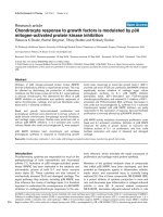
5 μM Lapatinib for 0 to 48 h (Fig. 2). Western blot
results showed a reduction in P-ERBB1 and P-ERBB2
upon Lapatinib treatment (Fig. 2), indicating the drug
is effective in inhibiting ERBB1/ERBB2 activity in
both NPC lines. Notably, both total ERBB1 and
ERBB2, but not ERBB3, were expressed at high levels
in both NPC lines and their expression remained at
high levels after Lapatinib treatment. FOXO3 has previously been shown to mediate the cytotoxic function
of Lapatinib through repressing FOXM1 expression.
The p38 and Jun N-terminal kinase (JNK) MAPKs
have also been demonstrated to phosphorylate and
activate FOXO3 in response to Lapatinib, while AKT
has been reported to be repressed by Lapatinib, leading to FOXO3 dephosphorylation (T32) and derepression. In agreement, western blot analysis showed that
Aimjongjun et al. BMC Cancer
Fig. 1 (See legend on next page.)
(2019) 19:1106
Page 5 of 17
Aimjongjun et al. BMC Cancer
(2019) 19:1106
Page 6 of 17
(See figure on previous page.)
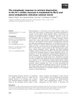
Fig. 1 Effects of Lapatinib on cell growth and proliferation of NPC cells. a Lapatinib sensitivity curves in NPC cells was determined by
sulforhodamine B staining. Cells (3 × 10³ / well) were seeded in 96-well plates and treated with increasing concentrations of Lapatinib for 72 h
before SRB staining and measurement of optical density at 492 nm. b Best-fit curves were generated to determine IC50 for each NPC cell lines
against Lapatinib. c NPC cells were seeded into 6-well plates (1 × 10³ cells / well) and treated with Lapatinib for 72 h. Colony formation was
monitored after 15 days, stained with crystal violet (representative images are shown) and absorbance at 592 nm determined (right panel). d Bestfit curves were generated to determine IC50 for each NPC cells against Lapatinib in terms of clonogenic assay. Numerical data represent the
average ± SEM of three independent experiments (**** P < 0.0001)
FOXO3 became dephoshorylated (T32) and activated
upon Lapatinib treatment in the sensitive 6-10B, but
remained phosphorylated (T32) and inactivated in the
resistant 5-8F cells. FOXO3 dephosphorylation was
also correlated with downregulation of FOXM1 expression in 6-10B but not in 5-8F cells, further confirming the role of FOXO3 in mediating Lapatinib
action. Interestingly, the induction of p38 and JNK
phosphorylation was not observed in the sensitive 610B cells upon Lapatinib treatment, suggesting p38
and JNK are unlikely to be instrumental in activating
FOXO3 in response to Lapatinib. FOXO3 activity can
also be enhanced by acetylation, which have been
shown to be promoted by EP300 and repressed by
the nuclear sirtuins, SIRT1, − 2 and − 6. Western blot
analysis showed EP300 was at low levels in the resistant, metastatic 5-8F cells but was induced upon Lapatinib treatment, whereas EP300 was expressed at high
levels in 6-10B and was downregulated in response to
Lapatinib. The results also showed SIRT1 and − 6
were expressed at comparable levels in both 5-8F and
6-10B cells, and their expression was not substantially
affected by Lapatinib treatment. The observed expression patterns of EP300, SIRT1 and SIRT6 suggested
that they are unlikely to be responsible for FOXO3
activation by Lapatinib, even though EP300, SIRT1
and SIRT6 could still modulate FOXO3 acetylation
and activity in these NPC cells. By contrast, upon
Lapatinib treatment the expression levels of SIRT2
remained constitutively high in the resistant 5-8F
cells, but were downregulated by Lapatinib in the sensitive 6-10B cells, suggesting that SIRT2 can potentially mediate Lapatinib response through modulating
FOXO3 acetylation and activity in these NPC cells.
These results suggest that highly metastatic and EBV
(Epstein Barr virus)-positive cell lines are more resistant to Lapatinib, probably due to their high levels of
FOXM1 and P-FOXO3 expression, suggesting that
SIRT2 induces Lapatinib resistance through FOXO3
acetylation and activity, which in turn led to reduction in FOXO3 activity.
FOXOs mediate the cytotoxic function of Lapatinib
FOXO proteins have previously been shown to be
functionally redundant and can compensate for one
another [27]. To verify the role of FOXO3 in mediating the cytotoxic response of Lapatinib, the effects of
Lapatinib treatment were investigated in wild-type
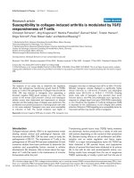
(WT) and foxo1/3/4 −/− MEFs. Western blot and
RTq-PCR analyses confirmed the Foxo1/3/4 knockout
in the foxo1/3/4 −/− MEFs and demonstrated EGFR/
HER2 overexpression in both wild-type (WT) and
foxo1/3/4 −/− MEFs (Fig. 3a, b), implying that they are
potentially sensitive to Lapatinib inhibition. Clonogenic assays showed that both wild-type (WT) and
foxo1/3/4 −/− MEFs are indeed sensitive to the antiproliferative functions of Lapatinib (Fig. 3c). Clonogenic assays also revealed that foxo1/3/4-deficient
MEFs displayed higher self-renewal ability and was
significantly less sensitive to the antiproliferative effects of Lapatinib compared with the WT MEFs (2way ANOVA, ****P < 0.0001) (Fig. 3c and d), confirming the key role of FOXOs in mediating the cytotoxic
functions of Lapatinib.
The clonogenic survival of NPC cells is sensitive to
chemical inhibitors of SIRTs
The anti-proliferative activity of FOXO3 is promoted by
acetylation which can be attenuated by nuclear SIRTs.
In order to investigate if the viability of the NPC cells
are also modulated by SIRTs, we subjected both the
Lapatinib resistant 5-8F and sensitive 6-10B NPC cells
to treatment with the SIRT1-specific (ie. EX527), SIRT2specific (ie. AK1 and AGK2) and pan-SIRT (ie. Sirtinol)
inhibitors individually for 72 h and their subsequent
long-term survival examined by clonogenic assays (Fig. 4a
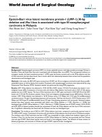
and b). The results showed that all four SIRT-inhibitors
decreased the clonogenic survival of both NPC cell lines,
with EX527, AK1 and AGK2 being more effective compared to Sirtinol, suggesting that the activity of SIRT1
and − 2 might have a role in modulating the long-term
viability of NPC cells.
SIRT2 inhibitors AK1 and AGK2 function cooperatively
with Lapatinib in NPC cells
Next, we tested if the SIRT inhibitors can act cooperatively with Lapatinib and enhance its antiproliferative functions. To this end, the resistant 5-8F and
sensitive 6-10B NPC cells were cultured with suboptimal levels of SIRT inhibitors (Fig. 2) or vehicle
Aimjongjun et al. BMC Cancer
(2019) 19:1106
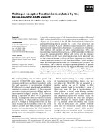
Fig. 2 Effects of Lapatinib on the expression and activity of proteins
involved in EGFR/ERBB2 signalling pathways in high and low
metastasis NPC cell lines. The high 5-8F and low 6-10B metastatic
NPC cells were treated with 5 μM Lapatinib for 0. 4, 8, 24, 48 h.
Protein lysates from whole-cell extracts were collected and then
analysed by western blotting using the antibodies against ERBB1, PERBB1, ERBB2, P-ERBB2, ERBB3, P-ERBB3, AKT, P-AKT, SIRT1, SIRT2,
SIRT6, p300, JNK, P-JNK, p38, P-p38, FOXO3, P-FOXO3(T32), FOXM1,
and β-tubulin. Molecular markers are shown on the right panel of
each blot. Representative results of three repeats are shown. Protein
levels were determined by using ImageJ and the protein levels
relative to β-tubulin are shown below protein bands as ratios to the
0 h Lapatinib controls
controls in the presence of increasing doses of Lapatinib and their cell viability examined by SRB assays
(Fig. 5a). Surprisingly, combining the pan-SIRT inhibitor Sirtinol with Lapatinib significantly increased the
overall viability of both NPC cell lines compared to
Page 7 of 17
Lapatinib treatment alone (2 way-ANOVA, ****P <
0.0001). Similarly, EX527 increased the viability of the
Lapatinib-treated 6-10B cells (2 way-ANOVA, **P =
0.0011) but had no significant effects on the viability
of the Lapatinib-treated 5-8F cells (2 way-ANOVA,
non-significant, P = 0.3667). By contrast, the SIRT2
inhibitor AGK2 demonstrated additive effects on the
cytotoxicity of Lapatinib (2 way-ANOVA, ****P <
0.0001, respectively) in both the sensitive 6-10B (IC50:
1.88 ± 0.26 to 5.94 ± 0.86 μM) and the resistant 5-8F
(10.55 ± 1.52 to 1.88 ± 0.26 μM) NPC lines (Fig. 5b).
Similarly, the SIRT2-inhibitor AK1 also functioned
additively with Lapatinib in the 5-8F cells (2 wayANOVA, *P = 0.0331, IC50, 11.77 ± 2.43 to 2.88 ±
0.57 μM) but had no significant positive effects on
Lapatinib in the sensitive 6-10F cells (2 way-ANOVA,
non-significant P = 0.3667, IC50, 5.80 ± 1.15 to = 6.50 ±
0.66 μM). AK1 did not demonstrate any additive effects with Lapatinib in 6-10B cells, probably because
of the predominant anti-proliferative function of
Lapatinib in the Lapatinib-sensitive cells. Moreover,
Lapatinib also functions to downregulate SIRT2 expression in 6-10B, and therefore, AK1 might be redundant in inhibiting SIRT2 after the downregulation
of SIRT2 in these cells by Lapatinib. However, overall
the SIRT2 inhibitors AGK2 and AK1, but not the
SIRT1 and pan-SIRT inhibitors, demonstrated additive
antiproliferative effects on Lapatinib in SRB cell proliferative assays, suggesting that SIRT2 plays a specific
role in limiting Lapatinib cytotoxicity and in promoting Lapatinib resistance in NPC cells.
To ascertain further the role of SIRT2 in enhancing
Lapatinib resistance in NPC cells, we performed clonogenic survival assays on the two NPC cell lines to study
the long-term antiproliferative effects of the two SIRT2
inhibitors AGK2 and AK1 on Lapatinib (Fig. 6a). In clonogenic assays, both SIRT2-inhibitors displayed additive
effects on Lapatinib in NPC cell lines (2 way-ANOVA,
****P < 0.0001, for all except **P < 0.0033 for AK1 in 610B), further reducing the overall clonogenic survival of
Lapatinib-treated cells (Fig. 6b). When combined with
Lapatinib, AGK2 showed additive effects on the antiproliferative functions of Lapatinib in both the Lapatinib
resistant 5-8F (IC50: 2.31 ± 1.06 to 0.21 ± 0.07 μM) and
sensitive 6-10B (IC50: 0.66 ± 0.08 to 0.21 ± 0.02 μM) NPC
cells (Fig. 6c). The SIRT2-inhibitor AK1 also functioned
additively with Lapatinib in both the resistant 5-8F and
(IC50: 1.07 ± 0.15 to 0.28 ± 0.03 μM) and sensitive 6-10B
(IC50: 0.66 ± 0.10 to 0.28 ± 0.05 μM) cells (Fig. 6c).
Silencing of SIRT2 enhances Lapatinib sensitivity of NPC
cells
To confirm further the role of SIRT2 in promoting
Lapatinib resistance, we next examined if siRNA-
Aimjongjun et al. BMC Cancer
Fig. 3 (See legend on next page.)
(2019) 19:1106
Page 8 of 17
Aimjongjun et al. BMC Cancer
(2019) 19:1106
Page 9 of 17
(See figure on previous page.)
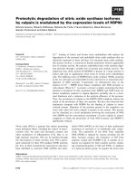
Fig. 3 FOXOs regulate Lapatinib sensitivity in mouse embryonic fibroblasts. a Western blot analysis was performed to analyse the protein levels
of P-EGFR, EGFR, P-ERBB2, ERBB2, FOXO3, FOXO1 in MEFs and Foxo1,3,4−/− MEFs. Representative western blot results are shown. Tubulin was used
as a protein loading control. b The mRNA levels of EGFR, ERBB2, FOXO3, FOXO1 and FOXO4 were assessed by real-time qPCR. The RPL19
housekeeping gene transcript was used as a control to normalise gene expression. c Wild-type (WT) MEFs and Foxo1,3,4−/− MEFs were treated
with increasing concentrations of Lapatinib for 3 days and colony formation was observed for 1 week. Cells were then stained with crystal violet
and absorbance measured at 592 nm (right panel). The colony formation capacity was analysed by two-way ANOVA and found to be significantly
different (***p < 0.00001) from one another. d Best-fit curves were generated to determine IC50 for each cell line against Lapatinib in terms of
clonogenicity. Numerical data represent the average ± SEM of three different experiments (**** P < 0.0001)
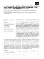
Fig. 4 SIRT inhibitors limit long-term cell growth and colony formation capacity of high and low metastatic NPC cells. a 5-8F and 6-10B cells
were seeded in 6-well plates and treated with the drug concentration indicated for 72 h. Then, cell culture media was removed and colony
formation observed for 15 days. At the end of this time period, cells were fixed with 4% paraformaldehyde and stained with crystal violet.
Representative pictures from triplicate experiments are shown. b The stain was solubilised with 33% acetic acid and absorbance at 592 nm were
obtained. Best fit-curves were generated to determine the IC50 of each drug in 5-8F and 6-10B cells
Aimjongjun et al. BMC Cancer
(2019) 19:1106
Page 10 of 17
Fig. 5 NPC cells show increased Lapatinib sensitivity when combined with AGK2 or AK1 but not Sirtinol or Ex527. a Cells (3 × 10³ / well) were
seeded in 96-well plates and treated with increasing concentrations of Lapatinib for 48 h before staining and measurement of optical density at
492 nm. Data were analysed by generating curves using Graph-pad Prism. b Best-fit curves were generated to determine the effect of AGK2 or
AK1 on reducing the IC50 of Lapatinib for each NPC cells. Data represent the average ± SEM of three different experiments (**** P < 0.0001)
depletion of SIRT2 can enhance the antiproliferative
effects of Lapatinib in the 5-8F and 6-10B NPC cells
using clonogenic assays. Knock-down of SIRT2 was
confirmed by western blotting analysis before the
NPC cells were subjects to clonogenic assays
(Fig. 7a). The results showed that despite the subtle
effects, depleting SIRT2 has additive effects on Lapatinib when compared with the non-silencing siRNA
controls in both the NPC cell lines (2 way-ANOVA,
****P < 0.0001, respectively) (Fig. 7b). Significantly,
the SIRT2-siRNA enhanced the anti-proliferative
functions of Lapatinib in both the resistant 5-8F and
(IC50: 4.64 ± 0.38 to 2.85 ± 0.41 μM) and sensitive 610B (IC50: 1.12 ± 0.10 to 0.90 ± 0.10 μM) cells
(Fig. 7c). In consequence, the results suggest that
SIRT2 knockdown can significantly enhance the
cytotoxic and cytostatic functions of Lapatinib in
both the sensitive and resistant NPC cells, consistent
with the findings from the SIRT2 pharmaceutical inhibitors, AK1 and AGK2. Collectively, these results
demonstrated that selective inhibition of SIRT2 can
promote Lapatinib sensitivity, indicating SIRT2 is a
potential target for overcoming Lapatinib resistance
in NPCs.
Inhibition and silencing of SIRT2 can combine with
Lapatinib to promote FOXO3 acetylation
To confirm that SIRT2 can modulate the ability of
Lapatinib to mediate FOXO3 acetylation, we next
determined if chemical inhibition or silencing of
SIRT2 can affect FOXO3 acetylation in the absence
or presence of Lapatinib treatment using co-
Aimjongjun et al. BMC Cancer
Fig. 6 (See legend on next page.)
(2019) 19:1106
Page 11 of 17
Aimjongjun et al. BMC Cancer
(2019) 19:1106
Page 12 of 17
(See figure on previous page.)
Fig. 6 Lapatinib in combination with AGK2/AK1 increase long term cell sensitivity compared to Lapatinib alone. a 5-8F and 6-10B cells were
seeded in 6-well plates and treated with the Lapatinib concentration indicated for 72 h. Five micromolar AGK2 and AK1 was used for
combination treatment. Cell culture media was then removed and colony formation observed for 15 days. At the end of this time period, cells
were fixed with 4% paraformaldehyde and stained with crystal violet. Representative pictures from triplicate experiments are shown. b The stain
was solubilised with 33% acetic acid and absorbance at 592 nm were obtained. Graphs were generated to show the effect of combination
treatment. c Best fit-curves were generated to determine the effect of AGK2/AK1 on the IC50 of Lapatinib in 5-8F and 6-10B cells.
immunoprecipitation (IP) assays (Fig. 8). Relative
FOXO3 acetylation levels were then determined by
comparing the ratio of acetylated to total FOXO3
co-precipitated. The computed results showed that
SIRT2-depletion by siRNA or -inhibition by AGK2
can induce FOXO3 acetylation in both the Lapatinib
resistant 5-8F and the sensitive 6-10B cell lines
(Fig. 8a and b). The chemical inhibitor AGK2 is
more effective in inducing FOXO3 acetylation than
SIRT2 siRNA probably because it is more effective
in inhibiting SIRT2 activity. Importantly, both chemical inhibition and siRNA-depletion of SIRT2 enhanced the FOXO3 acetylation induced by Lapatinib
in both the Lapatinib resistant 5-8F and sensitive 610B cell lines, except for the 6-10B cells treated with
SIRT2 siRNA. The low levels of acetylated FOXO3
precipitated from the sensitive 6-10B following combined Lapatinib and SIRT2-siRNA treatment is likely
to be due to the high levels of global protein degradation as a result of cell death caused by the combination of SIRT2-depletion and Lapatinib treatment.
Moreover, Lapatinib also downregulates SIRT2 in the
sensitive 6-10B cell line, rendering the effects of
SIRT2-siRNA redundant. Overall, these IP results
suggest that SIRT2 restricts the Lapatinib-mediated
FOXO3 acetylation in NPCs. In summary, our collective results reveal that SIRT2 has a role in modulating FOXO3 acetylation and Lapatinib response in
NPCs and that targeting SIRT2 can enhance the sensitivity of NPCs to Lapatinib and to overcome
Lapatinib-resistance in NPC.
Discussion
Metastasis and drug resistance are the characteristics
of aggressive cancers and the major causes for poor
survival in nasopharyngeal carcinoma (NPC) [12, 13].
An understanding of the mechanisms involved in the
development of NPC metastasis and drug resistance
will aid the development of early diagnostic biomarkers and the identification of potential therapeutic
targets. Previous studies have reported Lapatinib
could effectively induce cell death and autophagy in
NPC cells [12, 13]. Clinical studies also suggest that
Lapatinib alone or combined with standard chemotherapies are well-tolerated and promote patient survival in metastatic/recurrent head and neck squamous
cell carcinoma [28]; however, not all patients benefit
from Lapatinib-based treatments [29, 30]. In the
present study, we found that the highly metastatic
(C666–1 and 5-8F) NPC lines are significantly more
Lapatinib resistant compared to poorly metastatic
lines (6-10B, TW01 and HK-1), suggesting that the
molecular mechanisms involved in metastasis in NPC
may overlap with those responsible for the development of Lapatinib resistance.
FOXO3 is a tumour suppressive transcription factor
and an important modulator of sensitivity to chemotherapy [31, 32]. FOXO3 inhibits cell growth by driving the transcription of genes, such as Bim, FasL,
p27Kip1, p130 (RB2), essential for cell proliferative arrest, cell death and differentiation [31, 32]. Conversely, inactivation of FOXO3 is a crucial step for
oncogenic transformation and the development of
cytotoxic drug resistance [31, 32]. Previous work has
also suggested that FOXO3 mediates the cytotoxicity
of Lapatinib in breast cancer [33, 34]. In here, we
have shown definitively using foxo1/3/4-deficient fibroblasts that FOXO3 along with FOXO1 and − 4 are
involved in mediating the cytotoxic functions of Lapatinib. Consistent with this, recent evidence has
showed that natural products, such as curcumin, inhibits the growth and induces apoptosis in NPC cell
by increasing FOXO3 expression [35]. In addition, the
G-quadruplex ligand SYUIQ-5 also induces NPC autophagy by down-regulating Akt phosphorylation and
promoting FOXO3 nuclear translocation [36].
The activity, expression and subcellular localization of
FOXO3 are regulated by a diverse range of posttranslational modifications [37]. Phosphorylation by kinases, particularly Akt (also called PKB) FOXO3, ERK,
IKB kinase (IKK) and serum and glucocorticoidregulated kinase (SGK) can enhance FOXO3 nuclear to
cytoplasmic shuttling and its degradation [32, 37]. Conversely, other kinases, such as p38 MAPK [38], stress activated c-Jun-NH2-kinase (JNK) [39] have also been
demonstrated to promote FOXO3 activity and expression. Consistent with previous studies with EGFR1/
HER2(ERBB2)-targeted tyrosine kinase inhibitors (TKIs)
[34, 40], we found that Lapatinib can cause FOXO3dephosphorylation (T32) and the downregulation of its
target FOXM1 in Lapatinib-sensitive NPC cells. However, although EGFR1/HER2 inhibitors have previously
Aimjongjun et al. BMC Cancer
Fig. 7 (See legend on next page.)
(2019) 19:1106
Page 13 of 17
Aimjongjun et al. BMC Cancer
(2019) 19:1106
Page 14 of 17
(See figure on previous page.)
Fig. 7 Silencing of SIRT2 increases long-term Lapatinib cytotoxicity in NPC cells. a NPC cells were transfected with siNSC and siSIRT2 smartpools.
After 24 h both siNSC and siSIRT2-transfected cells were treated with DMSO or 5 μM Lapatinib for another 24 h. Forty-eight h after transfection,
cells were harvested and SIRT2 protein knockdown was confirmed by western blot analysis. Protein levels were determined by using ImageJ and
the protein levels relative to β-tubulin are shown below protein bands as ratios to the 0 h Lapatinib controls. b Forty-eight h after transfection,
1000 cells were seeded per well, into 6-well plates and then treated with the Lapatinib concentrations of 0, 0.01, 0.05, 0.1, 1 and 5 μM for 72 h
and colony formation observed for 15 days. At the end of this time period, cells were fixed with 4% paraformaldehyde and stained with crystal
violet. The stain was solubilised with 33% acetic acid and absorbance at 592 nm were obtained. Graphs were generated to show the effect of
silencing SIRT2 on Lapatinib sensitivity. c Best fit-curves were generated to determine the effect of SIRT2 knock down on the IC50 of Lapatinib in
5-8F and 6-10B cells. Numerical data represent the average ± SEM of three different experiments (**** P < 0.0001)
been shown to modulate p38 and JNK activity [41, 42],
we did not observe any substantial changes in p38 and
JNK phosphorylation/activity in NPC cells in response
to Lapatinib treatment, suggesting it is unlikely that
Lapatinib modulates FOXO3 activity via p38 and JNK in
these NPC cells. FOXM1 is a potent oncogene negatively
regulated by FOXO3 [32] and contributes to cancer drug
resistance through controlling many genes involved in
cell proliferation, survival, DNA repair, and tubulin
destabilization [32, 43, 44]. Consistent with a role for
FOXO3 in Lapatinib action in NPC cells, we found that
FOXM1 expression is repressed by Lapatinib in the sensitive cells but remains constitutively high in the resistant NPC cells, suggesting a role of FOXM1 in NPC
Lapatinib resistance. In agreement, FOXM1 has been
shown to be able to mediate paclitaxel resistance by
regulating the gene transcription of the ABCC5 drug efflux transporter in NPC [18]. In addition, overexpression
of FOXM1 is directly associated with metastasis in NPC,
and targeting FOXM1 with inhibitors or siRNA knockdown can effectively restrict the cell proliferation, migration, angiogenesis and survival of NPC cells [29, 45].
Besides phosphorylation, FOXO3 is also regulated
by other post-translational modifications such as
acetylation, methylation, ubiquitination and glycosylation (Zhao et al., 2011). The nuclear sirtuins SIRT1,
SIRT2 and SIRT6 have been shown to negatively
regulate FOXO3 acetylation and its activity [46–48].
In the present study, we found that SIRT2 specifically modulates the cytotoxicity of Lapatinib and is
linked to Lapatinib resistance using specific chemical
inhibitors and SIRT2 siRNAs. Although our findings
show that all four SIRT inhibitors (i.e. sirtinol,
EX527, AGK2 and AK1) can limit NPC cell proliferation, only the SIRT2 specific inhibitors AGK2 and
AK1 function cooperatively with Lapatinib, supporting the idea that SIRT2 specifically modulates Lapatinib response and resistance. Nevertheless, all four
SIRT inhibitors can restrict normal NPC cell viability
and clonogenicity, and this suggests that the nuclear
sirtuins SIRT1, − 2 and − 6 detected in NPC are all
potential oncogenes. In agreement, SIRT1 upregulation has been shown to be associated with tumour
progression and metastasis in NPC biopsies [49].
The pan-SIRT inhibitor sirtinol has previously been
shown to induce p53 acetylation and cell death
through targeting both SIRT1 and SIRT2 [50]. However, like EX527, the sirtinol concentrations
employed here preferentially inhibit SIRT1 and have
limited activities towards SIRT2 [50]. Therefore,
these observations support further the notion that
SIRT2 specifically moderates the cytotoxic action of
Lapatinib and mediates Lapatinib resistance, independently of SIRT1. The reason for the finding that
both the SIRT inhibitors sirtinol and EX527 oppose
the cytotoxic functions of Lapatinib is unclear. However, a context dependent tumour suppressive role
for SIRT1 has previously been proposed [26]. In
concordance, SIRT1-deficient cells have been shown
to be defective in the ability to normally upregulate
the p19(ARF) senescence mediator and its potent
downstream tumour suppressor p53 [26], and this
might account for the ability of sirtinol and EX527
to attenuate the antiproliferative function of Lapatinib. Inhibition of SIRT2 by chemical inhibitor or silencing significantly enhances the cytotoxicity of
Lapatinib not only in the sensitive but also in the resistant NPC cells, suggesting that SIRT2 not only
modulates the cytotoxic functions of Lapatinib in the
sensitive NPC cells, but it also mediates Lapatinib
resistance. This implies that targeting SIRT2 can enhance the cytotoxicity of Lapatinib and may represent a novel strategy for overcoming Lapatinib
resistance in NPC. This role of SIRT2 in modulating
Lapatinib response and sensitivity is confirmed by
SIRT2 depletion experiments showing silencing
SIRT2 can enhance the cytotoxicity in both Lapatinib sensitive and resistant NPC cells. Although the
siRNA depletion approach has less off-target effects,
it also relies on high delivery efficiencies; the incomplete SIRT2 depletion might account for the small
additional effects on Lapatinib.
In agreement, overexpression of SIRT2 has been shown
to essentially lengthen the M phase and defer mitotic exit
[15]. Moreover, SIRT2 can induce the cell proliferation of
leukaemia and resistance to apoptosis [51]. Conversely,
decreased SIRT2 activity can reduce glioma cell survival
by induced both necrosis and apoptosis [52] and limit
Aimjongjun et al. BMC Cancer
(2019) 19:1106
Page 15 of 17
Fig. 8 Inhibition of SIRT2 by AGK2 as well as silencing of SIRT2 in combination with Lapatinib upregulates acetylated FOXO3 levels in 5-8F and 610B cells. a NPC were treated with AGK2 alone or in combination with Lapatinib for 48 h. Proteins were obtained from whole cell extracts and
assessed by co-immunoprecipitation (co-IP) with an anti-FOXO3 antibody. Subsequent immunoblotting was performed using antibodies against
Ac-Lys and FOXO3. The anti-IgG was used as a negative control. Acetylated FOXO3 levels were determined by using ImageJ analysis and
representative bar diagram are shown (bottom panel after the blot). b NPC cells were transiently transfected with siNSC and siSIRT2 and treated
with 5 μM Lapatinib for 24 h. Proteins were obtained from whole cell extracts and assessed by Co-immunoprecipitation (Co-IP) with an antiFOXO3 antibody. Subsequent immunoblotting was performed using antibodies against Ac-Lys and FOXO3. Acetylated and total FOXO3 levels
were determined by using ImageJ analysis and representative bar diagrams demonstrating relative acetylated to total FOXO3 ratios are shown
(bottom panel after the blot).
Aimjongjun et al. BMC Cancer
(2019) 19:1106
melanoma cell growth and clonogenicity [53]. Furthermore, overexpression of SIRT1 and SIRT2 can also confer
resistance to chemotherapy such as paclitaxel [54].
Furthermore, our co-IP experiments also show that
the Lapatinib induces FOXO3 acetylation and that inhibition of SIRT2 by chemical inhibitor or silencing also
promotes FOXO3 acetylation in NPC cells. Importantly,
SIRT2 inhibition or silencing can combine with Lapatinib to cause further enhancement of FOXO3 acetylation
than Lapatinib treatment alone in both sensitive and resistant NPC cells, suggesting that SIRT2 can moderate
the cytotoxic functions of Lapatinib and promote resistance through limiting FOXO3 acetylation. In agreement,
FOXO3 deacetylation by SIRT2 has previously been reported to enhance FOXO3 ubiquitination and proteasomal degradation [55].
Conclusion
Collectively, these data suggest that the cytotoxic functions of Lapatinib are mediated through the acetylation
and activation of FOXO3, and that SIRT2 can specifically antagonise the cytotoxicity of Lapatinib through mediating FOXO3 deacetylation in both sensitive and
resistant NPC cells. The present findings also suggest
that SIRT2 can be an important biomarker for metastatic and Lapatinib resistant NPC and that targeting the
SIRT2-FOXO3 axis may provide a means to treat NPC
and to overcome NPC chemoresistance.
Abbreviations
EGFR: Epidermal growth factor receptor; FOX: Forkhead box;
NPC: Nasopharyngeal carcinoma; SIRT2: Sirtuin-2
Acknowledgements
Not Applicable
Authors’ contributions
Conceptualization, EW-FL, TJ, SA and ZM; Methodology, EW-FL, ZM and SA;
Validation, SA, ZM and EW-FL; Formal Analysis, SA and ZM; Investigation, SA,
ZM and EW-FL; Resources, EW-FL and TJ; Data Curation, ZM, SY, SA, YJ and
GA; Writing – Original Draft Preparation, EW-FL, SA and ZM; Writing – Review
& Editing, ZM, SY and GA; Supervision, EW-FL, TJ and EY; Project Administration, EW-FL and TJ. All authors read and approved the final manuscript.
Funding
Eric W.-F. Lam’s work is supported by MRC (MR/N012097/1), CRUK (C37/
A12011), Breast Cancer Now (2012MayPR070, 2012NovPhD016,
2014NovPhD326), the Cancer Research UK Imperial Centre, Imperial ECMC
and NIHR Imperial BRC. Tavan Janvilisri was supported by Thailand Research
Fund (BRG5980003). Glowi Alasiri is a recipient of a scholarship from the
Saudi Arabian Cultural Bureau in London (MSU434). Sathid Aimjongjun’s
work was supported by Newton Fund, Ph. D placement programme and The
Royal Golden Jubilee Ph.D. Program. Zimam Mahmud was supported by a
fellowship from the Commonwealth Scholarship Commission (BDCS-201563). The funding bodies had no role on the design, data collection, analysis
and manuscript writing of this study.
Availability of data and materials
Data sharing is not applicable to this article as no datasets were generated
or analysed during the current study.
Page 16 of 17
Ethics approval and consent to participate
Not applicable.
Consent for publication
Not applicable
Competing interests
The authors declare that they have no competing interests.
Author details
1
Department of Surgery and Cancer, Imperial College London, Hammersmith
Hospital Campus, London W12 0NN, UK. 2Graduate Program in Molecular
Medicine, Multidisciplinary Unit, Faculty of Science, Mahidol University,
Bangkok, Thailand. 3Department of Biochemistry, Faculty of Science, Mahidol
University, Bangkok, Thailand.
Received: 24 June 2019 Accepted: 29 October 2019
References
1. Wei WI, Sham JST. Nasopharyngeal carcinoma. Lancet. 2005;365(9476):2041–54.
2. Mahdavifar N, Ghoncheh M, Mohammadian-Hafshejani A, Khosravi B,
Salehiniya H. Epidemiology and inequality in the incidence and mortality of
nasopharynx cancer in Asia. Osong Public Health Res Perspect. 2016;7(6):
360–72.
3. Ma BBY, Chan ATC. Recent perspectives in the role of chemotherapy in the
management of advanced nasopharyngeal carcinoma. Cancer. 2005;103(1):
22–31.
4. Hsu CH, Chen CL, Hong RL, Chen KL, Lin JF, Cheng AL. Prognostic value of
multidrug resistance 1, glutathione-<i>S</i>−transferase-π and p53 in
advanced nasopharyngeal carcinoma treated with systemic chemotherapy.
Oncology. 2002;62(4):305–12.
5. Jiang R-D, Zhang L-X, Yue W, Zhu Y-F, Lu H-J, Liu X, et al. Establishment of a
human nasopharyngeal carcinoma drug-resistant cell line CNE2/DDP and
screening of drug-resistant genes; 2003. p. 337–45.
6. Cheung HW, Jin D-Y, Ling M-t, Wong YC, Wang Q, Tsao SW, et al. Mitotic
arrest deficient 2 expression induces chemosensitization to a DNAdamaging agent, cisplatin, in nasopharyngeal carcinoma cells. Cancer Res.
2005;65(4):1450.
7. Wang J, Wang H, Zhao L, Fan S, Yang Z, Gao F, et al. Down-regulation of Pglycoprotein is associated with resistance to cisplatin and VP-16 in human
lung cancer cell lines. Anticancer Res. 2010;30(9):3593–8.
8. Pan Y, Zhou F, Zhang R, Claret FX. Stat3 inhibitor stattic exhibits potent
antitumor activity and induces chemo- and radio-sensitivity in
nasopharyngeal carcinoma. PLoS One. 2013;8(1):e54565.
9. Ma BB, Poon TC, To KF, Zee B, Mo FK, Chan CM, et al. Prognostic
significance of tumor angiogenesis, Ki 67, p53 oncoprotein, epidermal
growth factor receptor and HER2 receptor protein expression in
undifferentiated nasopharyngeal carcinoma--a prospective study. Head
Neck. 2003;25(10):864–72.
10. Jin O, Chen S, Li G, Yao K. Expression of CerbB-2 and EGFR mRNA in human
nasopharyngeal carcinomas and pericarcinomatous tissues. Hunan Yi Ke Da
Xue Xue Bao. 1997;22(6):487–90.
11. Ma BB, Lui VW, Poon FF, Wong SC, To KF, Wong E, et al. Preclinical activity
of gefitinib in non-keratinizing nasopharyngeal carcinoma cell lines and
biomarkers of response. Investig New Drugs. 2010;28(3):326–33.
12. Liu L, Huang P, Wang Z, Chen N, Tang C, Lin Z, et al. Inhibition of eEF2 kinase sensitizes human nasopharyngeal carcinoma cells to Lapatinibinduced apoptosis through the Src and Erk pathways. BMC Cancer.
2016;16:813.
13. Lui VWY, Lau CPY, Ho K, Ng MHL, Cheng SH, Tsao S-W, et al. Anti-invasion,
anti-proliferation and anoikis-sensitization activities of Lapatinib in
nasopharyngeal carcinoma cells. Investig New Drugs. 2011;29(6):1241–52.
14. Wilson MSC, Brosens JJ, Schwenen HDC, Lam EW-F. FOXO and FOXM1 in
cancer: the FOXO-FOXM1 axis shapes the outcome of cancer
chemotherapy. Curr Drug Targets. 2011;12(9):1256–66.
15. Olmos Y, Brosens JJ, Lam EW. Interplay between SIRT proteins and tumour
suppressor transcription factors in chemotherapeutic resistance of cancer.
Drug Resist Updat. 2011;14(1):35–44.
16. Daitoku H, Sakamaki J-i, Fukamizu A. Regulation of FoxO transcription factors by
acetylation and protein–protein interactions. Mol Cell Res. 2011;1813(11):1954–60.
Aimjongjun et al. BMC Cancer
(2019) 19:1106
17. Jiang L, Wang P, Chen H. Overexpression of FOXM1 is associated with
metastases of nasopharyngeal carcinoma. Ups J Med Sci. 2014;119(4):324–32.
18. Hou Y, Zhu Q, Li Z, Peng Y, Yu X, Yuan B, et al. The FOXM1–ABCC5 axis
contributes to paclitaxel resistance in nasopharyngeal carcinoma cells. Cell
Death Dis. 2017;8(3):e2659.
19. Shou Z, Lin L, Liang J, Li J-L, Chen H-Y. Expression and prognosis of FOXO3a and
HIF-1α in nasopharyngeal carcinoma. J Cancer Res Clin Oncol. 2012;138(4):585–93.
20. Shu CH, Yang WK, Shih YL, Kuo ML, Huang TS. Cell cycle G2/M arrest and
activation of cyclin-dependent kinases associated with low-dose paclitaxelinduced sub-G1 apoptosis. Apoptosis. 1997;2(5):463–70.
21. Song LB, Yan J, Jian SW, Zhang L, Li MZ, Li D, et al. Molecular mechanisms
of tumorgenesis and metastasis in nasopharyngeal carcinoma cell sublines.
Ai Zheng. 2002;21(2):158–62.
22. Huang PY, Hong MH, Zhang X, Mai HQ, Luo DH, Zhang L. C-KIT
overexpression and mutation in nasopharyngeal carcinoma cell lines and
reactivity of Imatinib on these cell lines. Chin J Cancer. 2010;29(2):131–5.
23. Huang DP, Ho JH, Poon YF, Chew EC, Saw D, Lui M, et al. Establishment of a
cell line (NPC/HK1) from a differentiated squamous carcinoma of the
nasopharynx. Int J Cancer. 1980;26(2):127–32.
24. Cheung ST, Huang DP, Hui AB, Lo KW, Ko CW, Tsang YS, et al.
Nasopharyngeal carcinoma cell line (C666-1) consistently harbouring
Epstein-Barr virus. Int J Cancer. 1999;83(1):121–6.
25. van der Vos KE, Coffer PJ. The extending network of FOXO transcriptional
target genes. Antioxid Redox Signal. 2011;14(4):579–92.
26. Chua KF, Mostoslavsky R, Lombard DB, Pang WW, Saito S, Franco S, et al.
Mammalian SIRT1 limits replicative life span in response to chronic
genotoxic stress. Cell Metab. 2005;2(1):67–76.
27. Paik JH, Kollipara R, Chu G, Ji H, Xiao Y, Ding Z, et al. FoxOs are lineagerestricted redundant tumor suppressors and regulate endothelial cell
homeostasis. Cell. 2007;128(2):309–23.
28. Weiss JM, Bagley S, Hwang W-T, Bauml J, Olson JG, Cohen RB, et al.
Capecitabine and Lapatinib for the first-line treatment of metastatic/recurrent
head and neck squamous cell carcinoma. Cancer. 2016;122(15):2350–5.
29. Machiels J-PH, Haddad RI, Fayette J, Licitra LF, Tahara M, Vermorken
JB, et al. Afatinib versus methotrexate as second-line treatment in
patients with recurrent or metastatic squamous-cell carcinoma of the
head and neck progressing on or after platinum-based therapy (LUXHead & Neck 1): an open-label, randomised phase 3 trial. Lancet
Oncol. 2015;16(5):583–94.
30. Harrington KJ, Temam S, D'Cruz A, Jain MM, D'Onofrio I, Manikhas GM, et al.
Final analysis: a randomized, blinded, placebo (P)-controlled phase III study of
adjuvant postoperative Lapatinib (L) with concurrent chemotherapy and
radiation therapy (CH-RT) in high-risk patients with squamous cell carcinoma
of the head and neck (SCCHN). J Clin Oncol. 2014;32(15_suppl):6005.
31. Yao S, Fan LY, Lam EW. The FOXO3-FOXM1 axis: a key cancer drug target and
a modulator of cancer drug resistance. Semin Cancer Biol. 2018;50:77–89.
32. Myatt SS, Lam EW. The emerging roles of forkhead box (Fox) proteins in
cancer. Nat Rev Cancer. 2007;7(11):847–59.
33. Xia W, Bacus S, Hegde P, Husain I, Strum J, Liu L, et al. A model of acquired
autoresistance to a potent ErbB2 tyrosine kinase inhibitor and a therapeutic
strategy to prevent its onset in breast cancer. Proc Natl Acad Sci U S A.
2006;103(20):7795–800.
34. Karadedou CT, Gomes AR, Chen J, Petkovic M, Ho KK, Zwolinska AK, et al.
FOXO3a represses VEGF expression through FOXM1-dependent and
-independent mechanisms in breast cancer. Oncogene. 2012;31(14):1845–58.
35. Wu J, Tang QIN, Zhao S, Zheng F, Wu YAN, Tang GE, et al. Extracellular
signal-regulated kinase signaling-mediated induction and interaction of
FOXO3a and p53 contribute to the inhibition of nasopharyngeal carcinoma
cell growth by curcumin. Int J Oncol. 2014;45(1):95–103.
36. Zhou W-J, Deng R, Feng G-K, Zhu X-F. A G-quadruplex ligand SYUIQ-5
induces autophagy by inhibiting the Akt-FOXO3a pathway in
nasopharyngeal cancer cells; 2009. p. 1049–53.
37. Lam EW, Brosens JJ, Gomes AR, Koo CY. Forkhead box proteins: tuning forks
for transcriptional harmony. Nat Rev Cancer. 2013;13(7):482–95.
38. Ho KK, McGuire VA, Koo CY, Muir KW, de Olano N, Maifoshie E, et al.
Phosphorylation of FOXO3a on Ser-7 by p38 promotes its nuclear
localization in response to doxorubicin. J Biol Chem. 2012;287(2):1545–55.
39. Sunters A, Madureira PA, Pomeranz KM, Aubert M, Brosens JJ, Cook SJ,
et al. Paclitaxel-induced nuclear translocation of FOXO3a in breast
cancer cells is mediated by c-Jun NH2-terminal kinase and Akt. Cancer
Res. 2006;66(1):212–20.
Page 17 of 17
40. McGovern UB, Francis RE, Peck B, Guest SK, Wang J, Myatt SS, et al. Gefitinib
(Iressa) represses FOXM1 expression via FOXO3a in breast cancer. Mol
Cancer Ther. 2009;8(3):582–91.
41. Mora Vidal R, Regufe da Mota S, Hayden A, Markham H, Douglas J, Packham
G, et al. Epidermal growth factor receptor family inhibition identifies P38
mitogen-activated protein kinase as a potential therapeutic target in
bladder cancer. Urology. 2018;112:225 e1–7.
42. Gschwantler-Kaulich D, Grunt TW, Muhr D, Wagner R, Kolbl H, Singer CF.
HER specific TKIs exert their antineoplastic effects on breast cancer cell lines
through the involvement of STAT5 and JNK. PLoS One. 2016;11(1):e0146311.
43. Carr JR, Park HJ, Wang Z, Kiefer MM, Raychaudhuri P. FoxM1 mediates
resistance to herceptin and paclitaxel. Cancer Res. 2010;70(12):5054–63.
44. Miranda SCW, Jan JB, Helma DCS, Eric WFL. FOXO and FOXM1 in cancer:
the FOXO-FOXM1 axis shapes the outcome of cancer chemotherapy. Curr
Drug Targets. 2011;12(9):1256–66.
45. Jiang L, Wang P, Chen L, Chen H. Down-regulation of FoxM1 by
thiostrepton or small interfering RNA inhibits proliferation, transformation
ability and angiogenesis, and induces apoptosis of nasopharyngeal
carcinoma cells. Int J Clin Exp Pathol. 2014;7(9):5450–60.
46. Wang F, Nguyen M, Qin FX-F, Tong Q. SIRT2 deacetylates FOXO3a in response
to oxidative stress and caloric restriction. Aging Cell. 2007;6(4):505–14.
47. Brunet A, Sweeney LB, Sturgill JF, Chua KF, Greer PL, Lin Y, et al. Stressdependent regulation of FOXO transcription factors by the SIRT1
deacetylase. Science. 2004;303(5666):2011.
48. Khongkow M, Olmos Y, Gong C, Gomes AR, Monteiro LJ, Yague E, et al.
SIRT6 modulates paclitaxel and epirubicin resistance and survival in breast
cancer. Carcinogenesis. 2013;34(7):1476–86.
49. Hu C, Wei W, Chen X, Woodman CB, Yao Y, Nicholls JM, et al. A global view
of the oncogenic landscape in nasopharyngeal carcinoma: an integrated
analysis at the genetic and expression levels. PLoS One. 2012;7(7):e41055.
50. Peck B, Chen CY, Ho KK, Di Fruscia P, Myatt SS, Coombes RC, et al. SIRT
inhibitors induce cell death and p53 acetylation through targeting both
SIRT1 and SIRT2. Mol Cancer Ther. 2010;9(4):844–55.
51. Dan L, Klimenkova O, Klimiankou M, Klusman J-H, van den Heuvel-Eibrink
MM, Reinhardt D, et al. The role of sirtuin 2 activation by nicotinamide
phosphoribosyltransferase in the aberrant proliferation and survival of
myeloid leukemia cells. Haematologica. 2012;97(4):551–9.
52. He X, Nie H, Hong Y, Sheng C, Xia W, Ying W. SIRT2 activity is required for the
survival of C6 glioma cells. Biochem Biophys Res Commun. 2012;417(1):468–72.
53. Wilking-Busch MJ, Ndiaye MA, Liu X, Ahmad N. RNA interference-mediated
knockdown of SIRT1 and/or SIRT2 in melanoma: identification of
downstream targets by large-scale proteomics analysis. J Proteome. 2018;
170:99–109.
54. Matsushita N, Takami Y, Kimura M, Tachiiri S, Ishiai M, Nakayama T, et al. Role of
NAD-dependent deacetylases SIRT1 and SIRT2 in radiation and cisplatininduced cell death in vertebrate cells. Genes Cells. 2005;10(4):321–32.
55. Wang F, Chan CH, Chen K, Guan X, Lin HK, Tong Q. Deacetylation of FOXO3
by SIRT1 or SIRT2 leads to Skp2-mediated FOXO3 ubiquitination and
degradation. Oncogene. 2011;31:1546.
Publisher’s Note
Springer Nature remains neutral with regard to jurisdictional claims in
published maps and institutional affiliations.