Báo cáo y học: "Pathogenic organisms in hip joint infections"
Bạn đang xem bản rút gọn của tài liệu. Xem và tải ngay bản đầy đủ của tài liệu tại đây (1.01 MB, 7 trang )
Int. J. Med. Sci. 2009, 6
234
I
I
n
n
t
t
e
e
r
r
n
n
a
a
t
t
i
i
o
o
n
n
a
a
l
l
J
J
o
o
u
u
r
r
n
n
a
a
l
l
o
o
f
f
M
M
e
e
d
d
i
i
c
c
a
a
l
l
S
S
c
c
i
i
e
e
n
n
c
c
e
e
s
s
2009; 6(5):234-240
© Ivyspring International Publisher. All rights reserved
Review
Pathogenic organisms in hip joint infections
Udo Geipel
Institute of Medical Microbiology and Hygiene, University of Saarland Hospital, Homburg (GER)
Correspondence to: Udo Geipel, MD, Institute of Medical Microbiology and Hygiene, University of Saarland Hospital,
Kirrberger Strasse, Bldg 43, 66421 Homburg, Germany. Phone: +49-6841-162-3946; Fax: +49-6841-162-3985; Email:
Received: 2009.08.10; Accepted: 2009.08.28; Published: 2009.09.02
Abstract
Infections of the hip joint are usually of bacterial etiology. Only rarely, an infectious arthritis
is caused in this localization by viruses or fungi. Native joint infections of the hip are less
common than infections after implantation of prosthetic devices. Difficulties in prosthetic
joint infections are, (I) a higher age of patients, and, thus an associated presence of other
medical risk factors, (II) often long courses of treatment regimes depending on the bacte-
rium and its antibiotic resistance, (III) an increased mortality, and (IV) a high economic bur-
den for removal and reimplantation of an infected prosthetic device. The pathogenic
mechanisms responsible for articular infections are well studied only for some bacteria, e.g.
Staphylococcus aureus, while others are only partially understood. Important known bacterial
properties and microbiological characteristics of infection are the bacterial adhesion on the
native joint or prosthetic material, the bacterial biofilm formation, the development of small
colony variants (SCV) as sessile bacterial types and the increasing resistance to antibiotics.
Key words: arthritis, bacteria, diagnosis, prosthesis, therapy
Infectious arthritis
The infection of a joint can occur in different
ways, (I) via injection or during joint operation
through direct colonization, (II) by direct contact with
a neighboring infected site, or (III) by haematogenous
or lymphogenous seed of the pathogen. Another clas-
sification of bacterial arthritis distinguishes acute,
chronic and reactive forms, which differ in their type
of joint infection and their triggering bacteria. Reac-
tive arthritis is a postinfectious complication with no
need of presence for viable pathogens in the joint.
While reactive arthritis often simultaneously affect
several joints, the presence of polyarthritic types of
non reactive arthritis occur infrequently and then
mostly as a result of several bacteriaemic phases.
Among joint infections, the hip is the second
most frequent localization after the knee joint. Basi-
cally, there are no differences in the bacterial spec-
trum between hip joint infections and those of other
large joints. Hip joint infections, however, are aggra-
vated by the fact that they can exist over a long time
with only poor symptoms. An increased rate of infec-
tion occurs in the pre-damaged joint and is also asso-
ciated with particular predispositions of the patients
(Table 1) [1,2,3]. In particular, a joint prosthesis is a
high risk predisposition for an infection. Periopera-
tively the initial bacterial entry into the joints may
occur. On the other hand the implanted foreign mate-
rial causes in addition to the severe joint disease pre-
sent an additional reduction in local resistance, which
facilitates haematogenous infections. The prosthetic
materials are also additional binding sites for various
bacteria, and act as a starting point for prosthetic in-
fections. Thus, in addition to the local conditions, the
bacterial properties and their specific pathogenity
have to be considered for understanding the whole
mechanism of infection. Basically, a too late or not
Int. J. Med. Sci. 2009, 6
235
sufficiently cured joint infection can cause trophic and
functional limitations or can even be the starting point
of a progressive infection spreading in continuity,
lymphogenic or haematogenic. In general, the detec-
tion and treatment of acute infectious arthritis is an
acute emerging situation, in which a delay may pro-
gress to further septic inflammation [2].
Table 1: Predisposing factors
Bacteria responsible for (hip) joint infections
Some bacteria have preferences for certain infec-
tion routes and patterns. Infections not related to in-
juries or medical interventions (e.g. intraarticular
puncture, joint replacement) are mostly resulting
from often physiologic bacteriaemic periods. The
most frequently detected pathogens of joint infections
are staphylococci. Staphylococcus aureus has domi-
nance in acute purulent arthritis while coagu-
lase-negative staphylococci can be found mainly in
periprosthetic infections and after diagnostic arthro-
scopies. Other gram-positive bacteria as causative
agents for hip joint infections are streptococci, espe-
cially Streptococcus pyogenes, Enterococcus faecalis and
Corynebacteria species.
A large number of different gram-negative rods
act as infectious agents on joints. The group of en-
terobacteria contains a broad spectrum of pathogens.
Salmonella enterica, Shigella species, and Yersinia species
are classically described as pathogens for purulent
and reactive forms of arthritis. Pseudomonas aeruginosa
can be found more often in predisposed patients (e.g.
diabetics). In otherwise healthy people it is associated
with iatrogenic modes of infection during diagnostic
procedures. Campylobacter species, however, are classic
agents of reactive arthritis, as well as the obligate in-
tracellular bacteria Chlamydia trachomatis, Mycoplasma
pneumoniae, and Ureaplasma urealyticum. From the
spirochaetales only Borrelia burgdorferi sensu lato is
relevant. Less commonly identified organisms for
joint infections often accompanied with osteitis or
osteomyelitis are Brucella species and Mycobacterium
tuberculosis. Anaerobes, such as Bacteroides fragilis, are
rarely found and are usually part of a polymicrobial
infection. An overview is shown in Table 2.
Table 2: Bacteria responsible for (hip) joint infections
Pathogenesis
If bacteria reach into the joint they can bind to a
large number of different binding molecules. Espe-
cially fibrinogen, extracellular matrix proteins and
glycosaminoglycans, including fibronectin and
laminin, are components of the blood plasma, the
bacteria use adhesion to [4,5]. A large number of bac-
teria specific receptors (adhesins) and adhesion fac-
tors have been described. The intra- and interspecific
variation of the expression of these bacterial patho-
genity factors can be correlated with the inherent
bacterial virulence [6,7]. In this large microbiological
and infectiological field a lot of research is done for
Staphylococcus aureus. This is also substantiated by the
production of various cytotoxins and the presence of
highly effective signaling mechanisms [3]. Another
bacterial feature is the formation of a biofilm, a poly-
meric matrix of saccharides, primarily described in
the colonization of foreign material. Biofilm produc-
tion as an important mechanism of pathogenesis can
be detected for S. aureus, coagulase-negative staphy-
Int. J. Med. Sci. 2009, 6
236
lococci, Pseudomonas aeruginosa and other bacteria
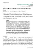
(Figure 1). It was also shown that bacteria inside the
biofilm have an increased resistance to the local host
defenses and to antibiotics [8]. Additionally, through
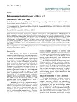
the conversion into so-called small colony variants
(Figure 2) additional modifications of bacterial cells
develop [9], while significantly reducing treatment
efficiency and triggering chronic processes. The for-
mation of an inflammatory response, with the local
generation of cytokines and reactive bacterial and
host specific metabolic products leads to a joint injury,
not seldom to irreversible destruction. Thus, effective
therapy strategies must combine calculated antibiotic
regimes with local surgical interventions.
Joint replacement surgery increases the risk of
infections due to intraoperative bacterial wound con-
tamination, leading to an early onset of the typical
symptoms. The material of the joint prosthesis is also
covered by host proteins such as fibrinogen and fi-
bronectin, which in turn facilitates bacterial coloniza-
tion, leading to a delayed type of PJI. The foreign
material allows not only a surface colonization; it is
also responsible for a reduction of the local defense
mechanisms. Among others, it leads to an apoptosis of
phagocytes surrounding the joint prosthesis; a
mechanism described as frustrane phagocytosis [10].
Reactive arthritis (RA), however, is aseptic and
usually not erosive. It results from a distant infection
(usually urethritis or enteritis). Often the reactive ar-
thritis is disseminated and involves multiple joints.
The immunological mechanism of the frequent asso-
ciation with HLA-B27 is not yet fully understood.
Using immunological and molecular biological pro-
cedures bacterial antigens can be detected in synovial
fluid and synovial membrane [11]. Although in some
studies viable bacteria could be detected [12], in an
overview of research results it can be concluded that
bacterial antigenetic material or antigen-antibody
complexes are haematogenous deposited in the joint.
This triggers a local inflammatory immune reaction,
even without local bacterial proliferation [3,13].
Figure 1: (left) Raster electron microscopy of Staphylococcus aureus from broth culture; (right) Staphylococcus aureus biofilm.
Images from S. Sailer und I. Chatterjee, Homburg/Saar.
Figure 2: Staphylococcus aureus as normal phenotype
and as small colony variant (SCV). Note the different
size and hemolysis (identical molecular pattern).
Int. J. Med. Sci. 2009, 6
237
Microbiologic procedures in joint infections
A differentiation of arthritis is practicable by
examination of typical laboratory medicine and mi-
crobiology characteristics in joint fluid. In addition to
the macroscopic parameters (e.g. color, viscosity) the
gram stain is the fastest test, giving a hint to the trig-
gering agent. Beside the inflammatory parameters,
such as erythrocyte sedimentation rate (ESR),
C-reactive protein (CRP), and white blood cell count,
which are nonspecific and do not provide loca-
tion-related information, the synovial fluid leukocyte
count is a simple, rapid and accurate test. The changes
in acute joint infections are often more pronounced.
For a targeted therapy the microbiologic examination
is the most important step [14]. Thus, joint fluid or
intraarticular tissue must be obtained before antibiotic
therapy is started. Swabs, also from intraoperative
sites should not be sent into the laboratory due to the
small quantity of carried material. Also swabs or tis-
sue from superficial wounds or fistula often show the
growth of skin flora and not the relevant bacteria.
Only isolation of S. aureus from sinus tracts is predic-
tive of the causative pathogen [15]. Especially in PJI an
operational approach is useful to obtain and examine
several materials. Although molecular techniques
have a high value as a diagnostic procedure, only
bacterial cultures can complete the diagnostic testing
by antibiograms. The incubation time of the bacterial
culture should last 7 days. Slow-growing bacteria as a
pathogen in question, the presence of bacteria modi-
ficated by antibiotic pretreatment, SCV or biofilm
production need an extended culture period [16].
Both, joint defect formation and chronification, if NJI,
are not treated quickly and efficiently, and increasing
mortality, high economic burden for removal of in-
fected joint prosthesis and implant renewing [17] al-
ways require the increased effort in sampling and
microbiological analysis.
Infection serology
In reactive arthritis the cultural detection of
pathogens is often not possible. This makes serologi-
cal analysis to a method principally necessary to de-
tect antibodies against the causing bacteria. The anti-
body detection has a great diagnostic value also due
to the reduced sensitivity of bacterial culture in joint
infections caused by Borrelia or Brucella. For a medi-
cally and economically adequate evaluation in sero-
logic diagnosis the sensitivity and specificity of the
various methods must be considered. An overview of
the bacteria that most commonly trigger reactive ar-
thritis is listed in Table 3.
Table 3: Serodiagnosis of common bacterial agents for postinfectious arthritis
Antibiotic therapy
The therapeutic approach has to be selected in
accordance with the mode of infection (NJI, PJI, RA),
the expected or found pathogens, and their resistance.
It should be remembered that the slowed growth of
bacteria in a biofilm on surfaces of joint prosthesis
may additionally reinforces antibiotic resistance
[18,19]. Responsible for such an increase against an-
tibacterial substances are changes in cell wall synthe-
sis, which limits the effect of beta-lactam antibiotics
and glycopeptides, and the occurrence of bacterial
variants with modifications of other metabolic activi-
ties, with implications for the action of quinolones,
aminoglycosides, and tetracyclines. In principle, the
spectrum of available antibiotics is limited by the
specific pharmacokinetic requirements in the treat-
ment of joint infections. This applies particularly to
chronic infections and prosthesis infections. For an
overview of common substances and therapeutic re-
gimes, see Table 4.
Int. J. Med. Sci. 2009, 6
238
Table 4: Antibiotics for therapy of infectious arthritis (all given dosages are for healthy adults of 70 kg with normal liver and
kidney function)