Báo cáo y học: "ISOLATION OF CHLAMYDIA PNEUMONIAE FROM SERUM SAMPLES OF THE PATIENTS WITH ACUTE CORONARY SYNDROME"
Bạn đang xem bản rút gọn của tài liệu. Xem và tải ngay bản đầy đủ của tài liệu tại đây (1.05 MB, 10 trang )
Int. J. Med. Sci. 2010, 7
181
I
I
n
n
t
t
e
e
r
r
n
n
a
a
t
t
i
i
o
o
n
n
a
a
l
l
J
J
o
o
u
u
r
r
n
n
a
a
l
l
o
o
f
f
M
M
e
e
d
d
i
i
c
c
a
a
l
l
S
S
c
c
i
i
e
e
n
n
c
c
e
e
s
s
2010; 7(4):181-190
© Ivyspring International Publisher. All rights reserved
Research Paper
ISOLATION OF CHLAMYDIA PNEUMONIAE FROM SERUM SAMPLES OF THE
PATIENTS WITH ACUTE CORONARY SYNDROME
Ivan M Petyaev
1
, Nayilia A Zigangirova
2
, Alexey M Petyaev
3
, Ulia P Pashko
2
, Lubov V Didenko
2
, Elena U
Morgunova
2
, Yuriy K Bashmakov
1
1. Cambridge Theranostics Ltd, Babraham Research Campus, Babraham, Cambridge, CB2 4AT, United Kingdom
2. Gamaleya Institute for Epidemiology and Microbiology RAMS, 18 Gamaleya Str., Moscow 123098, Russia
3. Rostov-on-Don Medical University. Nahichevanskii 37, Rostov-on-Don, Russia
Corresponding author: Dr Yuriy K Bashmakov, Cambridge Theranostics Ltd., Babraham Research Campus, Cambridge
CB2 4AT, United Kingdom. Telephone: +44-797-1598348, Fax: +44-122-3240340
Received: 2009.12.16; Accepted: 2010.06.07; Published: 2010.06.10
Abstract
BACKGROUND: Limited body of evidence suggests that lipopolysaccharide of C. pneu-
moniae as well as C. pneumoniae-specific immune complexes can be detected and isolated from
human serum. The aim of this study was to investigate the presence of viable elementary
bodies of C.pneumoniae in serum samples of patients with acute coronary syndrome and
healthy volunteers.
MATERIAL AND METHODS: Serum specimens from 26 healthy volunteers and 56 pa-
tients with acute coronary syndrome were examined subsequently by serological
(C.pneumoniae-specific IgA and IgG), PCR-based and bacteriological methods. Conventional,
nested and TaqMan PCR were used to detect C.pneumoniae genetic markers (ompA and 16S
rRNA) in DNA from serum specimens extracted with different methods. An alternative
protocol which included culturing high-speed serum sediments in HL cells and further
C.pneumoniae growth evaluation with immunofluorescence analysis and TaqMan PCR was
established. Pellet fraction of PCR-positive serum specimens was also examined by immu-
noelectron microscopy.
RESULTS: Best efficiency of final PCR product recovery from serum specimens has been
shown with specific C. pneumoniae primers using phenol-chloroform DNA extraction pro-
tocol. TaqMan PCR analysis revealed that human serum of patients with acute coronary
syndrome may contain genetic markers of C. pneumoniae with bacterial load range from 200 to
2000 copies/ml serum. However, reliability and reproducibility of TaqMan PCR were poor for
serum specimens with low bacterial copy number (<200 /ml). Combination of bacteriological,
immunofluorescence and PCR- based protocols applied for the evaluating HL cells infected
with serum sediments revealed that 21.0 % of the patients with acute coronary syndrome
have viable forms C.pneumoniae in serum. The detection rate of C.pneumoniae in healthy vo-
lunteers was much lower (7.7%). Immunological profile of the patients did not match accu-
rately C.pneumoniae detection rate in serum specimens. Elementary bodies of C.pneumoniae
with typical ultrastructural characteristics were also identified in serum sediments using
immunoelectron microscopy.
Conclusions: Viable forms C. pneumoniae with typical electron microscopic structure can be
identified and isolated from serum specimens of the patients with acute coronary syndrome
and some healthy volunteers. Increased detection rate of C. pneumoniae in serum among the
patients with an acute coronary syndrome may contribute towards enhanced
pro-inflammatory status in cardiovascular patients and development of secondary complica-
tions of atherosclerosis.
Key words: Chlamydia pneumoniae, PCR, human serum, acute coronary syndrome, cultured cells
Int. J. Med. Sci. 2010, 7
182
BACKGROUND
Despite unquestionable role of C. pneumoniae in
pathogenesis of respiratory infections there are many
questions about involvement of the pathogen in de-
velopment other human diseases including atheros-
clerosis (1), multiple sclerosis (2,3), Alzheimer’s dis-
ease (4), lymphogranuloma (5), reactive arthritis (6),
Guillain-Barre syndrome (7). The progress in that field
is substantially complicated by the lack of standar-
dized criteria for laboratory diagnostics of chronic C.
pneumoniae infection as well as contradictory infor-
mation about distribution of the pathogen throughout
of the tissues of human body.
Isolating and culturing of C. pneumoniae may
represent significant challenge for non-specialized
diagnostic labs. Several plasma serological markers
have been recently proposed based on the results of
proteomic analysis. In particular proteins encoded by
Omp11, the PmpG family, IncA and by CpPLD are
among promising candidates for immunological di-
agnostics of C. pneumoniae infection (8, 9). However,
changed antigenic profile of C. pneumoniae during
persistent colonization in human tissues (10, 11) un-
dermines the diagnostic value of serological markers.
Among molecular diagnostic criteria used for
detection of C. pneumoniae in human specimens are
polymerase chain reaction (PCR), in-situ hybridiza-
tion method and enzyme immunoassay protocols (12,
13). PCR-based approach usually targets parts of
chlamydial genome, in particular genes encoding 16S
rRNA, major outer membrane protein (OmpA), as
well as Pst1 (13).
However poor reproducibility limits signifi-
cantly the diagnostic importance of PCR and in-situ
hybridization for non-respiratory specimens. Detec-
tion of chlamydial lipopolysaccharide in serum is
claimed to improve reliability of molecular biology
methods when used in addition to PCR and in situ
hybridization protocols (12).
There are multiple reports validating the pres-
ence of C. pneumoniae in respiratory secretion fluid,
nasal, tracheal and lung tissues of the patients with
inflammatory lung disease (13, 14, 15). Moreover, C.
pneumoniae can efficiently propagate in blood cells, in
particular in mononuclear cells and lymphocytes
(16,17,18). The presence of C. pneumoniae in the blood
cells predetermines the possibility of pathogen dis-
semination from respiratory system to different or-
gans and tissues. Besides respiratory organs C. pneu-
moniae can be detected in specimens from atheroscle-
rotic plagues (1, 19), cerebrospinal fluid (2) and en-
dothelium (20).
In the present paper we report, that viable ele-
mentary bodies of C. pneumoniae with typical electron
microscopic structure can be isolated from the serum
samples of the patients with acute coronary syn-
drome. Furthermore, using combination of bacterio-
logical and PCR-based methods we show herein that
patients with acute coronary syndrome have higher C.
pneumoniae detection rate in serum as compared to
healthy volunteers.
MATERIAL AND METHODS
Cell lines and bacterial strains
HL cells (Washington Research Foundation,
Seattle, USA) as well as C. pneumoniae (strain Kajaani
6, K6) were kindly provided by Dr. P.Saikku (Univer-
sity of Oulu, Finland). HL cells were grown in RPMI
1640 supplemented with 10% FCS at 37° C in 5% CO
2
.
C.pneumoniae was initially propagated in HL cells and
elementary bodies (EB) were purified by Renografin
gradient centrifugation as widely described (21, 22).
EB of C. pneumoniae were used as a reference for
genetic and electron microscopy analysis.
Patients and serum specimens.
The study protocol was approved by the Ros-
tov-on- Don Medical University Ethics Committee.
All patients were informed about the purpose of the
study and have given written consent regarding par-
ticipation in the study. Initial observation has been
done on the group of 18 patients with acute coronary
syndrome (11 males and 7 females aged from 47 to
68). Once conditions for combined microbiologic and
nucleic acid amplification protocol were established,
38 more patients with acute coronary syndrome (21
males and 17 females, aged from 42 to 71) and 26
healthy volunteers with no indication of cardiovas-
cular disease were enrolled (major groups of the
study). Blood samples were collected into plastic
tubes, kept at 37° C for 20 minutes and centrifuged at
1000g, 4° C for 10 min. Resulting serum was imme-
diately separated and stored at - 80° C until assayed.
C.pneumoniae-specific IgA and IgG antibodies
were evaluated by using Chlamydia pneumo-
niae-IgG-ELISA medac plus and Chlamydia pneumo-
niae-IgA-ELISA plus commercial kits with high-
ly purified C.pneumoniae specific antigen without
LPS. (Medac, Hamburg, Germany).
Bacteriological assay.
Tubes containing 3 ml of frozen serum samples
were thawed on ice and subjected to the centrifuga-
tion on Beckman centrifuge AN (Beckman Coulter,
Int. J. Med. Sci. 2010, 7
183
Inc., USA) at 16000 g for 45 min at 4° C. Obtained se-
diments were gently resuspended with micropipette
in 1.0 ml of RPMI 1640 with 5% FCS, amphothericine
B (5 µg/ml) and gentamycin (4 µg/ml). Resulting
suspension was transferred to subconfluent mono-
layer of HL cells grown in 24- well plate. After inocu-
lation the plates were centrifuged at 1600g for 1 hour
at 30° C and incubated for 2 h at 37° C in 5% C0
2
. The
medium was removed and replaced with fresh RPMI
1640 supplemented with 1 µg/ml) of cycloheximide
and plates were cultivated for 72 hours at 37° C in 5%
CO
2
.
A 24 well plate rather than 96 well plates was
used in the study to avoid potential cross contamina-
tion. Each serum specimen inoculated into 24 well
dish was followed by two wells filled with incubation
medium alone. All manipulation with the plates were
done without agitation. Positive control plates were
set and examined by the end of each working day and
were kept in separate incubator. Each plate examina-
tion procedure was followed by careful disinfection of
the equipment. Positive findings were reconfirmed.
The plates were evaluated for chlamydial
growth by immunofluorescence microscopy with a
Chlamydia genus-specific antibody against LPS prior
to quantitative TaqMan- PCR for 16S rRNA of C.
pneumoniae. Each isolate was passaged up to 3 times.
Immunofluoresence staining.
Infected HL monolayers grown on coverslips in
24-well plates were fixed with methanol. Permebia-
lized cells were stained by direct immunofluoresence
using FITC – conjugated monoclonal antibody against
chlamydial lipopolysaccharide (NearMedic Plus, RF).
Inclusion-containing cells were visualized using Ni-
kon Eclipse 50i microscope fluorescence microscope at
x1350 magnification.
DNA isolation.
DNA isolation from whole serum
Briefly, 1.0 ml of whole serum was mixed with
0.5 ml of lysis buffer (0.2 M Tris-HCl buffer, pH 7,2
supplemented with 0.5 % SDS) with 0.25 mg/ ml
proteinase K (Promega, USA) and incubated for 2
hours at 56° C. DNA from the resulting lysates was
extracted using phenol-chloroform method as widely
described (23) and precipitated with absolute ethanol.
DNA pellet was finally resuspended in 25 µl of water.
For comparison purpose bacterial DNA was extracted
from the same volume of whole serum with QIAmp
Blood Midi Kit (QIAGEN, Valencia, CA) according to
the manual.
Bacterial DNA was also extracted from the bac-
terial particles trapped from the whole serum with
protein A from of Staph. aureus, insoluble (Sigma
P7155). 1.0 ml of whole serum was mixed with 0.15 ml
of protein A and incubated for 1 hour at 37 °C with
occasional gentle shaking. The mixture was centri-
fuged for 5 min at 5000 g and DNA was extracted
from the resulting pellet using QIAamp DNA Blood
Mini Kit (QIAGEN INC., Valencia, Calif.) according to
the manual.
DNA isolation from infected HL cells
Cells were harvested from 24 well plates and
resuspended in 200 µl of lysis buffer and DNA was
extracted using QIAamp DNA Blood Mini Kit
(QIAGEN INC., Valencia, Calif.) according to the
manual.
DNA isolation from C.pneumoniae reference strain
DNA was extracted from 100 µl of C. pneumoniae
purified EB using reagents and protocol from QIAmp
Blood Mini Kit (QIAGEN Inc., Valencia, Calif.).
PCR.
General Information
Numerous precautions were employed to ensure
validity of PCR protocols, especially nested PCR.
Different work areas/rooms, different sets of the pi-
pets, barrier-filter tips and scrupulous clean-
ing/decontamination procedures were used. All
samples were blinded for lab workers. Multiple con-
trols were used for PCR reactions. DNA extracted
from C. pneumoniae reference strain (low concentra-
tion) and/or DNA extracted from the serum sediment
of two C.pneumoniae infected patients were used as
positive control. Positive control specimens were se-
lected using electron microscopy and serological as-
say. Serum specimens from serologically negative
healthy volunteers with no C. pneumoniae EB detecta-
ble in serum sediments by electron microcopy were
used as a negative control. Each PCR set was accom-
panied by a reaction mix with all PCR components
except the target DNA. Positive findings were recon-
firmed.
Conventional qualitative PCR.
Briefly, 2 µl of DNA solution were transferred to
the reaction mixture containing 1x PCR buffer (Silex,
Moscow, RF) containing 10 mM Tris-HCl, pH 8,3 , 2.5
mM MgCl
2
, 200 µM of each dNTPs, 1 U
Taq-DNA-polymerase, 15 pmol of each primer. For-
ty-five cycles of amplification were performed on a
PCR Thermocycler Perkin Elmer. Each cycle consisted
of denaturation step at 94°C for 45 sec, primer an-
nealing at
63°C for 45 sec , primer extension at 72°C
for 45 sec. Amplified product (10 µl) was visualized
by electrophoresis
in a 1.5% agarose gel with ethidium
Int. J. Med. Sci. 2010, 7
184
bromide. Extracted DNAs were analyzed by PCR
with primers CPN90-CPN91 specific for C. pneumoniae
16S rRNA as described (24).
Nested PCR.
To ensure the specificity of PCR analysis a pro-
tocol for nested PCR for OmpA of C. pneumoniae was
employed. The outer (oCP1 – 5’
TTACAAGCCTTGCCTGTAGG 3’, oCP2 – 5’ GCGA
TCCCAAATGTTTAAGGC 3’) and nested (iCPC - 5’
TTATTAATTGATGGTACAATA 3’, iCPD - 5’
ATCTACGGCAGTAGTATAGTT 3’) primers were
used as published (24).
2 µl of DNA was added to reaction mixture
containing 1x PCR buffer (Silex, Moscow, RF) con-
taining 10 mM Tris-HCl, pH 8,3 , 2.5 mM MgCl
2
, 15
pmol of each primer, 200 µM of each of dNTPs and
1 U of Taq polymerase. First run of amplification was
conducted under cycling conditions consisting of an
initial denaturation at 95°C for 5 min, followed by
45 cycles of denaturation at 95°C for
30 sec, annealing
at 63°C for 30 sec, and extension for 30sec at
72°C. For
the second round of PCR, 2 µl of the first-round
product
was mixed with 23 µl of amplification mix-
ture containing primers for iCPC and iCPD and am-
plified using following cycling
conditions: 35 cycles of
denaturation at 95°C for
30 sec, annealing at 55°C for
30 sec, and extension for 30sec at
72°C. PCR products
were visualized by agarose electrophoresis with
ethidium bromide. Taq DNA polymerase and other
reagents for nested PCR were from Promega (UK).
Quantitative TaqMan-PCR.
For quantification purpose, Real-time PCR for
16S rRNA of C. pneumoniae was conducted. PCR
primers and TaqMan
probe for 16S rRNA (GenBank
accession
number AF131889) were designed using
Primer Express Software (Applied Biosystems,
Foster
City, CA, USA) and synthesized by Syntol (Moscow,
RF).
Designed primers and TaqMan probe (forward
primer СPN90, 5'-GGTCTCAACCCCATCCGT
GTCGG-3'; reverse primer СPN91,
5'-TGCGGAAAGCTGTATTTCTACAGTT-3'; and
TaqMan probe 557, 5'-TCCAGGTAAGGTCC
TTCGCGTTGCATCG-3') generated a PCR product of
predicted size (194 bp). The TaqMan probe
was la-
belled at the 5' end with 6-carboxyfluorescein
as the
reporter dye and at the 3' end with
6-carboxytetramethylrhodamine
as the quencher. An
additional BLAST search analysis was conducted to
unsure specificity of the primers and probe. Real-time
PCR was performed with the iCycler IQ ystem (Bio-
rad, USA). 2 µl of extracted DNA was analyzed with
the PCR mixture
in a total volume of 25 µl. The PCR
mixture consisted
of 10 mM Tris (pH 8.3), 50 mM KCl,
1,5 mM MgCl
2
,
200 µM of each dNTPs, 2,5 U of Ter-
mostar Taq DNA polymerase (Syntol, Moscow, RF);
and
5pmol of both forward and reverse
primers and
3,5 pmol probe. The real-time PCR
run was 10 min at
95°C, and 50 repeats
of 20 sec at 95°C and 50 sec at
62°C. All samples were analyzed in triplicates. A
sample was considered positive if three of three assay
results were positive in the triplicate test and
if the
average value for the PCR run was greater than or
equal
to 1.0.
Amounts of 16S rRNA are represented bellow in
16S rRNA genome equivalents per ml of serum. Cali-
brator standards were prepared using 194 bp 16S
rRNA DNA fragment of C pneumoniae cloned into the
pGEM-T plasmid vector (pVU56) using the TA clon-
ing kit (Invitrogen, San Diego, CA) similarly to Broc-
colo F (25).
The cycle
threshold (C
T
) values, defined as the
number of cycles at which
the fluorescence of the re-
porter dye first exceeds the calculated
background
level, were automatically estimated by the instrument
for each reaction. C
T
values for serum samples were
plotted
against calibrator standards of cloned DNA
fragment.
Electron Microscopy.
Thawed serum samples (10 ml) were spun at
16000g for 60 min. Resulting pellets were analyzed by
TaqMan PCR for C. pneumoniae 16S rRNA. Positive
specimens were fixed for 4 hours in phosphate buffer
(pH 7.8) containing 5% glutaraldehyde, post-fixed in
1% osmium tetroxide for 1 hour, dehydrated in etha-
nol and embedded in LR White resin (EMS, USA).
Stained ultrathin sections (200-300Аº) were evaluated
by electron microscopy using JEM-100B microscope
(Japan Electron Optics Laboratory Co., Tokyo, Japan).
Purified EB of C. pneumoniae reference strain were
used as positive control for electron microscopy stu-
dies. PCR-negative sediments of serum obtained from
healthy volunteers served as negative control.
Immunoelectron microscopy was performed in
specimens fixed with 2% paraformaldehyde and 0.1 %
glutaraldehyde in PBS (7.5) with further contrasting
with 2% uranyl acetate. Acetone-dehydrated speci-
mens were embedded into LR White Resin for ultra-
thin sectioning. The sections were blocked for 1 hour
with 0.5% bovine serum albumin in PBS and incu-
bated overnight with monoclonal antibody against
chlamydial lipopolysaccharide (NearMedic Plus, RF).
After washing in PBS sections were incubated for 2
hours with goat anti-mouse IgG conjugated with 10
nm colloid gold (Invitrogen, USA) and contrasted
with uranyl acetate. Sections were examined with a
Int. J. Med. Sci. 2010, 7
185
Joel 100B (Japan) electron microscope. Control sec-
tions were incubated with normal mouse IgG.
RESULTS
Initial observation took place when we obtained
sera from 18 patients with ACS and analyzed them for
presence of C. pneumoniae specific IgG and IgA using
ELISA Medac kit (Germany) as well as for presence of
genomic determinants of C. pneumoniae. As can be
seen from Table 1, 7 patients from the initial group
were positive for C. pneumoniae-specific IgG, whereas
4 patients had diagnostically relevant levels of IgA.
Simultaneous detection of increased titers of IgG and
IgA was documented only in 4 patients. Surprisingly,
when DNA specimens extracted from 1.0 ml of serum
aliquots were analyzed for presence of 16S rRNA by
conventional PCR, we have found that 5 patients with
ACS were positive for the genetic marker of C. pneu-
moniae. Finally, just 3 patients (out of 18) had in-
creased levels of two Ig isotypes and positive signal in
conventional PCR for 16S rRNA. Such inconsistency
between serologic and genetic markers of C. pneumo-
niae infection is well known and widely discussed (1).
However, detectability of the genetic marker of C.
pneumoniae in human serum appeared to be a rea-
sonably intriguing finding. Therefore, we decided to
optimize conventional PCR protocol for detection of
the C. pneumoniae genetic markers in serum.
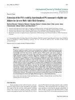
PCR-positive sera obtained from 2 randomly se-
lected ACS patients were used for this purpose. As
can be seen from Figure 1, there is an obvious increase
in the final recovery of 194 bp PCR product (16S
rRNA amplicon) when phenol-chloroform DNA ex-
traction protocol has been used. Somehow QIAmp
Midi Extraction kit (Qiagen) showed lower recovery
rate of final PCR product which can be explained by
lower efficiency of C. pneumoniae DNA extraction.
Sufficient recovery of final PCR product has been also
seen when protein A from Staph. aureus has been
used for isolation of C pneumoniae from whole serum.
This fact may suggest that extracted DNA originates
rather from intact chlamydial particles opsonized by
immunoglobulins, than remnants of C. pneumoniae
circulating in the blood.
To confirm the results obtained with conven-
tional PCR and insure its specificity we compared
side-by-side two amplification reactions with phe-
nol-chloroform extracted C. pneumoniae DNA. One
has been conducted with protocol using primers spe-
cific for 16S rRNA, another one – with the primers for
ompA in nested PCR format. As can be seen from
Figure 2, the sensitivity of PCR reaction was similar
regardless of the primer set used.
Table 1. C.pneumoniae positivity status assessment using serological, RT- PCR and bacteriological analysis of serum spe-
cimens
GROUPS
Total Number of individuals Positive in:
Serological assay TaqMan PCR in
serum
Bacteriological assay with
further PCR validation
IgA IgG
PRELIMINARY GROUP Patients with ACS 18 4 7 5 3
MAYOR GROUPS Healthy volunteers 26
1 4 - 2
Patients with ACS 38 6 13
- 8
Figure 1. Recovery of PCR product in DNA samples isolated from serum specimens using QIAamp DNA blood midi kit,
protein A and phenol-chloroform extraction method. 1 – molecular size standards; 2, 6 and 10 – PCR-positive serum from
patient M; 3, 7 and 11 – PCR-positive serum from patient P; 4, 8 and 12 – PCR negative serum from patient S; 5, 9 and 13 –
extraction control; 14 – negative control; 15 – positive control.