Báo cáo y học: " Non-syndromic multiple supernumerary teeth in a family unit with a normal karyotype: case report"
Bạn đang xem bản rút gọn của tài liệu. Xem và tải ngay bản đầy đủ của tài liệu tại đây (529.15 KB, 7 trang )
Int. J. Med. Sci. 2010, 7
378
I
I
n
n
t
t
e
e
r
r
n
n
a
a
t
t
i
i
o
o
n
n
a
a
l
l
J
J
o
o
u
u
r
r
n
n
a
a
l
l
o
o
f
f
M
M
e
e
d
d
i
i
c
c
a
a
l
l
S
S
c
c
i
i
e
e
n
n
c
c
e
e
s
s
2010; 7(6):378-384
© Ivyspring International Publisher. All rights reserved
Case Report
Non-syndromic multiple supernumerary teeth in a family unit with a
normal karyotype: case report
Francesco Inchingolo
1
, Marco Tatullo
2
, Fabio M. Abenavoli
3
,
Massimo Marrelli
4
, Alessio D. Inchingolo
5
,
Mattia Gentile
6
, Angelo M. Inchingolo
5
, Gianna Dipalma
7
1. Department of Dental Sciences and Surgery, University of Bari, Bari, Italy
2. Department of Medical Biochemistry, Medical Biology and Physics, University of Bari, Bari, Italy
3. Department of “Head and Neck Surgery”, Hospital “Fatebenefratelli”, Rome, Italy
4. Department of Maxillofacial Surgery, Calabrodental, Crotone, Italy
5. Department of Dental Sciences and Surgery, University of Milano, Milano, Italy
6. Department of Medical Genetic, Hospital “Di Venere”, Bari, Italy
7. Department of Maxillofacial Surgery, Calabrodental, Crotone, Italy
Corresponding author: Prof. Francesco INCHINGOLO, Piazza Giulio Cesare – Policlinico 70124 – Bari. E-mail:
; Tel.: 00390805593343 – Infoline: 00393312111104.
Received: 2010.09.22; Accepted: 2010.11.03; Published: 2010.11.05
Abstract
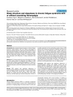
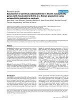
Introduction. Hyperdontia is an odontostomatologic anomaly characterized by an excess in
tooth number. It seems to occur more often in patients with hereditary factors concerning
this anomaly: this case represents a rare form of hyperdontia, with bilateral multiple super-
numerary teeth, with evident penetrance of the phenotype in the family unit engaged in the
present study. The karyotype determination excludes a pathogenesis on chromosomal basis.
Case report. A 3 0 y e a r s o l d p a t i e n t c a m e t o o u r o b s e r v a t i o n w i t h f i v e i m p a c t e d t e e t h ( 1 . 8 , 2 . 8 ,
3.8, 4.7 and 4.8), as well as with the presence of an impacted supernumerary tooth (disto-
m o l a r 4 . 9 ) . T h e p a t i e n t w a s s u g g e s t e d t o a l l o w u s t o p e r f o r m a r a d i o l o g i c s c r e e n i n g t o h i s t w o
sisters aged 17 and 13 years.
T h e X -r a y p h o t o g r a p h y s h o w e d t h a t t h e e l d e r s i s t e r h a d n i n e i m p a c t e d t e e t h ; t h e s e w e r e 1 . 8
– 1.9 – 2.8 – 2.9 – 2.10 – 3.8 – 3.9 – 4.8 – 4.9; while the youngest sister had four impacted
teeth, that is 1.8 – 1.9 – 2.8 – 2.9.
Conclusions. The value of the present case r e p o r t c a n b e u s e d a s a p a r a d i g m f o r t h e a s s e s s m e n t
of the hereditary factors predisposing the onset of hyperdontia, and for the consequent
management by oral surgeon of family units in which the odontostomatologic anomaly was
detected without any syndromic forms.
Key words: Hyperdontia, supernumerary teeth, impacted teeth.
Introduction
Hyperdontia is an odontostomatologic anomaly
characterized by an excess in tooth number, both
e r u p t e d a n d n o n -erupted. It can be described as “real”
if determined by an increased number of teeth, oth-
erwise it is “false” if caused by a delay in shedding of
deciduous teeth beyond the transition period
1, 2, 3, 5
.
In one of his studies, Tomes suggested a no-
menclature for teeth in excess
3
: they were defined as
“supplementary” if they present a normal morphol-
ogy and as “supernumerary” if they present mor-
phologic and volumetric anomalies. Supernumeraries
are classified according to morphology
3
into conical,
Int. J. Med. Sci. 2010, 7
379
tuberculate, supplemental and odontome; however,
the Literature also reports a classification according to
intraoral position of the supernumerary teeth: Mesio-
dens; Paramolar; Distomolar and Parapremolar
4
.
Hyperdontia is reported quite frequently
(males:females around 2:1)
2
, and it seems to occur
more often in patients with hereditary factors con-
cerning this anomaly
4
. A study, conducted on 30 pa-
tients with 41 “mesiodens”, anamnestically deter-
mined a familial predisposition in 31% of cases
6
. Su-
pernumerary teeth are frequently found in the supe-
rior maxillary bone and mainly in the premaxilla
(90-98%)
7
, they are often impacted (88,7%) and are
often present in the palatine area
8,9
.
The prevalence of multiple supernumerary teeth
ranges from 8 to 27% of cases
7,10
.
Hyperdontia is often occasional, but hereditary
factors can also be involved, especially in the most
serious cases, otherwise it can be associated to genetic
syndromes such as “Gardner Syndrome” or “Cleido-
cranial Dysplasia”; in these syndromic forms, hyper-
dontia is a sign of a clinical picture which is definitely
more complex, and further anomalies are always
present.
It follows that the ability to prematurely inter-
cept a clinical picture of hyperdontia is important also
for the possible association of this anomaly with in -
gravescent syndromic forms
10,11
; in order to make an
early diagnosis of any syndromic forms we can use
the G-ba n din g te c hni que
12
: G-banding is obtained
with Giemsa stain following digestion of chromo-
somes with trypsin. It yields a series of lightly and
darkly stained bands - the dark regions tend to be
heterochromatic, late-rep lic a tin g and AT r i ch. T he
light regions tend to be euchromatic, early-replicating
and GC rich.
The most frequent complication of having su-
pernumerary teeth is the dental malposition
1, 2, 3
of
teeth of the normal series (erupted or not) which in
turn leads to clinical consequences of orthodontic
and/or surgical nature; more rarely, impacted su-
pernumerary teeth are the cause of follicular cysts,
neuralgic manifestations, dysodontiasis of permanent
teeth.
1, 2, 3
The clinical situations that may indicate the
presence of supernumerary teeth are:
• absence of permanent teeth in the maxillary arch
10
,
• agenesia
13, 14, 15
,
• malposition of erupted permanent teeth
10, 16
,
• malocclusion
17, 18, 19
,
• wide interincisive diastema
20, 21
,
• positive familial anamnesis
4
,
• reabsorption of roots of the adjacent teeth
22
with
loss of their vitality
7
and symptomatology.
• Tumefaction on the vestibular or pala-
tine/lingual area.
Hyperdontia therapy depends on the area and
on the number of teeth in excess (erupted into proper
maxillary arch position, out of arch or impacted), and
also depends on the presence of pathologic processes
affecting the supernumerary teeth and/or the teeth of
the normal series which erupted, retained or impacted
1, 2
.
In cases where surgical therapy is recommend-
ed, an operation to prefer is germectomy of the su-
pernumerary tooth to be formed, in order to prevent
the onset of a malocclusion due to altered develop -
ment of the normal series; in case of completely
formed supernumerary teeth, a preoperatory evalua-
tion is necessary: any contiguity between the tooth
and the important anatomic structures will be inves-
tigated, and the operation will be planned with as
little trauma as possible, in order to preserve the hard
and soft surrounding structures.
Clinical case
A 30 years old Caucasian patient came to our
observation with five impacted teeth (1.8, 2.8, 3.8, 4.7
and 4.8, according with FDI World Dental Federation
notation), as well as with the presence of an impacted
supernumerary tooth (distomolar 4.9).
The patient reported localized pain and a slight ho -
molateral submandibular lymphadenopathy, without
functional limitations or fever. No occlusal hindrance
was caused by these supernumerary teeth. Alt h ou g h
anamnesis allowed to exclude stomatological pa-
thologies, congenital anomalies and genetic or syn -
dromic alterations, the patient reported a hereditary
etiology: her mother’s supernumerary teeth in the
posterior portion of the superior maxillary bone were
avulsed.
The patient was suggested to allow us to per-
form a radiologic screening to his two sisters aged 17
and 13 years, after giving their informed consent.
The X-ray photography showed that the pa-
tient’s sisters had a clinical picture of hyperdontia,
together with dental impaction, in a systemic and
non-syndromic form and with a normal psychophys-
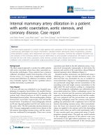
ical development. However, the karyotype determi-
nation was done by G banding technique (GTG)
(Seabright, 1971). The proband, the mother and the
elder sister had a normal male (46,XY) and normal
female (46,XX) karyotype, respectively (Figures 1, 2,
3).
The radiologic evaluation of the two sisters al-
lowed to determine as follows:
Int. J. Med. Sci. 2010, 7
380
The elder sister had nine impacted teeth; these
were 1.8 – 1.9 – 2.8 – 2.9 – 2.10 – 3.8 – 3.9 – 4.8 – 4.9
(Fig. 4), while the youngest sister had four impacted
teeth, that is 1.8 – 1.9 – 2.8 – 2.9 (Fig. 5).
In agreement with the international literature,
the Authors opted to leave the sisters’ supernumerary
teeth in situ, as there were not any signs or symptoms
justifying the extraction therapy. Whereas the patient
who first came to our attention was subjected to ex-
traction of teeth 4.7, 4.8 and 4.9, after a routine hema-
tological investigation and after the assessment of
radiographic exams, such as X-Ray Dental Panoramic
Tomogram and Denta-Sca n (Fig. 6) of the inferior
maxillary bone. Exodontia led to remission of the algic
symptomathology, without compromising somesthe-
sia in the treated region, although the extracted teeth
were in the close proximity of the inferior mandibular
canal.
At the end of surgery, one-week intramuscular
antibiotic and antiphlogistic therapy was scheduled
(cefazolin sodium 2g/day and ketoprofen lysine salt
200mg/day).
Figure 1: proband’s karyotype (G banding technique - GTG) (Seabright,1971)
Figure 2: mother’s karyotype (G banding technique - GTG) (Seabright,1971)
Int. J. Med. Sci. 2010, 7
381
Figure 3: elder sister’s karyotype (G banding technique - GTG) (Seabright,1971)
Figure 4: X-Ray Dental Panoramic Tomogram (elder sister)
Int. J. Med. Sci. 2010, 7
382
Figure 5: X-Ray Dental Panoramic Tomogram (younger sister)
Figure 6: Dental-Scan of the mandibular bone (proband)