The suppression of DUSP5 expression correlates with paclitaxel resistance and poor prognosis in basal-like breast cancer
Bạn đang xem bản rút gọn của tài liệu. Xem và tải ngay bản đầy đủ của tài liệu tại đây (1.25 MB, 10 trang )
Int. J. Med. Sci. 2018, Vol. 15
Ivyspring
International Publisher
738
International Journal of Medical Sciences
2018; 15(7): 738-747. doi: 10.7150/ijms.24981
Research Paper
The suppression of DUSP5 expression correlates with
paclitaxel resistance and poor prognosis in basal-like
breast cancer
Tieju Liu1,2, Huizhi Sun1, Shiqi Liu1, Zhao Yang1, Linqi Li1, Nan Yao1, Siqi Cheng1, Xueyi Dong1,2, Xiaohui
Liang1,2, Chen Chen1, Yi Wang1, Xiulan Zhao1,2
1.
2.
Department of Pathology, Tianjin Medical University, Tianjin 300070, China
Department of Pathology, General Hospital of Tianjin Medical University, Tianjin 300052, China
Corresponding author: Xiulan Zhao, Department of Pathology and General Hospital of Tianjin Medical University, Tianjin, China; E-mail:
; ; Tel:86-13602042200; Fax:86-22-83336813
© Ivyspring International Publisher. This is an open access article distributed under the terms of the Creative Commons Attribution (CC BY-NC) license
( See for full terms and conditions.
Received: 2018.01.16; Accepted: 2018.04.12; Published: 2018.05.16
Abstract
Basal-like breast cancer (BLBC) is resistant to endocrinotherapy and targeted therapy and new
molecular therapies are needed for BLBC. In this study, we evaluated the role of DUSP1 and DUSP5,
negative regulators of mitogen-activated protein kinase pathway, in the aggressiveness of BLBC.
MDA-MB-231 cells were given paclitaxel (PTX) treatment and subsequently PTX resistant cell
clones were established. Microarray analysis, real-time quantitative reverse transcription PCR
(qRT-PCR), and online analysis of large cohorts of breast cancer patients were performed. The PTX
resistant cells showed stronger cell proliferation ability by exhibiting the upregulation of CENPF,
CDC6, MCM3, CLSPN and SMC1A expression. Furthermore, DUSP1 and DUSP5 expression was
significantly downregulated in PTX resistant cells. In addition, in large breast cancer patients’
database, both DUSP1 and DUSP5 correlated negatively with higher histological grade. DUSP1 low
expression was obvious in HER2 positive and basal like while DUSP5 low expression was peculiar
for basal like compared with other subtypes. Remarkably, low expression of DUSP5, but not
DUSP1, was significantly correlated with poor survival of BLBC patients. In conclusion, our data
suggest that loss of DUSP5 expression results in PTX resistance and tumor progression, providing
a rationale for a therapeutic agent that restores DUSP5 in BLBC.
Key words: basal-like breast cancer; DUSP5; paclitaxel resistance
Introduction
Breast cancer has been considered as
heterogeneous disease with different expression of
hormone receptors (estrogen receptor, ER, and
progesterone receptor, PR) and human epidermal
growth factor receptor 2 (HER2)[1, 2]. The basal-like
breast cancer (BLBC) is composed of ER-PR-HER2(triple negative) tumors with high expression of basal
markers (such as keratins 5, 6, 14, 17, EGF receptor)
and proliferation markers[3]. BLBCs approximately
occupy 15-25% of breast cancers. Classically, BLBCs
are usually poorly differentiated tumors, with more
than 75% being high grade [4]. They display high
mitotic index, dramatic atypia, high nuclear/
cytoplasmic ratio, invading margins, and frequent
necrosis [1]. The prognosis of BLBC is usually poorer
than that of luminal A (ER+, PR ≥ 20% +, HER2-, Ki67
low expression), luminal B (ER+, PR < 20% + or Ki67
high expression, HER2-; or ER+, HER2+), and HER2
positive (ER-, PR-, HER2+) subtypes of breast cancer.
Treatment of BLBC has been challenging and the
lack of well-defined molecular targets in BLBC
renders these tumours insensitive to conventional
treatments targeting the hormone receptors or
HER2[5]. Given the poor prognosis of BLBC,
treatment with chemotherapy is often offered to most
patients. The frequent ER-negativity of BLBC as well
Int. J. Med. Sci. 2018, Vol. 15
as their high grade with high proliferative index
should theoretically confer them sensitivity to
chemotherapy, notably to drugs classically used in
breast cancer, such as paclitaxel (PTX). However,
despite this sensitivity to chemotherapy, BLBC are
associated with a relatively poor prognosis: this is the
“triple-negative paradox”[1]. Although these cancers
may initially respond to original treatment, they
become highly resistant to chemotherapy in the
metastatic and recurrent disease and thus traditional
chemotherapy is still associated with a high risk of
relapse and death in a large portion of patients[5-8].
All of these features of BLBC are of particular interest
in medicine and it implies that more personalised
interventions and the development of tailored
treatments for BLBC is urgently needed[9].
Dual-specificity phosphatases (DUSPs) belong to
a protein family responsible for dephosphorylating
threonine/serine and tyrosine residues on their
substrates. DUSPs selectively dephosphorylate the
components of the nuclear mitogen-activated protein
kinase (MAPK) pathway, and they can either act as
classical negative feedback regulators of MAPK
pathway or mediate cross talk between different
MAPK pathways and between MAPK pathway and
other intracellular signal molecules [10-12]. It has been
reported that there are currently 25 genes in the
DUSPs family designated as DUSPs, namely
DUSP1-28, with DUSP17, -20, and -23 redundantly
assigned as DUSP19, -18, and -25, respectively[13].
It is now clear that individual DUSPs can exhibit
either tumour suppressor function or can act as
oncogenes and this might be determined by the
expression levels of extracellular signal-regulated
kinase (ERK) that are either permissive for or provoke
cell proliferation or inversely cause cell cycle arrest or
cell death[11]. It has been reported that DUSP1
inhibits carcinogenesis in hepatocellular carcinoma
and head and neck squamous cell carcinoma as an
ERK inhibitor[14]. Recent work reveals a dynamic
pattern of DUSP1 expression within the tumor
microenvironment and loss of DUSP1 expression is a
characteristic of tumor-derived stem cells[15].
Decreased total DUSP1 protein levels may be
considered as a poor prognostic factor in breast
cancer[16]. Researchers have found that DUSP1
knockdown in sensitive non-small cell lung cancer
cells conferred chemotherapy resistance, but DUSP1
gene silencing in vivo significantly heightened
response to paclitaxel and increased apoptosis in
ovarian cancer[17, 18]. Moreover, there are also
reports that DUSP1 could promote carcinogenesis.
The increased expression of DUSP1 was found in
prostate, colon, bladder, and pancreatic cancer[14]. In
such condition, c-Jun N-terminal protein kinases
739
(JNK) activation would be inhibited following DUSP1
expression increase, which subsequently protects
cancer cells from JNK-induced apoptosis.
DUSP5, as one of four related mammalian
derivable nuclear DUSPs, has been found to act as a
negative feedback factor of Ras/ERK signaling which
could determine Ras pathway activity and functions
in Ras/ERK-related cancers[11, 19]. Furthermore, the
increased expression of DUSP5 in response to growth
factor stimulation is ERK dependent and it can inhibit
ERK activity by binding inactive ERK in the nucleus.
Therefore DUSP5 might act as a tumour suppressor
[19]. Loss of DUSP5 expression has been detected in
advanced gastric and prostate cancers, and is
associated with poor survival. Furthermore, the
exogenous expression of DUSP5 in gastric cancer cells
inhibited cell proliferation and colony forming ability
in vitro[20, 21]. Microarray analysis of gene expression
profiling has also demonstrated the reduced expression of DUSP5 in malignant transformation of breast
cancer[22]. DUSP5 was specifically upregulated in
luminal A MCF-7 cells treated with phorbol 12-myristate 13-acetate, the activator of MAPK phosphorylation, and this upregulation was correlated with the
shutdown of ERK pathway[22]. However, the role of
DUSP5 in basal-like breast cancer isn’t reported so far.
In this study, we found DUSP1 and DUSP5
downregulation in PTX resistant cells of MDA-MB231 using microarray analysis and quantitative realtime polymerase chain reaction (qRT-PCR), which
might be responsible for malignant progression and
chemotherapy resistance in BLBC. Specifically,
DUSP5 downregulation, rather than DUSP1 downregulation, was peculiar characteristic of BLBC and
showed close relationship with poor survival of BLBC
patients.
Results
Paclitaxel (PTX) treatment in MDA-MB-231
cells and generation of resistant cell clones
BLBC cell line MDA-MB-231 was given
paclitaxel (10 nM) treatment for 5 days. Then most
cells died in 2 weeks (Figure 1A, B). A small number
of residual cells survived and established
proliferation clones in 3-4 weeks (Figure 1C), and such
cells were considered to be PTX resistant cells.
DEG detection, validation, and functional
analysis of PTX resistant cell clones by
microarray
Analysis of GeneChip® Human Transcriptome
Array (HTA) data was performed using strict
statistical methods to detect the differentially
expressed genes (DEGs) in PTX resistant MDA-MB
Int. J. Med. Sci. 2018, Vol. 15
231 cells. The analysis identified 695 DEGs, of which
309 (44.5%) genes were upregulated and 386 (55.5%)
genes were downregulated.
Gene ontology (GO) enrichment analysis of
DEGs was carried out to detect the PTX resistancerelated biological process, molecular function, and
cellular component. Table S1 showed the top ten GO
functions of DEGs regulated in biological process
category in PTX resistant MDA-MB-231 cells (listed in
the order of significance from highest to lowest):
mitotic cell cycle, apoptosis, cell adhesion, DNA
replication, cellular nitrogen compound metabolic
process, response to drug, angiogenesis, cell cycle
checkpoint, nuclear mRNA splicing, via spliceosome,
RNA splicing. Intriguingly, PTX resistant MDA-MB231 cell clones exhibited stronger cell proliferation
ability. The proliferation markers of malignant cell
growth such as CENPF, CDC6, MCM3, CLSPN and
SMC1A were identified as significantly upregulated
genes in our microarray analysis and further
validated by qRT-PCR (Figure 2A). Many of the PTX
resistance-related genes were functionally connected
into interplay networks, as analyzed by the Search
Tool for the Retrieval of Interacting Genes/Proteins
(STRING) (Figure 2B). One large group of PTX
resistance-related genes was related to mitotic cell
cycle and apoptosis. The other group included many
genes involved in cell adhesion (Figure 2B). Taken
together, these results suggest that the biological
processes related to PTX resistance might be involved
in cell proliferation, apoptosis and adhesion.
DUSP1 and DUSP5 were downregulated in
PTX resistant BLBC cell clones
Among the identified DEGs, the top ten genes
(DUSP1, DUSP5, UGCG, CTGF, SAT1 and GPR110
were downregulated; CCL2, HNRNPM, CDH11 and
HIST1H1T were upregulated) were selected according to the absolute value of fold change for further
qRT-PCR validation (Figure 3A, B). The two members
of DUSPs family, DUSP1 and DUSP5 attract our
attention because of the deregulated DUSPs
expression in cancers and that DUSPs are a desirable
target for therapeutic use due to their small size and
740
their simple domain structure[13].
DUSP1 and DUSP5 expression levels were
significantly downregulated in PTX resistant
MDA-MB-231 cell clones compared with control cells
(fold change: 0.17; P < 0.001 and fold change: 0.23; P <
0.001, respectively) (Figure 3A). The other BLBC cell
line Hs578T cells also exhibited significant
downregulation for DUSP1 (fold change: 0.42; P <
0.001) and DUSP5 (fold change: 0.39; P < 0.001) in
survival cell clones after PTX treatment by using
qRT-PCR (Figure 3C). These findings suggest the
importance of declining expression of DUSP1 and
DUSP5 in PTX resistance of BLBC.
Validation of the downregulated DUSP5
expression in BLBC patients using
ONCOMINE and GOBO databases
To assess the expression of DUSP1 and DUSP5 in
large samples, we analyzed breast cancer data from
ONCOMINE database that can categorize the samples
to PAM50 subtypes. The expression of DUSP1 was
lower in luminal B (n = 492; P < 0.001), HER2 (n = 240;
P < 0.001), and basal (n = 331; P < 0.001) subtypes
when compared with luminal A (n =721) (Figure 4A),
suggesting that the reduced expression of DUSP1
correlated significantly with the molecular subtypes.
Luminal A exhibited the highest DUSP1 expression,
while the decreasing expression order was observed
in luminal B, basal subtype, and HER2 subtype being
the lowest (Figure 4A).
Expression of DUSP1 was also assessed by using
data from GOBO for 1881 cases of breast cancers[23].
Using the PAM50 subtypes, DUSP1 expression was
significantly lower in basal (n = 304), HER2 (n = 240)
and luminal B (n = 471) subtypes compared with
luminal A (n = 465) (P < 0.001), consistent with
ONCOMINE data. However basal subtype showed
the lowest expression level of DUSP1 in this cohort
(Figure 4C). DUSP1 expression was also correlated
negatively with higher histological grade being the
lowest in grade 3 cases (n = 239 for grade 1, n = 677 for
grade 2, and n = 495 for grade 3, P < 0.001) (Figure
4C).
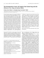
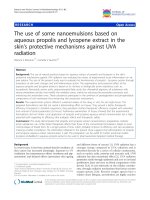
Figure 1. The generation of PTX resistant cell clones. (A) MDA-MB-231 cells in normal culture. (B) Most cells died in 2 weeks after 5 d of PTX exposure. (C) PTX
resistant cell clone was established.
Int. J. Med. Sci. 2018, Vol. 15
741
Meantime, DUSP5 expression was also correlated
negatively with higher histological grade being the
lowest in grade 3 cases (Figure 5E, P < 0.001).
Reduced expression of DUSP5 correlates with
poor prognosis in BLBC patients
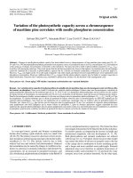
Figure 2. PTX resistance-related genes were functionally connected. (A)
CENPF, CDC6, MCM3, CLSPN and SMC1A expression was upregulated in PTX
resistant MDA-MB-231 cells by qRT-PCR. (B) The functional association
networks of PTX resistance-related genes were analyzed using the STRING
database, with subgroups marked by their functions.
Interestingly, the pattern of DUSP5 expression in
PAM50 subtypes was different from DUSP1 in
ONCOMINE and GOBO data sets. In the cohort of
ONCOMINE data, the expression of DUSP5 was
slightly higher in luminal B (P = 0.002) and in HER2 (P
= 0.044) when compared with luminal A (Figure 5A,
B). However, a noticeable decrease of DUSP5
expression was shown in basal subtype compared
with the other three subtypes (Figure 5A, B, P < 0.001).
The identical expression pattern of DUSP5 was
also found in GOBO data set using the PAM50
subtypes. DUSP5 expression was significantly
reduced in ER-negative (n = 395) tumors compared
with ER-positive (n = 1225, P < 0.001) (Figure 5C).
Compared with luminal A, luminal B and HER2
subtypes, basal subtype showed an obvious decrease
in DUSP5 expression (Figure 5D, P < 0.001). These
results suggested that the downregulated DUSP5
expression might be peculiar for BLBC patients.
In order to analyze the relationship of DUSP1
and DUSP5 expression with survival, Kaplan-Meier
(KM) Plotter (www.kmplot.com)[24], which contained gene expression data and survival information of
5143 clinical breast cancer patients downloaded from
GEO, EGA and TCGA, was used. To analyze the
prognostic value of DUSP1 and DUSP5, patient
samples were split into two groups according to lower
quartile expression (high vs. low expression) and
assessed by a KM survival plot, with the hazard ratio
(HR) with 95% confidence intervals (CI) and logrank
P value.
The KM survival analysis showed that although
DUSP1 expression wasn’t significantly associated
with overall survival (OS) of patients with breast
cancer (Figure S1A) (n = 1402, HR = 0.85 (0.66 - 1.08),
logrank P = 0.19), it was significantly associated with
relapse free survival (RFS) (Figure S1B) (n = 3951, HR
= 0.85 (0.75 - 0.96), logrank P = 0.0075). Interstingly,
DUSP5 expression was not only associated with OS of
patients with breast cancer (Figure S1C) (n = 1402, HR
= 0.76 (0.6 - 0.96), logrank P = 0.021), but also significantly associated with RFS (Figure S1D) (n = 3951, HR
= 0.67 (0.59 - 0.75), logrank P < 0.001). The breast
cancer patients with lower mRNA levels of DUSP1 or
DUSP5 were predicted to have poor RFS while the
lower mRNA levels of DUSP5 alone was poor
prognostic marker for OS in breast cancer patients.
Next we evaluated the prognostic value of
DUSP1 and DUSP5 expression in basal subtype in this
cohort of KM database (Figure 6A-D). DUSP1
expression was neither associated with OS (n = 241,
HR = 1.29 (0.67 - 2.48), logrank P = 0.44) (Figure 6A)
nor with RFS (n = 618, HR = 1.19 (0.91 - 1.55), logrank
P = 0.21) (Figure 6B). Remarkably, DUSP5 expression
was significantly associated with RFS (n = 618, HR =
0.58 (0.44 - 0.76), logrank P < 0.001) (Figure 6D). The
median RFS of BLBC patients with low DUSP5
expression (14.13 months) was shorter than that of
patients with high DUSP5 expression (26 months).
Moreover, the median OS of BLBC patients with low
DUSP5 expression (34.49 months) was much shorter
than that of patients with high DUSP5 expression
(80.64 months), and this difference almost reached
statistically significant effect (n = 241, HR = 0.59 (0.35 1.01), logrank P = 0.053) (Figure 6C). These results
suggested that the lower mRNA levels of DUSP5,
rather than DUSP1, might be poor prognostic marker
for BLBC patients and play roles in PTX resistance.
Int. J. Med. Sci. 2018, Vol. 15
742
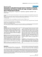
Figure 3. DUSP1 and DUSP5 were downregulated in PTX resistant BLBC cell clones. (A-B) The expression of the top ten genes in microarray data was validated by
qRT-PCR, and DUSP1 and DUSP5 expression was downregulated in PTX resistant MDA-MB-231 cells compared with control cells. (C) Similarly, DUSP1 and DUSP5
expression was downregulated in PTX resistant Hs578T cells compared with control cells.
Figure 4. DUSP1 expression in BLBC patients. (A-B) The expression of DUSP1 was lower in luminal B, HER2 and basal subtypes when compared with luminal A, and
HER2 subtype being the lowest by analyzing ONCOMINE data. (C) The expression of DUSP1 was lower in luminal B, HER2 and basal subtypes when compared with
luminal A, and basal subtype being the lowest by analyzing GOBO data. (D) DUSP1 expression correlated negatively with higher grade.
Discussion
BLBC patients usually present with aggressive
clinical features, such as metastasis to the lung and
brain, high histologic grade and have a poor
prognosis and thus need chemotherapy[25].
However, after the chemotherapy, residual cancer
cells mostly survive and provoke tumor growth,
which contributes to cancer recurrence and mortality.
During the last decades, molecular targeted therapies
has been vigorously advocated by precision medicine
model [26]. Therefore identifying markers involved in
the progression of BLBC would allow the
development of targeted therapies.
A growing body of evidence suggests that
DUSPs may provide prognostic and predictive utility
in several cancers including breast cancer[14, 16]. This
Int. J. Med. Sci. 2018, Vol. 15
study demonstrated, for the first time, the mRNA
expression and prognostic value of DUSP1 and
DUSP5 in basal like breast cancer. Herein, we
demonstrated that DUSP1 and DUSP5 expression was
significantly downregulated in PTX-resistant BLBC
cell lines, suggesting their association with resistance
to chemotherapy. Moreover, microarray data, GO
analysis and STRING analysis provided evidence that
743
the PTX-resistant BLBC cells was associated with a
highly aggressive phenotype, and proliferative
markers of malignant cell growth such as CENPF,
CDC6, MCM3, CLSPN and SMC1A showed elevated
expression in PTX-resistant BLBC cells, suggesting the
role of PTX-resistant cells with DUSP1 and DUSP5
downregualtion in BLBC progression.
Figure 5. The downregulated DUSP5 expression in BLBC patients. (A-B) Basal subtype showed the lowest expression level of DUSP5 compared with luminal A,
luminal B and HER2 subtypes by analyzing ONCOMINE data. (C-D) DUSP5 expression was significantly lower in ER-negative tumors compared with ER-positive (C),
and basal subtype showed an obvious decrease in DUSP5 expression compared with luminal A, luminal B and Her2 subtypes (D) by analyzing GOBO data. (E) DUSP5
expression correlated negatively with higher grade.
Int. J. Med. Sci. 2018, Vol. 15
744
Figure 6. The prognostic value of DUSP1 and DUSP5 expression in BLBC patients. (A-B) DUSP1 expression was neither associated with OS (A) nor with RFS (B)
of BLBC patients. (C) DUSP5 expression was almost significantly associated with OS of BLBC patients and the P value was close to 0.05. (D) DUSP5 expression was
significantly associated with RFS of BLBC patients.
To confirm the data of the cell line studies, we
performed analysis in breast cancer patients by using
publically available gene expression database.
Analyses had been performed using ONCOMINE
cohorts, GOBO Affymetrix-based data sets and KM
database. These analyses demonstrated the
association of low DUSP1 and DUSP5 expression with
high histological grade. Although low DUSP1
expression wasn’t significantly associated with OS of
patients with breast cancer, it was significantly
associated with RFS. Low DUSP5 expression was not
only associated with OS of patients with breast cancer
but also significantly associated with RFS. Therefore
these findings together suggested that DUSP1 and
DUSP5 functioned as tumor suppressors and might
inhibit the progression of breast cancer.
In addition, we found that DUSP1 expression
pattern in four subtypes of breast cancer was
discrepant in these large publically databases, with it
being lowest in HER2 subtype in ONCOMINE
database and in basal subtype in GOBO database.
These results were consistent with the study of He J et
al.[27], which showed that DUSP1 expression was
significantly lower in ER-negative breast cancer cell
lines (basal like and HER2) than in ER-positive breast
cancer cell lines (luminal A and luminal B) by the
integrated analysis of GEO database. Taken together,
these consistent results suggest that DUSP1
expression may be associated with ER status and
could be considered as a potential target gene for the
treatment of ER-negative breast cancer.
Importantly, we demonstrated that lower
expression of DUSP5 was seen in basal like than in
non-basal like breast cancer, with consistent
ONCOMINE and GOBO data analysis. DUSP5
expression was lowest in basal like cancers,
underlining DUSP5 expression levels were correlated
with the basal like subtype of breast cancer and the
possible role of DUSP5 in aggressive process of BLBC.
Remarkably, further analysis demonstrated the
significant correlation of low DUSP5 levels with
shorter OS and RFS in BLBC. However, the data
revealed that there were no significant differences in
OS and RFS between high and low DUSP1 expression.
Therefore, it is DUSP5, not DUSP1, may have
potential to be a useful biomarker for BLBC and
Int. J. Med. Sci. 2018, Vol. 15
additional efforts to explore its clinical significance in
BLBC patients are needed in future.
Rushworth LK et al. have demonstrated that
DUSP5 is a nonredundant regulator of both nuclear
ERK activation and localization, and DUSP5 functions
as a tumor suppressor and may play a part in
restraining tumor aggressiveness in different
cancers[19]. Yan X et al. has found DUSP5 expression
is positively correlated with E-cadherin expression,
but negatively correlated with N-cadherin and
vimentin expression, suggesting it may be involved in
regulation of epithelial-to-mesenchymal transition
program and tumor progression in advanced
colorectal cancer[28]. They also found that high risk
stage patients receiving chemotherapy with high
DUSP5 expression appeared to have a significantly
better survival than those with low DUSP5
expression[28]. The study from Boeckx C et al.[29] also
showed chemotherapy resistant cancer cells exhibited
low expression of DUSP5 and concomitant ERK
signaling activation in head and neck squamous cell
carcinoma, suggesting DUSP5 expression as an ERK
inhibitor might be a new strategy for overcoming
chemotherapy resistance. In our study, the dramatic
reduction of DUSP5 expression appeared in
PTX-resistant basal like breast cancer cells and basal
subtype of breast cancer, suggesting that DUSP5 may
participate in various cancer-related biological
processes, and loss of DUSP5 expression contributed
to drug resistance and tumor progression of BLBC.
In summary, we demonstrate that DUSP5
expression is characteristically downregulated in
basal like subtype compared with other subtypes of
breast cancer, and may be associated with malignant
development of BLBC. We identify DUSP5 expression
can serve as a useful prognostic biomarker for BLBC
patients. Moreover, we suggest DUSP5 expression is
correlated with PTX resistance in basal like cancer
cells, which may partly explain its prognostic effect on
BLBC patients since most BLBC patients should be
given chemotherapy. Overall, these findings
collectively demonstrate that DUSP5 has great
potential to be translated into clinical practice and
induced DUSP5 upregulation could be a promising
strategy to overcome PTX acquired resistance in
BLBC.
Materials and Methods
Cell culture and Paclitaxel treatment
The human breast cancer cell lines MDA-MB-231
were obtained from the American Type Culture
Collection. Hs578T cells were provided by the Cell
Bank of Type Culture Collection of the Chinese
Academy of Sciences, Shanghai, China. These cells
745
were cultured in Dulbecco’s Modified Eagle’s
Medium supplemented with 10% fetal bovine serum
(Hyclone) in a humidified 5% CO2 incubator at 37°C.
Paclitaxel (Selleckchem) treatment for cancer
cells was performed as previously described[30].
Briefly, 1×10⁶ cells were plated and cultured in
100-mm dishes for 24 h and then treated with 10 nM
paclitaxel for 5 days. Cells were then washed with
PBS and maintained in drug-free culture with media
replacement every 48 h until resistant cell clones
established.
RNA extraction and microarray analysis
Total RNA was extracted using Trizol reagent
(Tiangen Biotech, Beijing, China), and sent to
Oebiotech (Shanghai, China) for Affymetrix GeneChip® Human Transcriptome Array 2.0 analysis. The
microarray data have been deposited in NCBI’s Gene
Expression Omnibus (GEO) (Liu et al., 2016) and are
accessible through GEO Series accession number
GSE90145 ( />y/acc.cgi?acc=GSE90145).
QRT-PCR
QRT-PCR was performed as previously
described[30, 31]. Briefly, 2 μg of total RNA was
reverse-transcribed into cDNA using a Reverse
Transcription Kit (Takara, RR037A). QRT-PCR
analyses were performed with Power SYBR Green
(Takara, RR820A) in 7500HT Real-Time PCR System
(Applied Biosystems, Foster City, CA). GAPDH
internal control was used as an endogenous control,
and fold changes were presented by using the 2–ΔΔCt
method using the equation (ΔΔCT = (Ct gene of
interest - Ct GAPDH) treated sample - (Ct gene of
interest – Ct GAPDH) control sample). All qRT-PCR
reactions were performed in triplicates. The fold
change > 2 or < 0.5 was considered as significant.
ONCOMINE analysis
ONCOMINE gene expression array datasets
(www. oncomine.org), an online cancer microarray
database[32], was used to analyze the expression
levels of DUSP1 and DUSP5 in breast cancers. Breast
cancer patients were classified into four different
subtypes (luminal A, luminal B, HER2-enriched, or
basal) based on the PAM50 signature. The expression
level of DUSP1 and DUSP5 in luminal B, HER2enriched, and basal was acquired and compared with
luminal A breast cancers by using Students’t-test.
GOBO analysis
DUSP1 and DUSP5 expression levels for 1881
breast cancer patients were analyzed based on
molecular subtypes and other clinicopathological
parameters (stage, grade, nodal status) by using the
Int. J. Med. Sci. 2018, Vol. 15
data sets from the gene expression-based outcome for
breast cancer online algorithm (GOBO). Clinical
characteristics of individual data sets were described
previously[23].
The kaplan-meier plotter
746
3.
4.
5.
The prognostic value for survival was evaluated
using an online database, Kaplan-Meier Plotter
(www.kmplot.com)[24]. Only the JetSet best probe set
of DUSP1 and DUSP5 were chosen to obtain
Kaplan-Meier plots.
7.
Statistical analysis
9.
Data analysis was performed with the SPSS16.0
software package (IBM). All P values were two-sided,
and statistical significance was measured at the 0.05
level.
Abbreviations
BLBC, basal-like breast cancer; CI, confidence
intervals; DEGs, differentially expressed genes;
DUSPs, Dual-specificity phosphatases; ER, estrogen
receptor; ERK, extracellular signal-regulated kinase;
GEO, Gene Expression Omnibus; GO, gene ontology;
GOBO, gene expression-based outcome for breast
cancer online algorithm; HER2, human epidermal
growth factor receptor 2; HR, hazard ratio; HTA,
Human Transcriptome Array; JNK, c-Jun N-terminal
protein kinases; KM, Kaplan-Meier; MAPK, mitogenactivated protein kinase; OS, overall survival; PR,
progesterone receptor; PTX, paclitaxel; QRT-PCR,
quantitative real-time polymerase chain reaction; RFS,
relapse free survival; STRING, search tool for the
retrieval of interacting genes/proteins.
6.
8.
10.
11.
12.
13.
14.
15.
16.
17.
18.
19.
Supplementary Material
20.
Figure S1. />Table S1. />
21.
Acknowledgment
This work was partly supported by a grant from
The National Natural Science Foundation of China
(No. 81672870 to T. Liu and No. 81572872 to X. Zhao),
and National Undergraduate Training Program for
Innovation and Entrepreneurship (No. 201510062001
to H. Sun).
22.
23.
24.
Competing Interests
25.
The authors have declared that no competing
interest exists.
26.
References
1.
2.
Bertucci F, Finetti P, Birnbaum D. Basal breast cancer: a complex and deadly
molecular subtype. Current molecular medicine. 2012; 12: 96-110.
Kennecke H, Yerushalmi R, Woods R, Cheang MC, Voduc D, Speers CH, et al.
Metastatic behavior of breast cancer subtypes. Journal of clinical oncology :
official journal of the American Society of Clinical Oncology. 2010; 28: 3271-7.
27.
28.
Rane C, Senapedis W, Baloglu E, Landesman Y, Crochiere M, Das-Gupta S, et
al. A novel orally bioavailable compound KPT-9274 inhibits PAK4, and blocks
triple negative breast cancer tumor growth. Scientific reports. 2017; 7: 42555.
Dai X, Li T, Bai Z, Yang Y, Liu X, Zhan J, et al. Breast cancer intrinsic subtype
classification, clinical use and future trends. American journal of cancer
research. 2015; 5: 2929-43.
Leidy J, Khan A, Kandil D. Basal-like breast cancer: update on
clinicopathologic, immunohistochemical, and molecular features. Archives of
pathology & laboratory medicine. 2014; 138: 37-43.
Milioli HH, Tishchenko I, Riveros C, Berretta R, Moscato P. Basal-like breast
cancer: molecular profiles, clinical features and survival outcomes. BMC
medical genomics. 2017; 10: 19.
Carey L, Winer E, Viale G, Cameron D, Gianni L. Triple-negative breast
cancer: disease entity or title of convenience? Nature reviews Clinical
oncology. 2010; 7: 683-92.
Sorolla A, Ho D, Wang E, Evans CW, Ormonde CF, Rashwan R, et al.
Sensitizing basal-like breast cancer to chemotherapy using nanoparticles
conjugated with interference peptide. Nanoscale. 2016; 8: 9343-53.
Denkert C, Liedtke C, Tutt A, von Minckwitz G. Molecular alterations in
triple-negative breast cancer-the road to new treatment strategies. Lancet.
2017; 389: 2430-42.
Caunt CJ, Keyse SM. Dual-specificity MAP kinase phosphatases (MKPs):
shaping the outcome of MAP kinase signalling. The FEBS journal. 2013; 280:
489-504.
Kidger AM, Keyse SM. The regulation of oncogenic Ras/ERK signalling by
dual-specificity mitogen activated protein kinase phosphatases (MKPs).
Seminars in cell & developmental biology. 2016; 50: 125-32.
Low HB, Zhang Y. Regulatory Roles of MAPK Phosphatases in Cancer.
Immune network. 2016; 16: 85-98.
Huang CY, Tan TH. DUSPs, to MAP kinases and beyond. Cell & bioscience.
2012; 2: 24.
Shen J, Zhang Y, Yu H, Shen B, Liang Y, Jin R, et al. Role of DUSP1/MKP1 in
tumorigenesis, tumor progression and therapy. Cancer medicine. 2016; 5:
2061-8.
Mills BN, Albert GP, Halterman MW. Expression Profiling of the MAP Kinase
Phosphatase Family Reveals a Role for DUSP1 in the Glioblastoma Stem Cell
Niche. Cancer microenvironment : official journal of the International Cancer
Microenvironment Society. 2017.
Hou MF, Chang CW, Chen FM, Wang SN, Yang SF, Chen PH, et al. Decreased
total MKP-1 protein levels predict poor prognosis in breast cancer. World
journal of surgery. 2012; 36: 1922-32.
Kang Y, Nagaraja AS, Armaiz-Pena GN, Dorniak PL, Hu W, Rupaimoole R, et
al. Adrenergic Stimulation of DUSP1 Impairs Chemotherapy Response in
Ovarian Cancer. Clinical cancer research : an official journal of the American
Association for Cancer Research. 2016; 22: 1713-24.
Lin YC, Lin YC, Shih JY, Huang WJ, Chao SW, Chang YL, et al. DUSP1
expression induced by HDAC1 inhibition mediates gefitinib sensitivity in
non-small cell lung cancers. Clinical cancer research : an official journal of the
American Association for Cancer Research. 2015; 21: 428-38.
Rushworth LK, Kidger AM, Delavaine L, Stewart G, van Schelven S, Davidson
J, et al. Dual-specificity phosphatase 5 regulates nuclear ERK activity and
suppresses skin cancer by inhibiting mutant Harvey-Ras (HRasQ61L)-driven
SerpinB2 expression. Proceedings of the National Academy of Sciences of the
United States of America. 2014; 111: 18267-72.
Shin SH, Park SY, Kang GH. Down-regulation of dual-specificity phosphatase
5 in gastric cancer by promoter CpG island hypermethylation and its potential
role in carcinogenesis. The American journal of pathology. 2013; 182: 1275-85.
Cai C, Chen JY, Han ZD, He HC, Chen JH, Chen YR, et al. Down-regulation of
dual-specificity phosphatase 5 predicts poor prognosis of patients with
prostate cancer. International journal of clinical and experimental medicine.
2015; 8: 4186-94.
Nunes-Xavier CE, Tarrega C, Cejudo-Marin R, Frijhoff J, Sandin A, Ostman A,
et al. Differential up-regulation of MAP kinase phosphatases MKP3/DUSP6
and DUSP5 by Ets2 and c-Jun converge in the control of the growth arrest
versus proliferation response of MCF-7 breast cancer cells to phorbol ester.
The Journal of biological chemistry. 2010; 285: 26417-30.
Ringner M, Fredlund E, Hakkinen J, Borg A, Staaf J. GOBO: gene
expression-based outcome for breast cancer online. PloS one. 2011; 6: e17911.
Gyorffy B, Lanczky A, Eklund AC, Denkert C, Budczies J, Li Q, et al. An online
survival analysis tool to rapidly assess the effect of 22,277 genes on breast
cancer prognosis using microarray data of 1,809 patients. Breast cancer
research and treatment. 2010; 123: 725-31.
Chung S, Jin Y, Han B, Qu Y, Gao B, Giuliano AE, et al. Identification of
EGF-NF-kappaB-FOXC1 signaling axis in basal-like breast cancer. Cell
communication and signaling : CCS. 2017; 15: 22.
Feng YZ, Zhang QY, Fu MT, Zhang ZF, Wei M, Zhou JY, et al. Low expression
of PinX1 is associated with malignant behavior in basal-like breast cancer.
Oncology reports. 2017; 38: 109-19.
He J, Yang J, Chen W, Wu H, Yuan Z, Wang K, et al. Molecular Features of
Triple Negative Breast Cancer: Microarray Evidence and Further Integrated
Analysis. PloS one. 2015; 10: e0129842.
Yan X, Liu L, Li H, Huang L, Yin M, Pan C, et al. Dual specificity phosphatase
5 is a novel prognostic indicator for patients with advanced colorectal cancer.
American journal of cancer research. 2016; 6: 2323-33.
Int. J. Med. Sci. 2018, Vol. 15
747
29. Boeckx C, Op de Beeck K, Wouters A, Deschoolmeester V, Limame R,
Zwaenepoel K, et al. Overcoming cetuximab resistance in HNSCC: the role of
AURKB and DUSP proteins. Cancer letters. 2014; 354: 365-77.
30. Liu T, Sun H, Zhu D, Dong X, Liu F, Liang X, et al. TRA2A Promoted Paclitaxel
Resistance and Tumor Progression in Triple-Negative Breast Cancers via
Regulating Alternative Splicing. Molecular cancer therapeutics. 2017; 16:
1377-88.
31. Sun H, Liu T, Zhu D, Dong X, Liu F, Liang X, et al. HnRNPM and CD44s
expression affects tumor aggressiveness and predicts poor prognosis in breast
cancer with axillary lymph node metastases. Genes, chromosomes & cancer.
2017; 56: 598-607.
32. Rhodes DR, Yu J, Shanker K, Deshpande N, Varambally R, Ghosh D, et al.
ONCOMINE: a cancer microarray database and integrated data-mining
platform. Neoplasia. 2004; 6: 1-6.