- Trang chủ >>
- Nông - Lâm - Ngư >>
- Thú y
Anepidemiological study of feline and canine dermatophytoses in japan
Bạn đang xem bản rút gọn của tài liệu. Xem và tải ngay bản đầy đủ của tài liệu tại đây (688.21 KB, 6 trang )
Med. Mycol. J.
Med.
Vol. 60 (No. 2) , 2019
Vol.
60, Mycol.
39- 44, J.
2019
ISSN 2185-6486
39
Review
An Epidemiological Study of Feline and Canine Dermatophytoses
in Japan
1
2
Shigeo Yamada , Kazushi Anzawa and Takashi Mochizuki
1
2
2
Yamada Animal Hospital
Department of Dermatology, Kanazawa Medical University
ABSTRACT
In a 2012-2014 epidemiological study of feline and canine dermatophytoses in Japan, we investigated the prevalence of fungi
among 296 cats and 170 dogs treated at a veterinary clinic and 51 cats and dogs at an animal shelter at Fukui City in Japan.
Microsporum canis was isolated from only one cat out of the 517 animals. Also, from 2012 to 2017, we analyzed isolates from 76
cats and 15 dogs with dermatophytoses at 14 veterinary clinics across 10 prefectures in Honshu and Shikoku. M. canis was the
cause for 85 of the cases and Microsporum gypseum for the other six. M. canis infection routes in cats are thought to include stray
cats as well as breeding facilities and pet shops, whereas for dogs, only breeding facilities and pet shops. Tinea was found in 18.7
% (14/75) of the owners of these animals. We showed that microsatellite genotyping is useful for molecular epidemiological
investigations such as determination of infection routes of M. canis.
Key words : cats, dermatophytoses, dogs, epidemiology, Japan, Microsporum canis
Introduction
Most dermatophytes infecting pet cats and dogs are
Microsporum canis, Microsporum gypseum (new classification: Nannizzia gypsea), and some zoophilic species of
1-3)
Trichophyton mentagrophytes and Trichophyton benhamiae ,
all of which can cause tinea in humans. The prevalence of
feline and canine dermatophytoses in Japan has been falling in
recent years due to better rearing environments and measures
4, 5)
implemented by municipalities against stray cats and dogs .
Nevertheless, measures to control dermatophytoses have
become important as raising dogs and cats indoors has become
popular, making contact between pet animals and humans
more common. Herein, we discuss the results of our recent
investigation into the prevalence of dermatophytes in cats and
dogs and the results of another investigation into outbreaks of
dermatophytoses at 14 veterinary clinics in Honshu and
Shikoku. We also discuss the application of microsatellite
(MS) polymorphism analysis as a molecular biological marker
for differentiation of M. canis strains.
Address for correspondence: Shigeo Yamada, DVM
Yamada Animal Hospital, Ohmachi 2-1112, Fukui, Fukui 918-8116, Japan
Received: 25, October 2018, Accepted: 19, December 2018
E-mail:
Prevalence of symptomless feline and canine
dermatophytoses
Between November 2012 and December 2014, we
examined 296 cats and 170 dogs taken to a veterinary clinic
run by one of the authors (SY) in Fukui City (Fukui prefecture,
Hokuriku, Fig. 1). None of the animals had symptoms of
dermatophytoses. Skin swabs taken with cotton wool sticks
6)
from all 466 animals were cultured for fungus . Briefly, using
a sterile cotton swab moistened by sterile saline, almost the
whole body surface of an animal was rubbed carefully, then
the tip of the swab was pressed onto the agar plate and the
cotton part of each swab was cut and placed onto the same
agar plate. The medium used in the study was Sabouraud’s
dextrose agar with cycloheximide and chloramphenicol
®
(Mycosel agar , Eiken Chemical Co. Ltd., Tochigi, Japan)
supplemented with gentamicin sulfate (50 µg/ml). The
inoculated plates were aerobically incubated at 25ºC for up to
14 days. Growing fungi were identified by morphological
characteristics. The animals’ origin, living conditions, and age
were also analysed (Table 1). During the same period, swab
samples from 46 cats and 5 dogs at a Fukui Health and
40
Medical Mycology Journal Volume 60, Number 2, 2019
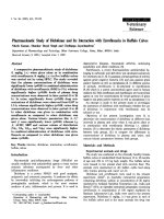
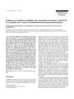
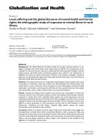
Fig. 1. Origins of animals with dermatomycoses in the present survey.
Shaded areas denote regions examined.
Numbers in parentheses denote number of isolates.
Table 1. Background of healthy animals examined in a veterinary clinic6)
Species
No. of animals
Cat
296
Dog
170
Animal origin, exposure to environment and age
No. of identified (%)
Kept indoors constantly
95 (32.1)
Stray cat, aged < 6 months old
95 (32.1)
Reside indoors but allowed to wander outside
45 (15.2)
Stray cat, aged ≧6 months old
37 (12.5)
Purchased from pet shop within 2 weeks
24 (8.1)
Purchased from pet shop within 2 weeks
95 (55.9)
Kept indoors and not taken for walks on grass
43 (25.3)
Kept indoors but taken for walks on grass
24 (14.1)
Kept outdoors
Welfare Center in Fukui City were also obtained and cultured
6)
using the same methods . The cats were assumed to be at least
6-months old if their permanent teeth had already fully
developed.
Table 2 shows our results along with those of recent studies
indicating prevalence rates of dermatophytoses among cats
and dogs in Japan. Although we investigated 517 symptomless
animals, one was positive for M. canis, which was isolated
from a swab taken from a pedigree cat immediately after
purchase from a pet shop. The prevalence of dermatophytes
among cats and dogs in and round Fukui City was found to be
8 (4.7)
very low, irrespective of animal living conditions, origin, and
age.
These results agree with reports of 0% prevalence among 32
5)
house cats in Kanto (Tokyo and Kanagawa prefectures) , 0%
among 177 dogs in an animal shelter in Kanto (Saitama
7)
prefecture) , and 1.1% among 180 domestic cats (geographic8)
al areas not given) . Therefore, in recent years, the prevalence
of dermatophytes among pet cats and dogs and stray cats in
Japan has been extremely low. However, an analysis of the
source of infection of 25 people diagnosed with M. canis at a
dermatology clinic in Kyushu (Kumamoto prefecture) showed
Med. Mycol. J. Vol. 60 (No. 2) , 2019
41
Table 2. Recent studies of prevalence of dermatophytes among healthy cats and dogs in Japan
First author
(Reference No.)
Region
(Prefecture)
Yamada6)
Hokuriku (Fukui)
Yamada6)
Kano5)
7)
Animal origin
Prevalence rate of dermatophytes
(positive/examined)
2015
Household cats and dogs,
Stray cats
Cat: 0.34% (1/296): M. canis
Dog: 0% (0/170)
Hokuriku (Fukui)
2015
Shelter cats and dogs
Cat: 0% (0/46)
Dog: 0% (0/5)
Kanto (Tokyo, Kanagawa)
2008
Household cats reared indoors
Cat: 0% (0/32)
Year of report
Sakaki
Kanto (Saitama)
2017
Shelter dogs
Dog: 0% (0/177)
Itoh8)
Not listed
2017
Household cats
Cat: 1.1%: (2/180): M. canis
Itoh13)
Not listed
2017
Cats raised in pet shops
Cat: 3% (3/99): M. canis
Chiba14)
Kanto (Tokyo)
2015
Cats raised by animal-handling
business
Cat: 21.5% (29/135)
Microsporum sp.: 20.7% (28/135)
Trichophyton sp.: 0.7% (1/135)
that 12 probably contracted it from stray cats they had
adopted, and three probably contracted it from cats bought at
9)
pet shops . And an analysis of 21 cases of dermatophytosis
diagnosed at a dermatology clinic in Hokuriku (Ishikawa
prefecture) showed that two contracted it from dogs and 15
from cats, among which 14 cats had probably contracted it
4)
through contact with other animals outdoors . There may be
large differences in the incidence rates of infections by isolates
per year. In a seven-year survey at a veterinary clinic in the
same area, the majority of cases occurred in just two of the
4)
seven years . Therefore, it is speculated that infection
reservoirs exist outdoors locally and for limited periods.
Survey of cases of dermatophytoses in animals
We conducted a survey of animals diagnosed with
dermatophytoses at 14 veterinary clinics in Honshu, which is
the largest island in Japan, and Shikoku (2 in Kanto, 7 in
Hokuriku, 2 in Kinki, 2 in Chugoku, and 1 in Shikoku)
between September 2012 and April 2017. We investigated
causative fungi, age, origin and living condition of infected
animals, and whether transmission occurred indoors. A total of
91 animals were infected, 77 cats and 14 dogs, of which 35
were in Kanto, 31 in Hokuriku, 3 in Kinki, 7 in Chugoku, and
15 in Shikoku (Fig. 1).
We asked the owners about the source of infection, their
living conditions, and whether there was an infected human in
their household. Owners who had tinea lesions took samples
of themselves by pressing sticky tape over the lesion after
giving their informed consent to participate in this study. The
harvested scales were cultivated, and the fungus was
identified. The results are shown in Table 3.
The only dermatophytes isolated were M. canis and M.
gypseum. The former was much more common, accounting for
96.1% of the incidences associated with cats and 78.6% with
dogs. These findings were similar to those of previous reports
10, 11)
in Japan
. Most cats under 6 months old infected with M.
canis were either stray cats, from animal shelters, or had just
been bought from a pet shop. Older cats over 6 months old
infected with M. canis were house pets in almost half the
cases, the others being, in decreasing numbers, stray cats, pet
cats that often went outdoors, and cats from pet shops. Of the
14 dogs infected with M. canis, nine probably contracted it in
pet shops or at grooming service providers. All the animals
infected with M. gypseum were thought to have contracted it
outdoors: the cats with M. gypseum were strays or housed in
animal shelters, and the dogs with M. gypseum habitually
came into contact with soil. Tinea infected 14 (18.7%) of 75
households that own animals, and M. canis was isolated from
skin samples of owners in all 14 cases. The transmission rate
4, 12)
from pet to owner was similar to that reported previously .
Itoh et al. reported 3% prevalence of dermatophytes among
99 young cats aged 1 to 6 months old housed in 8 pet shops
13)
14)
(geographical areas not given) . In contrast, Chiba et al.
reported dermatophyte prevalence at 16 facilities among
animal-handling businesses in Tokyo, and categorized it as a
high rate (21. 5%). Therefore, in Japan, there may be
concentrations of animals infected with M. canis in some
insanitary pet shops and facilities where pets are reared in
large numbers such as at pet-breeding establishments, which
may be one of the routes of transmission to humans and other
animals. Outdoor transmission is another major infection
route, but the prevalence of dermatophytes might not be high
even among stray cats and pet cats that go outdoors. However,
because outdoor reservoirs of infection may emerge regionally, we should continuously investigate the status quo of
parasitic fungi. In contrast, the most likely route of infection
for dogs is the pet shop, because most dogs in Japan, unlike
cats, are confined indoors. Indeed, municipalities have
publicized animal-rearing methods, and the risk of dogs
42
Medical Mycology Journal Volume 60, Number 2, 2019
Table 3. Analysis of 91 cats and dogs with dermatophytoses
Species
No.
of animals
Age
(months)
No. (%) of
M. canis isolated
Cat
77
Total
74 (96.1)
<6
39
No. (%) of
Lifestyle and route of infection
M. gypseum isolated
No. (%) of
animals
3 (3.9)
Stray cat or shelter cat
28 (71.8)
Immediately after purchase from pet shop
10 (25.6)
Infected at house where other cat is infected
≧6
35
Cohabiting infection
3
Dog
14
Total
11 (78.6)
<6
7
≧6
1 (2.6)
17 (48.6)
Stray cat or shelter cat
8 (22.9)
Household cat going out
3 (8.6)
Immediately after purchase from pet shop
2 (5.7)
After shampoo treatment at pet shop
2 (5.7)
Long-term steroid treatment in progress
1 (2.9)
Owner touched infected cat at other residence
1 (2.9)
Unidentified (Full indoor rearing, suffering from
diabetes mellitus)
1 (2.9)
Stray cat or shelter cat
3 (100.0)
Immediately after purchase from pet shop
6 (85.7)
Cohabiting infection
1 (14.3)
After shampoo treatment at pet shop
3 (75.0)
Cohabiting infection
1 (25.0)
Outdoor rearing
1 (33.3)
Displays digging behavior
1 (33.3)
Walking around the lawn for long-term steroid
treatment
1 (33.3)
3 (21.4)
4
3
contracting M. canis outdoors has probably dramatically fallen
due to decreasing numbers of stray dogs. As M. gypseum is
geophilic, it will probably be isolated as a causative fungus of
canine and feline dermatophytoses at a low rate in the future.
Extra caution is especially needed for dogs on long-term
immuno-suppressants when they come into contact with soil.
In addition, although the genus Trichophyton was not isolated
in this study, T. mentagrophytes was isolated from a kitten
housed in an animal shelter in Fukui prefecture in 2018
(unpublished data). It is inferred that this fungus also infects at
a low rate.
Application of microsatellite analysis to molecular
epidemiology
Molecular markers for detecting intraspecific variation and
for strain discrimination have been studied to elucidate the
15-19)
17)
status quo of M. canis infection . Sharma et al. and
18)
Pasquetti et al. reported that the most sensitive biomolecular
marker is microsatellite DNA polymorphism. It is even useful
for molecular epidemiological studies of M. canis, which
19)
shows little intraspecific variation . Hereafter, we introduce
results of MS analysis of the isolated Japanese strains and
discuss its future use.
18)
Using the MS marker method of Pasquetti et al. ,
20)
Watanabe et al. analyzed 70 M. canis strains in Japan, of
which 59 were isolated from humans and 11 from cats, and
divided them into 20 genotypes (A to T). They found the same
genotype of M. canis isolated from pet cats and confirmed catto-human transmission in five families (Table 4). Using
Watanabe’s method to show that M. canis isolated from tinea
on the face of a 2-month-old baby had the same genotype as an
isolate from a symptomless cat that lived with the baby’ s
grandparents, we were able to confirm that the baby must have
21)
contracted the infection while staying at her grandparents
(Table 4).
MS analysis is therefore a useful method for investigating
routes and sources of infections. In the future, analyzing
isolates from animals could be used to monitor the spread of
infections from pet shops and for investigating reservoirs of
infection outdoors. By looking at genotype variance, we can
Med. Mycol. J. Vol. 60 (No. 2) , 2019
43
Table 4. Studies of genotypes, identified by multilocus microsatellite typing (MLMT),
in infected cohabitants
First author
(Reference No.)
Case No.
Source
Genotype of the
isolates by MLMT
Watanabe21)
1
Human
A
2
3
4
5
4)
Takeda
1
see the number of infection sources within stray cats and
thereby get a picture of the habitat conditions of M. canis
outdoors.
Conclusion
There continues to be many incidences of feline and canine
dermatophytoses in Japan. Determining the prevalence and
routes of infection of M. canis could inform a prevention
policy. There appears to be two main routes in cats, namely,
pet shops and outdoors due to strays, whereas for dogs, the
main route is thought to be through pet shops. Regarding the
outdoor route for cats, M. canis reservoirs may form locally in
certain regions and then spread sporadically. Therefore, when
cases of outdoor-related infections, such as infected stray cats
or pet cats with dermatophytoses known to be among a stray
cat community, are diagnosed by dermatologists or veterinarians, attention should be paid to the probable existence of
reservoirs of infection and the risk of subsequent outbreaks.
MS analysis is a useful method for elucidating the status quo
of infections.
Acknowledgment
These studies were partially supported by the Research
Program on Emerging and Re-emerging Infectious Diseases
from the Japan Agency for Medical Research and Development, AMED (JP17fk0108208). We would like to thank the
Cat
A
Human
A
Cat-1
A
Cat-2
A
Human-1
I
Human-1
I
Cat-1
I
Cat-2
I
Human
R
Cat
R
Human
S
Cat
S
Cat
A
Human
A
Fukui Health and Welfare Center (Fukui Prefecture) who
helped with the collection of strains and cases and the
following 13 veterinary hospitals: Kanto Region: Taguchi
Animal Hospital, Hidamari Animal Clinic; Hokuriku Region:
Yoshida Animal Hospital, Tamura Animal Hospital, Animal
Hospital Pia, Rurbannomori Animal Clinic, Harue Animal
Hospital, Inaba Animal Clinic; Kinki Region: Fujimura
Animal Hospital, Ai Animal Hospital; Chugoku Region:
Sanyo Animal Medical Center, Nonaka Animal Hospital;
Shikoku Region: Central City Animal Hospital
Conflicts of interest
None to declare.
References
1) Miller WH, Griffin CE, Campbell KL: Fungal and algal skin
diseases. Muller & Kirk’s Small Animal Dermatology 7th ed.,
pp. 223-283, Elsevier Inc., St. Louis, 2013.
2) Moriello KA, Coyner K, Paterson S, Mignon B: Diagnosis and
treatment of dermatophytosis in dogs and cats: clinical
consensus guidelines of the world association for veterinary
dermatology. Vet Dermatol 28: 266-e68, 2017.
3) Kano R, Iyori K, Harada K, Murayama N, Yamasaki M,
Makimura K, Tsuboi R, Yamagishi K, Murai T, Nishifuji K,
Hasegawa A, Nagata M: Canine and feline dermatophytosis: a
guideline for the antifungal therapy. Jpn J Vet Dermatol 24: 912, 2017. [Article in Japanese]
44
4) Kobayashi H, Yoshioka M, Anzawa K, Mochizuki T: Cases of
Microsporum canis infection between 2005 and 2011 in a
dermatology clinic in the southern part of Kanazawa City. Skin
Research 12: 219-223, 2013. [Article in Japanese]
5) Kano R: Animal cutaneous mycoses in Japan. Med Mycol J 53:
19-23, 2012. [Article in Japanese]
6) Yamada S, Anzawa K, Mochizuki T: Survey of dermatophyte
infection in cats and dogs in the Fukui region of Japan. Skin
Research 14: 166-170, 2015. [Article in Japanese]
7) Sakaki Y, Arai H, Kobayashi C, Narusawa T, Usui A, Saeki E:
Prevalence of dermatophytes of dogs in an animal shelter.
Abstract book for 20th Annual Meeting of Japanese Society of
Veterinary Dermatology, p108, 2017. [Article in Japanese]
8) Itoh N, Kato H, Ito Y, Oozasa N, Kimura Y, Kanai K:
Prevalence of dermatophytes in private household cats. Jpn J
Vet Dermatol 23: 9-12, 2017. [Article in Japanese]
9) Sakae H, Noguchi H, Ichinokawa Y, Hiruma M: Analysis of 25
cases of Microsporum canis infection encountered at a
dermatology clinic in Kumamoto during a recent 3-year period.
Med Mycol J 52: 139-144, 2011. [Article in Japanese]
10) Ikeshoji T, Takahashi M, Igami, N, Kubo S, Matsuda M,
Takata M, Ishikawa M: Distribution of dermatophytes carriers
in cats. J Jpn Vet Med Ass 45: 430-431, 1992. [Article in
Japanese]
11) Nagata M, Nankou H: Microsporum canis infection in animals.
MB Derma 45: 29-34, 2001. [Article in Japanese]
12) Nagata M: Human and animal common infections seen from
dogs and cats. Rinsho Derma 54: 329-335, 2012. [Article in
Japanese]
13) Itoh N, Suda M, Iijima Y, Ito Y, Totsapon P, Kimura Y:
Medical Mycology Journal Volume 60, Number 2, 2019
14)
15)
16)
17)
18)
19)
20)
21)
Prevalence of dermatophytes in pet shop young cats. Jpn J Vet
Dermatol 23: 69-72, 2017. [Article in Japanese]
Chiba T, Takahashi Y, Uehara S: Examination and analysis of
zoonotic fungi in Tokyo. Ann Rep Tokyo Metr Inst Pub Health
67: 39-48, 2016. [Article in Japanese]
Dobrowolska A, Debska J, Kozlowska M, Staczek P: Strains
differentiation of Microsporum canis by RAPD analysis using
(GACA) 4 and (ACA) 5 primers. Pol J Microbiol 60: 145148, 2011.
Cano J, Rezusta A, Solé M, Gil J, Rubio MC, Revillo MJ,
Guarro J: Inter-single-sequence-repeat-PCR typing as a new
tool for identification of Microsporum canis strains. J Dermatol
Sci 39: 17-21, 2005.
Sharma R, de Hoog S, Presber W, Gräser Y: A virulent
genotype of Microsporum canis is responsible for the majority
of human infections. J Med Microbiol 56: 1377-1385, 2007.
Pasquetti M, Peano A, Soglia D, Min AR, Pankewitz F, Ohst T,
Gräser Y: Development and validation of a microsatellite
marker-based method for tracing infections by Microsporum
canis. J Dermatol Sci 70: 123-129, 2013.
Mochizuki T, Takeda K, Anzawa K: Molecular markers useful
for intraspecies subtyping and strain differentiation of
dermatophytes. Mycopathologia 182: 57-65, 2017.
Watanabe J, Anzawa K, Mochizuki T: Molecular epidemiology of Japanese isolates of Microsporum canis based on
multilocus microsatellite typing fragment analysis. Jpn J Infect
Dis 70: 544-548, 2017.
Takeda K, Anzawa K, Mochizuki T, Yamada S, Kobayashi H,
Kimura S: Infant case of tinea faciei caused by Microsporum
canis. J Dermatol 45: e187-e188, 2018.