Giải phẫu khuôn mặt của người châu Á - Ứng dụng trong tiêm filler và botox
Bạn đang xem bản rút gọn của tài liệu. Xem và tải ngay bản đầy đủ của tài liệu tại đây (22.09 MB, 188 trang )
Clinical Anatomy
of the Face for
Filler and Botulinum
Toxin Injection
Hee-Jin Kim
Kyle K. Seo
Hong-Ki Lee
Jisoo Kim
123
Clinical Anatomy of the Face for Filler
and Botulinum Toxin Injection
Hee-Jin Kim • Kyle K. Seo
Hong-Ki Lee • Jisoo Kim
Clinical Anatomy
of the Face for Filler
and Botulinum Toxin
Injection
Hee-Jin Kim
Yonsei University College of Dentistry
Seoul
Republic of Korea
Kyle K. Seo
Modelo Clinic
Seoul
Republic of Korea
Hong-Ki Lee
Image Plastic Surgery Clinic
Seoul
Republic of Korea
Jisoo Kim
Dr. Youth Clinic
Seoul
Republic of Korea
Illustrations by Kwan-Hyun Youn.
Extended translation from the Korean language edition: 보툴리눔 필러 임상해부학
by Hee-Jin Kim, Kyle K. Seo , Hong-Ki Lee, Jisoo Kim
Copyright © 2015. All Rights Reserved.
ISBN 978-981-10-0238-0
ISBN 978-981-10-0240-3
DOI 10.1007/978-981-10-0240-3
(eBook)
Library of Congress Control Number: 2016938223
© Springer Science+Business Media Singapore 2016
This work is subject to copyright. All rights are reserved by the Publisher, whether the whole or
part of the material is concerned, specifically the rights of translation, reprinting, reuse of
illustrations, recitation, broadcasting, reproduction on microfilms or in any other physical way,
and transmission or information storage and retrieval, electronic adaptation, computer software,
or by similar or dissimilar methodology now known or hereafter developed.
The use of general descriptive names, registered names, trademarks, service marks, etc. in this
publication does not imply, even in the absence of a specific statement, that such names are
exempt from the relevant protective laws and regulations and therefore free for general use.
The publisher, the authors and the editors are safe to assume that the advice and information in
this book are believed to be true and accurate at the date of publication. Neither the publisher nor
the authors or the editors give a warranty, express or implied, with respect to the material
contained herein or for any errors or omissions that may have been made.
Printed on acid-free paper
This Springer imprint is published by Springer Nature
The registered company is Springer Science+Business Media Singapore Pte Ltd.
Preface
First, I would like to thank my friend, Dr. Kyle Seo, for organizing all the
extremely important clinical information and tips. I also wish to thank Dr.
Hong-Ki Lee for his insightful inquisitions and questions that made coming
up of creative contents possible. Also, I give my thanks to Dr. Jisoo Kim, who
played a strong role in the planning of cadaver dissection workshops and in
other works related to organizing necessary contents. Without the efforts and
sacrifice of the above individuals in providing clinical manuscripts and in
revising all of the visuals despite their busy clinical schedules, this book’s
text and artwork would not have been able to shine. As such, I send infinite
thanks to Dr. Kwan-Hyun Youn for providing all of the visuals for this book.
I believe that Dr. Youn, an art major graduate with a PhD in Anatomy, has
raised our country’s medical illustrations to that of world class. Many thanks
to the effort of the Medart team led by Dr. Youn to make this book to have
many clear, simple, and creative visual contents to be possible.
In the Fall of 2011, my research on clinical anatomy research in relation to
aesthetics—and through this, teachings on clinical anatomy—started after
receiving advice from John Rogers, a US neurology specialist and medical
director of the Pacific Asian region for Allergan Inc., who visited my anatomy lab. Rogers, who had no particular interest in aesthetic treatments,
enabled me to devote myself more to this field. Through regional and international educations, I had presented basic information on new methods regarding aesthetic treatment guidelines based on anatomy in order to avoid
complications. Then, after hearing that many regional doctors were following
anatomic guidelines based on Western research, the coauthors and I designed
this book to introduce new methods to fit for Asians, who have slightly different anatomic features. For instance, Asians possess different locations of the
modiolus, different directions and changes of facial arteries, and different
attachment regions for muscles unlike to Caucasians. All of these and more
are explained in detail in this book using research papers presented during my
lectures as foundational information. Through this, new injection techniques
are described in the book.
Current medical techniques are rapidly changing due to the development
of science. As a result, this trend is giving way to a new slogan for medicine
such as “borderless” and “above and beyond the border” for a movement
working to dismantle academic borders. Biocompatible fillers and botulinum
toxin injection development have started to create a new medical field of noninvasive aesthetic plastic surgery, referred to as ‘Beauty Plastic Surgery’, and
v
Preface
vi
the desire for new medical techniques is bringing about developments in
clinical anatomy. Likewise, I feel that it is right for clinical doctors from all
fields to come together as a virtuous group to jump over the wall of traditional
medicine for the development of medical practices. And, as a health personnel studying basic medicine, I feel immense responsibility and a sense of
worth in being a part of this movement.
This book includes various images and pictures for simpler understanding
of anatomy from ‘Plastic and Reconstructive Surgery’ and other 80 research
papers from acknowledged journals in relation to clinical anatomy. In addition, we worked to include various documents about Koreans so that it may
be utilized as a useful document in other areas. It is my wish that, through this
book, readers are able to learn clinical techniques related to aesthetic treatments and to grow in knowledge regarding the prevention of complications.
I also thank Professor Kyungseok Hu and my graduate student Sang-Hee
Lee, You-Jin Choi, Hyung-Jin Lee, Jung-Hee Bae, Liyao Cong, and Kyuho
Lee from Yonsei University College of Dentistry who actively helped search
for visual information and aided in other revision works for this book. Lastly,
I would like to thank Dr. Yoonjung Hwang, Mr. Sanghoon Kwon, Juyong
Lee, Yongwoong Lee and Ms. Hwieun Hur, and Young-Gyung Kim in translating the Korean manuscript of this textbook.
On the behalf of the authors,
Seoul, South Korea
November, 2015
Hee-Jin Kim
Contents
1
General Anatomy of the Face and Neck. . . . . . . . . . . . . . . . . . .
1.1 Aesthetic Terminology . . . . . . . . . . . . . . . . . . . . . . . . . . . . .
1.1.1 Basic Aesthetic Terminology . . . . . . . . . . . . . . . . . .
1.2 Layers of the Face . . . . . . . . . . . . . . . . . . . . . . . . . . . . . . . . .
1.2.1 Layers of the Skin . . . . . . . . . . . . . . . . . . . . . . . . . . .
1.2.2 Thickness of the Skin . . . . . . . . . . . . . . . . . . . . . . . .
1.3 Muscles of Facial Expressions and Their Actions . . . . . . . .
1.3.1 Forehead Region . . . . . . . . . . . . . . . . . . . . . . . . . . . .
1.3.2 Temporal Region (or Temple). . . . . . . . . . . . . . . . . .
1.3.3 Orbital Region. . . . . . . . . . . . . . . . . . . . . . . . . . . . . .
1.3.4 Nose Region . . . . . . . . . . . . . . . . . . . . . . . . . . . . . . .
1.3.5 Perioral Muscles . . . . . . . . . . . . . . . . . . . . . . . . . . . .
1.3.6 Platysma Muscle . . . . . . . . . . . . . . . . . . . . . . . . . . . .
1.4 SMAS Layer and Ligaments of the Face . . . . . . . . . . . . . . .
1.5 Nerves of the Face and Their Distributions . . . . . . . . . . . . .
1.5.1 Distribution of the Sensory Nerve . . . . . . . . . . . . . .
1.5.2 Distribution of the Motor Nerve . . . . . . . . . . . . . . . .
1.5.3 Upper Face . . . . . . . . . . . . . . . . . . . . . . . . . . . . . . . .
1.5.4 Midface . . . . . . . . . . . . . . . . . . . . . . . . . . . . . . . . . . .
1.5.5 Lower Face . . . . . . . . . . . . . . . . . . . . . . . . . . . . . . . .
1.6 Nerve Block . . . . . . . . . . . . . . . . . . . . . . . . . . . . . . . . . . . . .
1.6.1 Supraorbital Nerve Block (SON Block) . . . . . . . . . .
1.6.2 Supratrochlear Nerve Block (STN Block) . . . . . . . .
1.6.3 Infraorbital Nerve Block (ION Block) . . . . . . . . . . .
1.6.4 Zygomaticotemporal Nerve Block (ZTN Block) . . .
1.6.5 Mental Nerve Block (MN Block) . . . . . . . . . . . . . . .
1.6.6 Buccal Nerve Block (BN Block) . . . . . . . . . . . . . . .
1.6.7 Inferior Alveolar Nerve Block (IAN Block). . . . . . .
1.6.8 Auriculotemporal Nerve Block (ATN Block). . . . . .
1.6.9 Great Auricular Nerve Block (GAN Block) . . . . . . .
1.7 Facial Vessels and Their Distribution Patterns . . . . . . . . . . .
1.7.1 Facial Branches of the Ophthalmic Artery . . . . . . . .
1.7.2 Facial Branches of the Maxillary Artery. . . . . . . . . .
1.7.3 Facial Artery . . . . . . . . . . . . . . . . . . . . . . . . . . . . . . .
1.7.4 Frontal Branch of the Superficial Temporal Artery .
1.7.5 Facial Veins. . . . . . . . . . . . . . . . . . . . . . . . . . . . . . . .
1.7.6 Connections of the Vein . . . . . . . . . . . . . . . . . . . . . .
1
2
2
5
5
6
7
8
10
11
13
14
20
21
23
24
24
24
25
26
28
28
28
28
29
29
29
31
31
31
32
34
35
35
37
38
42
vii
Contents
viii
1.8 Facial and Skull Surface Landmarks . . . . . . . . . . . . . . . . . .
1.9 Characteristics of Asian (Korean) Skull and Face . . . . . . . .
1.10 Anatomy of the Aging Process . . . . . . . . . . . . . . . . . . . . . . .
1.10.1 Aging Process of the Facial Tissue . . . . . . . . . . . . . .
1.10.2 The Complex Changes of the Facial
Appearance with Aging . . . . . . . . . . . . . . . . . . . . . .
Suggested Reading. . . . . . . . . . . . . . . . . . . . . . . . . . . . . . . . . . . . .
Physical Anthropological Traits in Asians . . . . . . . . . . . . . .
Muscles of the Face and Neck . . . . . . . . . . . . . . . . . . . . . . .
Vessels of the Face and Neck . . . . . . . . . . . . . . . . . . . . . . . .
Peripheral Nerves of the Face and Neck. . . . . . . . . . . . . . . .
2
3
42
45
48
49
50
51
51
52
52
53
Clinical Anatomy for Botulinum Toxin Injection . . . . . . . . . . .
2.1 Introduction. . . . . . . . . . . . . . . . . . . . . . . . . . . . . . . . . . . . . .
2.1.1 Effective Versus Ineffective Indications
of Botulinum Toxin for Wrinkle Treatment . . . . . . . .
2.1.2 Botulinum Rebalancing . . . . . . . . . . . . . . . . . . . . . .
2.2 Botulinum Wrinkle Treatment . . . . . . . . . . . . . . . . . . . . . . .
2.2.1 Crow’s Feet (Lateral Canthal Rhytides) . . . . . . . . . .
2.2.2 Infraorbital Wrinkles. . . . . . . . . . . . . . . . . . . . . . . . .
2.2.3 Horizontal Forehead Lines . . . . . . . . . . . . . . . . . . . .
2.2.4 Glabellar Frown Lines . . . . . . . . . . . . . . . . . . . . . . .
2.2.5 Bunny Lines . . . . . . . . . . . . . . . . . . . . . . . . . . . . . . .
2.2.6 Plunged Tip of the Nose . . . . . . . . . . . . . . . . . . . . . .
2.2.7 Gummy Smile, Excessive Gingival Display . . . . . . .
2.2.8 Nasolabial Fold . . . . . . . . . . . . . . . . . . . . . . . . . . . . .
2.2.9 Asymmetric Smile, Facial Palsy . . . . . . . . . . . . . . . .
2.2.10 Alar Band . . . . . . . . . . . . . . . . . . . . . . . . . . . . . . . . .
2.2.11 Purse String Lip . . . . . . . . . . . . . . . . . . . . . . . . . . . .
2.2.12 Drooping of the Mouth Corner . . . . . . . . . . . . . . . . .
2.2.13 Cobblestone Chin . . . . . . . . . . . . . . . . . . . . . . . . . . .
2.2.14 Platysmal Band . . . . . . . . . . . . . . . . . . . . . . . . . . . . .
2.3 Botulinum Facial Contouring . . . . . . . . . . . . . . . . . . . . . . . .
2.3.1 Masseter Hypertrophy. . . . . . . . . . . . . . . . . . . . . . . .
2.3.2 Temporalis Hypertrophy . . . . . . . . . . . . . . . . . . . . . .
2.3.3 Hypertrophy of the Salivary Gland . . . . . . . . . . . . . .
Suggested Reading. . . . . . . . . . . . . . . . . . . . . . . . . . . . . . . . . . . . .
Muscles of the Face and Neck . . . . . . . . . . . . . . . . . . . . . . .
Peripheral Nerves of the Face and Neck. . . . . . . . . . . . . . . .
Others . . . . . . . . . . . . . . . . . . . . . . . . . . . . . . . . . . . . . . . . . .
55
56
Clinical Anatomy of the Upper Face for Filler Injection . . . . .
3.1 Forehead and Glabella . . . . . . . . . . . . . . . . . . . . . . . . . . . . .
3.1.1 Clinical Anatomy . . . . . . . . . . . . . . . . . . . . . . . . . . .
3.1.2 Injection Points and Methods . . . . . . . . . . . . . . . . . .
3.1.3 Side Effects . . . . . . . . . . . . . . . . . . . . . . . . . . . . . . . .
3.2 Sunken Eye and Pretarsal Roll . . . . . . . . . . . . . . . . . . . . . . .
3.2.1 Clinical Anatomy . . . . . . . . . . . . . . . . . . . . . . . . . . .
3.2.2 Injection Points and Methods . . . . . . . . . . . . . . . . . .
3.2.3 Side Effects . . . . . . . . . . . . . . . . . . . . . . . . . . . . . . . .
93
94
94
94
100
103
103
105
109
56
56
58
58
62
63
63
69
70
71
71
72
75
75
75
80
81
84
84
88
89
91
91
92
92
Contents
ix
3.3
Temple. . . . . . . . . . . . . . . . . . . . . . . . . . . . . . . . . . . . . . . . . .
3.3.1 Clinical Anatomy . . . . . . . . . . . . . . . . . . . . . . . . . . .
3.3.2 Injection Points and Methods . . . . . . . . . . . . . . . . . .
3.3.3 Side Effects . . . . . . . . . . . . . . . . . . . . . . . . . . . . . . . .
Suggested Reading. . . . . . . . . . . . . . . . . . . . . . . . . . . . . . . . . . . . .
Muscles of the Face and Neck . . . . . . . . . . . . . . . . . . . . . . .
Vessels of the Face and Neck . . . . . . . . . . . . . . . . . . . . . . . .
Peripheral Nerves of the Face and Neck. . . . . . . . . . . . . . . .
109
111
113
116
118
118
118
118
4
Clinical Anatomy of the Midface for Filler Injection . . . . . . . .
4.1 Tear Trough. . . . . . . . . . . . . . . . . . . . . . . . . . . . . . . . . . . . . .
4.1.1 Clinical Anatomy . . . . . . . . . . . . . . . . . . . . . . . . . . .
4.1.2 Injection Points and Methods . . . . . . . . . . . . . . . . . .
4.2 Nasojugal Groove . . . . . . . . . . . . . . . . . . . . . . . . . . . . . . . . .
4.2.1 Clinical Anatomy . . . . . . . . . . . . . . . . . . . . . . . . . . .
4.2.2 Injection Points and Methods . . . . . . . . . . . . . . . . . .
4.3 Palpebromalar Groove . . . . . . . . . . . . . . . . . . . . . . . . . . . . .
4.3.1 Clinical Anatomy . . . . . . . . . . . . . . . . . . . . . . . . . . .
4.3.2 Injection Points and Methods . . . . . . . . . . . . . . . . . .
4.4 Nasolabial Fold . . . . . . . . . . . . . . . . . . . . . . . . . . . . . . . . . . .
4.4.1 Clinical Anatomy . . . . . . . . . . . . . . . . . . . . . . . . . . .
4.4.2 Injection Points and Methods . . . . . . . . . . . . . . . . . .
4.5 Hollow Cheek . . . . . . . . . . . . . . . . . . . . . . . . . . . . . . . . . . . .
4.5.1 Clinical Anatomy . . . . . . . . . . . . . . . . . . . . . . . . . . .
4.5.2 Insertion Points and Methods . . . . . . . . . . . . . . . . . .
4.6 Subzygoma Depression. . . . . . . . . . . . . . . . . . . . . . . . . . . . .
4.6.1 Clinical Anatomy . . . . . . . . . . . . . . . . . . . . . . . . . . .
4.6.2 Injection Points and Methods . . . . . . . . . . . . . . . . . .
4.7 Nose . . . . . . . . . . . . . . . . . . . . . . . . . . . . . . . . . . . . . . . . . . .
4.7.1 Clinical Anatomy . . . . . . . . . . . . . . . . . . . . . . . . . . .
4.7.2 Injection Points and Methods . . . . . . . . . . . . . . . . . .
Suggested Reading. . . . . . . . . . . . . . . . . . . . . . . . . . . . . . . . . . . . .
Physical Anthropological Traits in Asians . . . . . . . . . . . . . .
Muscles of the Face and Neck . . . . . . . . . . . . . . . . . . . . . . .
Vessels of the Face and Neck . . . . . . . . . . . . . . . . . . . . . . . .
Peripheral Nerves of the Face and Neck. . . . . . . . . . . . . . . .
119
120
120
123
124
124
127
128
128
128
128
128
131
135
135
135
138
138
139
139
139
148
150
150
150
151
151
5
Clinical Anatomy of the Lower Face for Filler Injection . . . . .
5.1 Lip . . . . . . . . . . . . . . . . . . . . . . . . . . . . . . . . . . . . . . . . . . . . .
5.1.1 Clinical Anatomy . . . . . . . . . . . . . . . . . . . . . . . . . . .
5.1.2 Injection Points and Methods . . . . . . . . . . . . . . . . . .
5.1.3 Side Effects . . . . . . . . . . . . . . . . . . . . . . . . . . . . . . . .
5.2 Chin. . . . . . . . . . . . . . . . . . . . . . . . . . . . . . . . . . . . . . . . . . . .
5.2.1 Clinical Anatomy . . . . . . . . . . . . . . . . . . . . . . . . . . .
5.2.2 Injection Points and Methods . . . . . . . . . . . . . . . . . .
5.2.3 Side Effects . . . . . . . . . . . . . . . . . . . . . . . . . . . . . . . .
5.3 Perioral Wrinkles . . . . . . . . . . . . . . . . . . . . . . . . . . . . . . . . .
5.3.1 Clinical Anatomy . . . . . . . . . . . . . . . . . . . . . . . . . . .
5.3.2 Injection Points and Methods . . . . . . . . . . . . . . . . . .
5.3.3 Side Effects . . . . . . . . . . . . . . . . . . . . . . . . . . . . . . . .
153
154
154
154
157
160
160
160
162
165
165
166
166
Contents
x
5.4
Marionette Line and Jowl . . . . . . . . . . . . . . . . . . . . . . . . . . .
5.4.1 Clinical Anatomy . . . . . . . . . . . . . . . . . . . . . . . . . . .
5.4.2 Injection and Methods. . . . . . . . . . . . . . . . . . . . . . . .
5.4.3 Side Effects . . . . . . . . . . . . . . . . . . . . . . . . . . . . . . . .
5.5 Anatomical Considerations of the Symptoms
That May Accompany Filler Treatment . . . . . . . . . . . . . . . .
5.5.1 Vascular Compromise . . . . . . . . . . . . . . . . . . . . . . . .
5.5.2 Suggested Methods to Reduce Vascular
Problems Related with Filler Injection . . . . . . . . . . .
Suggested Reading. . . . . . . . . . . . . . . . . . . . . . . . . . . . . . . . . . . . .
Physical Anthropological Traits in Asians . . . . . . . . . . . . . .
Muscles of the Face and Neck . . . . . . . . . . . . . . . . . . . . . . .
Vessels of the Face and Neck . . . . . . . . . . . . . . . . . . . . . . . .
Peripheral Nerves of the Face and Neck. . . . . . . . . . . . . . . .
166
166
168
168
Index . . . . . . . . . . . . . . . . . . . . . . . . . . . . . . . . . . . . . . . . . . . . . . . . . . .
175
169
169
172
173
173
173
173
174
1
General Anatomy of the Face
and Neck
Hee-Jin Kim (Illustrated
by Kwan-Hyun Youn)
© Springer Science+Business Media Singapore 2016
H.-J. Kim et al., Clinical Anatomy of the Face for Filler and Botulinum Toxin Injection,
DOI 10.1007/978-981-10-0240-3_1
1
1
2
1.1
Aesthetic Terminology
Inconsistencies exist between anatomical and
aesthetic terminology. We attempt to redefine
common clinical terms according to anatomical
regions (Fig. 1.1).
1.1.1
Basic Aesthetic Terminology
Facial Creases
Facial creases are deep, shallow creases caused
by changes in the structural integrity of the skin.
It occurs due to loss of skin and muscle fiber elasticity caused by repetitive facial movements and
changes in facial expressions. Creases are generally termed wrinkles and lines. Other terms such
as furrow, groove, and sulcus are used in the clinical fields.
General Anatomy of the Face and Neck
Skin Folds
Skin folds occur due to sagging, loss of
tension, and gravity. Representative skin folds
are the nasolabial fold, the labiomandibular
fold, etc.
Baggy Lower Eyelids (or Cheek Bags,
Malar Bags)
Baggy lower eyelids occur due to a drooping of
the adipose tissue underneath the orbicularis
oculi m. This should be distinguished from the
festoon since the baggy lower eyelid occurs inferior to the orbital margin.
Blepharochalasis
Blepharochalasis occurs due to sagging of the
eyelid skin.
Horizontal forehead lines
Glabellar frown lines
Blepharochalasis
Tear trough
Palpebromalar groove
Glabellar transverse lines
Crow’s feet
Baggy lower eyelid
Nasojugal groove
Preauricular lines
Horizontal upper lip line
Mentolabial creases
Platysmal band
Midcheek furrow
Bunny lines
Nasolabial fold
Marionette line
Jowl
Labiomandibular fold
Horizontal neck lines
Fig. 1.1 Aging facial creases and wrinkles (Published with kind permission of ࿈ Kwan-Hyun Youn 2016. All rights
reserved)
1.1
Aesthetic Terminology
Bunny Line
The bunny line is the oblique nose furrows lateral
to the nose bridge that is pronounced by various
facial expressions. The levator labii superioris
alaeque nasi m. below the skin and the medial
muscular band of the orbicularis oculi m. participate in the formation of the bunny line.
Commissural Lines
Commissural lines are short, vertical lines
appearing on each sides of the mouth corner.
Occasionally, deep creases may form starting
from the perioral regions.
Crow’s Feet (Lateral Canthal Wrinkles)
Crow’s feet are thin, bilateral wrinkles at the lateral sides of the eyes formed by the orbicularis
oculi m.
Festoon
Festoon is the bulged appearance of the lower
eyelids caused by a sagging of the skin and of the
orbicularis oculi m. and by a protrusion of the
inferior orbital fat compartment underneath the
orbital septum.
Horizontal Forehead Lines (Worry Lines)
Horizontal forehead lines are horizontal lines
across the forehead region where the frontalis m.
is located.
Glabellar Frown Lines (Glabellar Creases
or Lines)
Glabellar frown lines are vertical creases along
the glabellar region caused by the corrugator
supercilii muscle fibers.
Glabellar Transverse Lines
Glabellar transverse lines are horizontal lines on
the radix that are typically produced during facial
distortion. They occur perpendicular to the fibers
of the procerus m.
3
Gobbler Neck (Platysmal Bands)
The gobbler neck appears as bilateral vertical
skin bands on the neck along the anterior cervical
and submental region. This occurs due to sagging
of the medial border of the platysma muscle.
Horizontal Neck Lines
Horizontal neck lines are horizontal skin folds on
the anterior cervical region. They are produced
by a combination of platysmal muscle fibers and
sagging neck skin.
Horizontal Upper Lip Lines (Transverse
Upper Lip Lines)
Horizontal upper lip lines are 1–2 horizontal
lines located at the philtrum on the upper lip.
Jowl (Jowl Sagging)
Jowl is the protrusion and sagging of the subcutaneous adipose tissue along the mandibular border. The anterior border of the prejowl sulcus
clearly signifies the existence of mandibular
retaining ligaments.
Oral Commissure
The labial commissure is the region where the
upper and lower lips join on each lateral side. The
joining point is referred to as the cheilion.
Labiomandibular Fold
The labiomandibular fold spans from the corner
of the mouth to the mandibular border and
becomes prominent with age. The depressor
anguli oris m. (DAO) defines the fold’s medial
and lateral borders. The attachment of the mandibular retaining ligament causes the labiomandibular fold to be located more anteriorly and
medially.
Marionette Line
The marionette line is a long, vertical line that
proceeds inferiorly from the corner of the mouth.
4
1
General Anatomy of the Face and Neck
It occurs commonly with age but with unknown
causes. It is more pronounced in people with less
fat tissues than in those with more fat tissues.
This line is also called the “disappointment line.”
muscles, and the zygomaticus major m. into the
skin in this area. In addition, the facial area tends
to lie underneath the nasolabial fold with variable
depths.
Mentolabial Creases (or Furrows)
Mentolabial creases are horizontal creases (one
or more) between the lower lip and the chin
(mentum). These creases lie between the orbicularis oris m. and the mentalis m.
Palpebromalar Groove
The palpebromalar groove is the border between
the lower lid and the malar region.
Midcheek Furrow (Indian Band)
The midcheek furrow is a downward and lateral
band, or furrow, that extends the nasojugal groove
from the lateral aspect of the nose to the region
superior to the anterior cheek. This band may
carry on inferior to the cheek. With age, the cheek
and the midface droop inferiorly and medially,
and the band forms along the inferior margin of
the zygomatic bone at the same height where the
zygomatic cutaneous ligament attaches to the
skin in this region.
Nasojugal Groove
The nasojugal groove is formed at the border
between the lower lid and the cheek and runs
inferolaterally from the medial canthus. The
nasojugal groove region corresponds with the
lower border of the orbicularis oculi m. and
becomes more pronounced with the existence of
the medial muscular band of the orbicularis oculi
m. With age, this groove obliquely continues
downward to the midcheek furrow.
Nasolabial Fold (or Nasolabial Groove)
The nasolabial fold starts from the side of the
nasal ala and extends obliquely between the
upper lip and the cheek. With age, the subcutaneous adipose tissue of the anterior cheek sags,
causing the fold to deepen and move downward.
The adipose tissue of the anterior cheek cannot
descend inferior to the nasolabial fold due to
compact attachment of the fascia, the skin, the
cutaneous insertions of upper lip elevator
Preauricular Lines
Periauricular lines are several vertical skin lines
located near the tragion, the ear lobule, and the
anterior region of the auricles.
Ptotic Chin
The ptotic chin is a flat and contracted chin associated with a deepened submental crease.
Tear Trough
The tear trough is a line originating from the
medial canthus and proceeding inferolaterally
along with the infraorbital margin. With age,
the inferior and medial portions of the orbit
sink due to contraction of the soft tissues (skin,
muscle, and fat) covering the area. The tear
trough has various forms according to how the
medial part of the orbicularis retaining ligament and the fibers of the medial muscular
band of orbicularis oculi m. come into contact
with the skin.
Temple Depression
Temporal depression is the gradual decrease in
volume of the soft tissues of the temporal region
expressed with age. The bone structure of the
temporal crest becomes more pronounced.
Vertical Lip Line
As aging is processed, the tooth is lost and
alveolar bone is absorbed. It leads perioral muscle and lip contracts, so the vertical lip line
appears along the vermilion border.
5
1.2 Layers of the Face
1.2
Layers of the Face
1.2.1
Layers of the Skin
Basic facial soft tissues are composed with five
layers: (1) skin, (2) subcutaneous layer, (3) superficial musculoaponeurotic system (SMAS),
(4) retaining ligaments and spaces, and (5) periosteum and deep fascia. Facial skin can move
over the loose areolar connective tissue layer
with the exception of the auricles and the nasal
ala, which are supported by the cartilage under
the skin. Facial skin contains numerous sweat
and sebaceous glands (Fig. 1.2a, b).
a
Skin
Subcutaneous layer
Facial mm. & superficial
musculoaponeurotic system
(SMAS)
Retaining ligament and space
Periosteum and deep fascia
b
c
Superficial layer of SMAS
Deep temporal fascia
SMAS
Temporal branch of facial n.
Innominate fascia
Parotid gland
Fig. 1.2 Anatomical layers of the face. (a) Basic five layers of the face, (b) SMAS (superficial musculoaponeurotic system), (c) reflected SMAS at the lateral aspect of
the face (Published with kind permission of © Hee-Jin
Kim, Kwan-Hyun Youn and Joo-Heon Lee 2016. All
rights reserved)
1
6
Among the subcutaneous fat tissue of the face,
superficial fat is divided into malar, nasolabial
fat, and so on. However, the boundary is not visible to the naked eye and the superficial fat may
seem to cover the whole face. Deep fat is placed
in the deeper part of the facial muscle and is
demarcated by dense connective tissues such as
the capsules or retaining ligaments. The color
and properties of the deep fat show different
characteristics from the superficial fat.
Suborbicularis oculi fat (SOOF), retro-orbicularis
oculi fat (ROOF), buccal fat, and deep cheek fat
are included in the deep fat of the face. Fibrous
connective tissues pass through facial fat tissues
and play in role in connecting the fat tissue, facial
muscles, dermis, and bone (Figs. 1.3 and 1.4).
The superficial fascia, or subcutaneous connective tissue, contains an unequal amount of fat
tissue, and these fat tissues smoothen the facial
contour between facial musculatures. In some
areas, fat tissues are broadly distributed. The buccal fat pad forms the bulged cheek and continues
to the scalp and the temple region. The facial v.,
the trigeminal nerve, the facial nerve, and the
superficial facial muscle are contained within the
subcutaneous tissue (Fig. 4.27).
The SMAS (superficial muscular aponeurotic system) is the superficial facial structure
General Anatomy of the Face and Neck
composed of muscle fibers and superficial facial
fascia. It is a continuous fibromuscular layer
investing and interlinking the facial m. The
SMAS extends from the platysma to the galea
aponeurotica and is continuous with the
temporoparietal fascia (TPF, superficial temporal fascia) and the galea layer. It is known that
the SMAS consists of three distinct layers: a
fascial layer superficial to the muscles, a layer
intimately associated with the facial m., and a
deep layer extensively attached to the periosteum of facial bones (Fig. 1.2c).
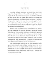
1.2.2
Thickness of the Skin
The general thickness of the facial skin is
described in the figure below. When treating in
areas with thin layers of skin, a filler injection
should be cautiously performed while trying to
avoid shallow filler placement. Upper and lower
eyelids, glabellar regions, and nasal regions have
an exceptionally thin skin layer. On the other
hand, the skin layer of the anterior cheek and the
mental region are relatively thicker. During filler
treatment, the skin’s flexibility and internal space
should also be considered along with its thickness (Fig. 1.5).
Forehead fat compartment
Lateral orbital fat compartment
Retaining ligaments
Palpebral portion of
orbicularis oculi m.
Medial muscular band
Malar fat compartment
Buccal fat pad
Nasolabial fat compartment
Prejowl fat compartment
Fig. 1.3 Superficial fat and superficial muscles of the face (Published with kind permission of ࿈ Kwan-Hyun Youn
2016. All rights reserved)
1.3
7
Muscles of Facial Expressions and Their Actions
Retro-orbicularis oculi fat
(ROOF)
Subprocerus galeal fat
Suborbicularis oculi fat (SOOF)
Deep medial cheek fat
Buccal fat pad
Fig. 1.4 Deep fat compartments of the face (Published with kind permission of ࿈ Kwan-Hyun Youn 2016. All rights
reserved)
1.3
1.08 mm
0.38 mm
0.77 mm
0.83 mm
1.06 mm
1.25 mm
0.86 mm
1.23 mm
0.86 mm
1.19 mm
0.58 mm
Fig. 1.5 Average skin thickness of the face (Published
with kind permission of ࿈ Kwan-Hyun Youn 2016. All
rights reserved)
Muscles of Facial Expressions
and Their Actions
Facial mm. are attached to the facial skeleton, or
membranous superficial fascia, beneath the skin,
or subcutaneous tissue. The topography of the
facial m. varies between males and females and
between individuals of the same gender. It is
important to define muscle shapes, their associations with the skin, and their relative muscular
actions in order to explain the unique expressions
people can make.
The face divides into nine distinct areas: (1)
the forehead including glabella from eyelids to
hair line, (2) temple or temporal region anterior
to the auricles, (3) orbital region, (4) nose region,
(5) zygomatic region, (6) perioral region and lips,
(7) cheek, (8) jaws, and (9) auricle.
These muscles are distributed in different
locations and (1) direct the openings of the orifices as dilators or sphincters and (2) form various facial expressions. These facial muscles,
located within the superficial fascia, or subcuta-
1
8
neous tissue layers, originate from the facial bone
or fascia and attach to the facial skin. They reveal
various expressions such as sadness, anger, joy,
fear, disgust, and surprise.
Facial mm. are widely distributed in different
regions of the face. However, they are generally
categorized different regions such as the forehead, the orbital, the nose, and other perioral
regions. The platysma m., which is involved in
the movement of the perioral region, is also considered a facial muscle (Fig. 1.6).
1.3.1
Forehead Region
The occipitofrontalis m. is a large, wide muscle
underlying the forehead and the occipital area. It
is divided into the frontal belly of the forehead
General Anatomy of the Face and Neck
region and the occipital belly of the occipital
region. Clinically, the frontal belly of the occipitofrontalis m. is referred to as the “frontalis muscle” and arises from the galea aponeurosis and
inserts into the orbicularis oculi m. and the frontal
skin above the eyebrow. The width and contraction of the frontalis m. vary between individuals;
during an individual’s anxiety and surprise, this
muscle produces transverse wrinkles on the
forehead.
The frontalis m. is rectangular and possesses
bilateral symmetry. Its muscle fibers are vertically oriented and join the orbicularis oculi and
the corrugator supercilii m. near the superciliary
arch of the frontal bone. The frontalis m. lies
beneath the skin of the forehead (3–5 mm in average), though depth can differ considerably
(27 mm) between individuals (Fig. 1.7).
a
Frontalis m.
Depressor supercilii m.
Orbicularis oculi m.
Zygomaticus major m.
Zygomaticus minor m.
Levator labii superioris m.
Risorius m.
Corrugator supercilii m.
Levator labii
superioris alaque
nasi m.
Levator anguli oris m.
Orbicularis oris m.
Depressor anguli oris m.
Depressor labii inferioris m.
Mentalis m.
Platysma m.
Fig. 1.6 Facial muscles. (a) Frontal view, (b) lateral view, (c) oblique view (Published with kind permission of
࿈ Kwan-Hyun Youn 2016. All rights reserved)
1.3
Muscles of Facial Expressions and Their Actions
b
Frontalis m.
Orbicularis oculi m.
Levator labii superioris alaque nasi m.
Nasalis m.
Levator labii superioris m.
Zygomaticus minor m.
Zygomaticus major m.
Orbicularis oris m.
Deressor labii inferioris m.
Depressor anguli oris m.
Mentalis m.
Risorius m.
Platysma m.
c
Frontalis m.
Orbicularis oculi m.
Levator labii superioris
alaque nasi m.
Nasalis m.
Levator labii superioris m.
Zygomaticus minor m.
Zygomaticus major m.
Risorius m.
Depressor anguli oris m.
Orbicularis oris m.
Depressor labii inferioris m.
Mentalis m.
Platysma m.
Fig. 1.6 (continued)
9
1
10
a
General Anatomy of the Face and Neck
b
Frontalis m.
Frontalis m.
Fig. 1.7 Frontalis muscle of the forehead (a, b) (Published with kind permission of ࿈ Hee-Jin Kim and Kwan-Hyun
Youn 2016. All rights reserved)
1.3.2
Temporal Region (or Temple)
The temporal region is confined within the
boundary of the temporal fossa. Within the temporal fossa, a fan-shaped temporalis and its vessels and nerves occupy this concavity. The
temporalis m. is divided into two layers: superficial and deep. A majority of the temporalis
belong to the deep layer and arise from the broad
temporal fossa, whereas the superficial layer of
the temporalis m. arises from the internal aspect
of the deep temporal fascia (temporalis muscle
fascia). The deep temporal fascia (temporalis
muscle fascia) is the tenacious fascia attached
superiorly to the superior temporal line and inferiorly to the upper margin of the zygomatic arch.
Though the superficial layer of the temporalis
developed in four-legged animals, the superficial
layer in human seems very thin and rudimentary.
All the temporalis muscle fibers converge as a
tendon and attach to the tip of the coronoid process and to the anteromedial side of the mandibular ramus. The temporalis holds a flat, fan
shape due to its broader origin and narrower
attachment.
There is a region in which the muscle fibers
transition into tendons. The upper half of the
temporalis superior to the zygomatic arch is composed only of the muscle belly, and the lower half
(roughly two- or three-digit widths) is occupied
by a converged tendon and a part of the deep
layer of the temporalis that is covered by the aponeurotic structure.
The temporalis m. is divided into three
parts: anterior, middle, and posterior temporalis m. While its anterior temporalis fibers proceed almost vertically, the fibers of the
posterior temporalis run almost horizontally.
The main functions of the temporalis differ
according to muscular orientation. A whole
temporalis m. raises the mandible for mouth
closing, providing tension to prevent the mouth
from opening against gravity. The temporalis
m. is innervated by the anterior, middle, and
posterior deep temporal nerves from the mandibular n. It is supplied by the anterior and
posterior deep temporal arteries for the anterior 2/3 of the temporalis and by the middle
temporal a. for the posterior 1/3 region as well
(Figs. 1.8 and 3.26).
1.3
Muscles of Facial Expressions and Their Actions
a
11
b
Temporalis m.
Masseter m.
Fig. 1.8 Temporalis muscle of the temporal region (a, b) (Published with kind permission of ࿈ Hee-Jin Kim and
Kwan-Hyun Youn 2016. All rights reserved)
1.3.3
Orbital Region
The shape of the eyes is well framed by moving
muscles that surround it, which determine basic
facial expressions. Orbicularis oculi m. is a broad,
flat, elliptical muscle composed of an orbital part
and a palpebral part. The palpebral part is then
divided again into a superficial portion (ciliary
bundle) and a deep portion (lacrimal part).
The main function of the orbicularis oculi m. is
to mediate eye closure. The orbicularis oculi m. has
many neighboring muscles (e.g., corrugator supercilii m., procerus m., frontalis m., zygomaticus
major m., and zygomaticus minor m.), and various
direct and indirect muscular connections exist
between the orbicularis oculi m. and the surrounding musculature. These connections may participate in the formation of various facial expressions.
In Asians, the lateral muscular band and the medial
muscular band of the orbital portion of the orbicularis oculi m. are observed in 54 % and 66 % of the
cases, respectively (Figs. 1.9, 1.10, 2.4, and 2.5).
Furthermore, it is observed that 89 % of Asians possess direct muscular connections between the zygomaticus minor m. and the orbicularis oculi m.
The corrugator supercilii m. originates from
the periosteum of the frontal bone on the medial
side of the superciliary arch, proceeds superiorly
and laterally, and then merges with the frontalis
m. It consists of two distinct bellies—the transverse and oblique belly. The origin of the transverse belly of the corrugator supercilii m. is
superior and more lateral than the origin of the
oblique belly, and most of them attach to the frontalis m. (Fig. 1.11) and to the superolateral orbital
part of the orbicularis oculi m. The transverse
belly is located deeper and proceeds in a more
horizontal direction than the oblique belly. This
muscle makes narrow, vertical wrinkles on the
glabellar region and presents an aged appearance
by producing these wrinkles with the frontalis m.
The depressor supercilii m. is a fan-shaped or
triangular-shaped muscle that originates from the
frontal process of the maxilla and from the nasal
portion of the frontal bone above the medial
palpebral ligament. The depressor supercilii m.
proceeds through the glabellar region while being
mixed with the corrugator supercilii m., and it
intermingles with medial fibers of the orbicularis
oculi m. (Fig. 1.10).
1
12
Fig. 1.9 Orbicularis
oculi muscle of the
orbital region. (a)
Frontal view, (b)
lateral view (Published
with kind permission
of ࿈ Kwan-Hyun
Youn and ByungHeon Kim 2016. All
rights reserved)
a
General Anatomy of the Face and Neck
b
Lateral muscular
band of
orbicularis oculi m.
Palpebral portion
of orbicularis oculi m.
Orbital portion
of orbicularis oculi m.
Fig. 1.10 Medial muscular
band of the orbicularis oculi
muscle and upper lip elevators
(Published with kind
permission of ࿈ Hee-Jin Kim
2016. All rights reserved)
Depressor supercilii m.
Palpebral portion
of orbicularis oculi m.
Medial muscular band of the
orbicularis oculi m.
Oblique band of the Transverse band of the
corrugator supercilii m. corrugator supercilii m.
Fig. 1.11 Corrugator
supercilii muscle
(Published with kind
permission of ࿈ Hee-Jin
Kim 2016. All rights
reserved)
1.3
Muscles of Facial Expressions and Their Actions
1.3.4
Nose Region
The nose is a dynamic structure that moves nasal
cartilages and plays an important role in the nasal
physiology. Muscles of the nose and the nose
region contain of the procerus m., the nasalis m.,
and the depressor septi nasi m., along with several other muscles attached to the nasal ala.
The procerus m. is a small muscle that originates from the nasal bone, proceeds superiorly,
and attaches to the skin of the radix. Fibers of the
frontalis m. at the insertion point are cross-locked.
This muscle makes a horizontal line on the radix
below the glabella by pulling the medial side of
the eyebrow down (Fig. 1.12).
The nasalis consists of a transverse part and an
alar part. The transverse part is a C-shaped, triangular muscle raised from the maxilla and the
canine fossa to the nasal ala. The transverse part
Nasalis m.
(transverse part)
Procerus m.
13
extends from the superficial layer of the levator
labii superioris alaeque nasi m. The alar part is a
small rectangular muscle arising from the maxilla superior to the maxillary lateral incisor and
inserting into the deep skin layer of the alar facial
crease of the alar cartilage. The transverse part
compresses and decreases the size of the naris,
while the alar part serves to enlarge the size of the
naris (Fig. 1.13).
The depressor septi nasi m. is located on the
deep part of the lip. This muscle arises from the
incisive fossa (between the central and lateral
incisors) and inserts into the moving part of the
nasal septum. It pulls the nose tip inferiorly to
enlarge the size of the naris (Fig. 1.12).
Furthermore, it was observed that all of the
LLSAN m., 90 % of the LLS m., and 28 % of the
additional fibers of the zygomaticus minor m.
were attached to the nasal ala.
Orbicularis
oculi m.
a
Nasalis m.
(transverse part)
Nasalis m.
(alar part)
b
levator labii superioris
alaque nasi m.
nasalis m.
(alar part)
depressor septi m.
Fig. 1.12 Perinasal muscles (a, b) (Published with kind permission of ࿈ Hee-Jin Kim and Kwan-Hyun Youn 2016.
All rights reserved)
1
14
General Anatomy of the Face and Neck
Levator labii superioris alaeque nasi m.
Levator labii superioris
Lateral
crus
AC
Nasalis m. (transverse part)
Nasalis m. (alar part)
*
Fig. 1.13 The alar part of the nasalis in the posterior
aspect (left side of the specimen). The N-alar is located
anterior to the transverse part of the nasalis and is inserted
into the alar facial crease and its adjacent deep surface of
the external alar skin (AC accessory alar cartilage, * point
between the alar facial crease and the alar groove)
(Published with kind permission of ࿈ Hee-Jin Kim and
Kwan-Hyun Youn 2016. All rights reserved)
1.3.5
and medially proceeding inferiorly to the
orbicularis oris muscle fibers. The inferior band,
unlike other bands, continues bilaterally to the
median plane of the mandible (Fig. 1.15).
Perioral Muscles
1.3.5.1 Intrinsic Muscles of the Lip
and Cheek (Fig. 1.14)
Orbicularis Oris Muscle (OOr)
The orbicularis oris m. is a mouth constrictor
surrounding the mouth region. Most muscle
fibers are continuations from various muscles
in the mouth region. Intrinsic orbicularis oris
muscle fibers originate from the alveolar bone
of the maxillary and mandibular incisors. This
muscle works to close the mouth and pucker
the lips.
Buccinator Muscle
The buccinator m. originates from the lateral side
of the alveolar portion of maxillary and mandibular molars and from the anterior border of the
pterygomandibular raphe. The buccinators consist of four bands: the first band (the superior
band) originating from the maxilla, the second
band originating from pterygomandibular raphe,
the third band originating from the mandible, and
the fourth band (the inferior band) originating
inferiorly to the third band, extending inferiorly,
1.3.5.2 Dilators of the Lips
Muscles Inserted into the Modiolus
Zygomaticus Major Muscle (ZMj)
The zygomaticus major m. originates from the
facial side of the zygomatic bone, proceeds
inferiorly and medially, joins the orbicularis oris
m., and attaches to the modiolus. Thus, the
well-known function of the ZMj is elevating the
mouth corner. However, the insertion pattern
varies, and the fiber running deeper than the
levator anguli oris m. is always observed. These
fibers insert into the anterior region of the
buccinators (Fig. 1.16).
Levator Anguli Oris Muscle (LAO)
The levator anguli oris m. originates from the
canine fossa inferior to the infraorbital foramen,
joins the orbicularis oris m., and attaches to the
modiolus. It serves to elevate the mouth corner
(Figs. 1.16 and 1.17).